Roles of presynaptic NMDA receptors in neurotransmission and
plasticity
Abhishek Banerjee1,*,5, Rylan S. Larsen2,*, Benjamin D. Philpot3,†, and Ole Paulsen4,†
1Department of Brain and Cognitive Sciences, Massachusetts Institute of Technology,
Cambridge, MA, USA.
2Allen Institute for Brain Science, Seattle, WA, USA.
3Department of Cell Biology and Physiology, Neuroscience Center, & Carolina Institute for
Developmental Disabilities University of North Carolina, Chapel Hill, NC, USA.
4Department of Physiology, Development and Neuroscience, University of Cambridge,
Cambridge, UK.
Abstract
Presynaptic NMDA receptors (preNMDARs) play pivotal roles in excitatory neurotransmission and synaptic plasticity. They facilitate presynaptic neurotransmitter release and modulate mechanisms controlling synaptic maturation and plasticity during formative periods of brain development. There is an increasing understanding of the roles of preNMDARs in experience-dependent synaptic and circuit-specific computation. In this review, we summarize the latest understanding of compartment-specific expression and function of preNMDARs, and how they contribute to synapse-specific and circuit-level information processing.
Keywords
NMDA receptors; Cortex; Synaptic Plasticity; Development
PreNMDARs influence synaptic transmission and short- and long-term
plasticity
N-methyl-D-aspartate receptors (NMDARs) are ligand-gated ionotropic glutamate receptors,
critical for functions ranging from excitatory synaptic transmission and coincidence detection in Hebbian plasticity at a cellular level to the complexities of information processing, learning, and memory at a systems level in the mammalian central nervous
†Addresses for correspondence: Benjamin D. Philpot, Ph.D., Department of Cell Biology and Physiology, University of North Carolina, Chapel Hill, NC 27599-7545, U.S.A., [email protected] OR Ole Paulsen, M.D., Ph.D., Department of Physiology, Development and Neuroscience, University of Cambridge, Cambridge, CB2 3EG, U.K., [email protected].
*These authors contributed equally
5Current Address: Brain Research Institute, University of Zürich, Switzerland
Publisher's Disclaimer: This is a PDF file of an unedited manuscript that has been accepted for publication. As a service to our
HHS Public Access
Author manuscript
Trends Neurosci. Author manuscript; available in PMC 2017 January 01.
Published in final edited form as:
Trends Neurosci. 2016 January ; 39(1): 26–39. doi:10.1016/j.tins.2015.11.001.
Author Manuscript
Author Manuscript
Author Manuscript
system [1–3]. NMDARs are traditionally thought to be located postsynaptically, and the existence and functional roles of such receptors on the presynaptic side of the synapse have been less well studied. However, anatomical and physiological evidence demonstrates the presence of presynaptic NMDARs (preNMDARs) that powerfully shape synaptic
transmission and plasticity [4, 5]. While a role for preNMDARs in neurotransmitter release was first suggested more than 20 years ago [6, 7], important questions regarding the synapse-specific expression and functions of preNMDARs remain.
PreNMDARs can influence short-term plasticity and neurotransmitter release, and, like their postsynaptic counterparts, are critical in mediating certain forms of long-term synaptic plasticity. PreNMDARs are required for the induction of spike timing-dependent long-term depression (t-LTD) [8–12] and a novel form of presynaptic spike pattern-dependent LTD (p-LTD) in sensory cortices [13], underscoring their potential importance in synaptic pruning and circuit homeostasis. PreNMDARs are also required for long-term potentiation (LTP) at cortical projections to the amygdala and striatum [14, 15], and may contribute to the presynaptic enhancement of glutamate release at CA3-to-CA1 synapses that occurs after LTP induction [16]. The general properties of preNMDARs and their discovery from a historical perspective have previously been reviewed [4, 5, 17, 18]. Here, we summarize the latest understanding of compartment-specific expression and function of preNMDARs and how they contribute to synapse-specific and circuit-level computations.
PreNMDARs have synapse-specific effects on neurotransmitter release and plasticity
Unlike postsynaptic NMDARs, which are found at most excitatory synapses in the brain, preNMDARs influence presynaptic release and plasticity only in certain brain regions and only at subsets of synapses [19–23]. Similar to many other presynaptic ionotropic and metabotropic receptors, preNMDARs have synapse-specific presynaptic expression in a number of brain regions including layer (L)4-L2/3 synapses of mouse [10, 24] and rat somatosensory cortex [8, 21], L4-L2/3 synapses of mouse visual cortex [9], thick-tufted L5 neurons in rat visual cortex [11, 22], rat cerebellum [25, 26], mouse cortico-striatal synapses [15], rat spinal primary afferent terminals [27], and in the mouse amygdala [14]. This expression pattern suggests that preNMDARs are important in regulating synapse-type-specific plasticity [28]. Input- and target-synapse-type-specific activation of preNMDARs presumably depolarizes synaptic terminals [29] and may influence presynaptic release and short-term plasticity, which may, in turn, adjust input coherence.
Consequences of synapse- and pathway-specific expression of preNMDARs have been dissected at excitatory L2/3 synapses in rat somatosensory cortex, where L4-L2/3 synapses, but not L2/3-L2/3 synapses, specifically express preNMDARs [19, 21]. In the developing neocortex, this synapse-specific expression of preNMDARs results in two mechanisms for t-LTD induction at excitatory L2/3 synapses: preNMDAR-mediated t-t-LTD at L4 inputs and postsynaptic NMDAR-mediated LTD at L2/3 inputs [19, 20, 30, 31]. The difference in t-LTD mechanism at these inputs results in distinct integration time windows, with
preNMDAR-mediated t-LTD at L4-L2/3 synapses having a wider time window of delay between posbefore-presynaptic activity for successful induction [8, 19]. Induction of t-LTD at L4-L2/3 synapses requires preNMDARs during early life, but shifts with experience
Author Manuscript
Author Manuscript
Author Manuscript
during development to require postsynaptic NMDARs instead [9, 20, 32]. The input-specific expression of preNMDARs may dictate the timing-requirements for t-LTD induction at L4-L2/3 synapses in sensory cortices, and their presence might lengthen the integration time-window for uncorrelated inputs to induce LTD and subsequent synapse elimination in immature circuits [19, 33].
At synapses onto L2/3 pyramidal neurons, preNMDARs appear to most strongly influence neurotransmitter release at inter-laminar afferents from L4 neurons, but in other cortical layers preNMDARs also influence plasticity and neurotransmitter release at recurrent synapses within a cortical layer. In L5 neurons of the visual cortex, preNMDARs in presynaptic pyramidal neurons influence glutamate release within the same layer specifically onto other pyramidal cells and somatostatin-positive Martinotti cells, but not parvalbumin-positive basket cells [11, 22]. In contrast, whether preNMDARs typically influence glutamate release at L4-L4 excitatory synapses is unclear and may depend on the cortical area examined. In the somatosensory cortex, preNMDARs do not appear to affect evoked glutamate release evoked between L4-L4 excitatory synapses and preNMDARs do not enhance spontaneous release at these L4 neurons [21, 34]. However, at L4-L4 visual cortical synapses, brief bursts of action potentials elicited every 10 seconds produce preNMDAR-dependent LTD at these synapses in early development (called slow-wave LTD), and preNMDAR-dependent slow-wave LTP with the same protocol in later development [35]. Additionally, preNMDARs influence spontaneous glutamate release at these L4 visual cortical neurons [9]. This suggests that preNMDARs at L4-L4 synapses may only be expressed in specific sensory cortical areas or may only be activated by different stimuli than those that activate preNMDARs at L4-L2/3 synapses. Broadly, the synapse-specific expression of preNMDARs indicates that these receptors have spatially and functionally restricted roles in controlling information flow in specific neocortical microcircuits.
Molecular mechanisms for preNMDAR-mediated regulation of neurotransmitter release
PreNMDARs impact spontaneous transmitter release and evoked neurotransmission, and influence both short- and long-term plasticity [4, 7]. PreNMDARs may regulate
neurotransmitter release by causing Ca2+ influx to directly trigger vesicle exocytosis, by depolarizing terminals to activate voltage-gated Ca2+ channels and indirectly trigger transmitter release, or by modulating downstream intracellular signaling cascades, possibly through metabotropic effects that occur independent of ion flux [36]. Like postsynaptic NMDARs, the subunit composition of preNMDARs influences their intrinsic regulatory mechanisms and expression. Unique preNMDAR subunit compositions may influence the voltage-sensitive Mg2+ block of Ca2+ permeable NMDARs, the varied range of temporal activation/deactivation kinetics, and the association of preNMDARs with intracellular scaffolding proteins and signalling molecules [37]. Therefore, preNMDAR subunit composition is likely an important factor for regulating where and how preNMDARs influence neurotransmitter release.
PreNMDARs regulate spontaneous neurotransmitter release—Emerging evidence indicates that preNMDARs exert a tonic facilitatory effect on both spontaneous
Author Manuscript
Author Manuscript
Author Manuscript
glutamate [9, 11, 21] and GABA release [25, 38, 39] at subsets of synapses throughout the brain. Interestingly, preNMDARs appear to tonically facilitate spontaneous neurotransmitter release that occurs in the absence of action potential-driven depolarization [40], suggesting that they are activated by ambient glutamate and that relief of the Mg2+ block of the receptor may not always be necessary for preNMDAR activation [4]. Interestingly, in some
instances, this may result from the incorporation of preNMDAR subunits with reduced Mg2+ sensitivity such as GluN2C, GluN2D, or GluN3A subunits [24, 41, 42]. Alternatively, preNMDARs may activate a signaling cascade that alters Ca2+ dynamics in the presynaptic terminal. For example, in cerebellar molecular layer interneurons, it has been reported that preNMDARs modulate Ca2+ release from ryanodine-sensitive intracellular stores, increasing spontaneous GABA release [39, 43]. In contrast, preNMDARs in the visual cortex influence spontaneous glutamate release through a distinct mechanism involving GluN3A subunit-containing receptors and protein kinase C, without requiring activation of ryanodine-sensitive intracellular stores [41, 44]. Interestingly, at these synapses preNMDARs also appear to promote Ca2+-independent spontaneous glutamate release [44]. While it is likely that preNMDARs predominantly influence presynaptic release through Ca2+-dependent mechanisms [45, 46], this finding suggests that preNMDARs may also regulate Ca2+ -independent spontaneous release. The contribution of preNMDARs to spontaneous release also requires activation of c-Jun N-terminal kinase (JNK) pathway [47], presumably through JNK2 interacting with the scaffold protein JIP1 [48]. Generally, since miniature synaptic events likely help regulate dendritic protein synthesis [49], the influence of preNMDARs on spontaneous release suggests that preNMDARs may play important roles in modulating protein synthesis and stabilizing nascent synapses.
PreNMDARs regulate evoked neurotransmitter release—In addition to modulating spontaneous neurotransmitter release, preNMDARs also regulate evoked action potential-driven neurotransmitter release [11, 21, 43]. However, preNMDAR expression does not typically determine whether a synapse is facilitating or depressing [22], suggesting that preNMDARs influence the properties of evoked neurotransmission at a given synapse in concert with other factors. In contrast to their influence on spontaneous release, which by nature does not reflect repetitive neurotransmitter release events at the same synapse, preNMDARs appear to most strongly influence evoked neurotransmission at specific frequencies of presynaptic neuron firing. At hippocampal CA3-to-CA1 synapses, for example, preNMDARs seem tuned to specifically enhance glutamate release at theta (5 Hz) frequency stimulation [16]. In contrast, preNMDARs are activated most effectively at frequencies above 10 Hz in the visual cortex [11, 20] and at frequencies between 40 Hz and 1 kHz at cerebellar parallel fiber-to-Purkinje cell synapses [50]. Interestingly, at neocortical synapses, NMDAR antagonists reduce presynaptic release following the first action potential within a high frequency train [11, 20, 21, 41, 51], indicating that glutamate release and auto-activation of preNMDARs are not the sole mechanisms determining how
preNMDARs influence evoked neurotransmission. These properties of preNMDARs enable them to help maintain reliable synaptic transmission during high-frequency presynaptic firing. Indeed, preNMDARs facilitate frequency-dependent di-synaptic inhibition at synapses between pyramidal neurons and GABAergic Martinotti cells during high-frequency excitatory neuronal activity [22, 52].
Author Manuscript
Author Manuscript
Author Manuscript
PreNMDARs regulate long-term synaptic plasticity—The mechanism by which preNMDARs influence long-term synaptic plasticity is only beginning to be understood. An influential study demonstrated that the expression of presynaptic t-LTD at L4-L2/3 synapses requires coincident activation of preNMDARs along with an additional requirement of voltage-sensitive calcium channels, group I metabotropic glutamate receptors (mGluRs), IP3 receptors, and calcium release from intracellular stores [8]. This Ca2+-dependent signaling cascade results in retrograde endocannabinoid signaling functioning as a second coincidence detector [8] (Figure 1). These experiments and others suggest that t-LTD mediated by preNMDARs at L4-L2/3 and L5 cortical synapses also requires endocannabinoid signaling [8, 11, 12, 53]. However, subsequent studies of t-LTD and p-LTD at L4-L2/3 mouse barrel cortex synapses suggest that LTD mediated by preNMDARs does not always require endocannabinoid or retrograde signaling [13, 24, 54]. Therefore, the exact requisite conditions for the involvement of endocannabinoid signaling in t-LTD still remain to be elucidated. When endocannabinoid signaling is involved, however, even the cellular location of the relevant endocannabinoid CB1 receptors involved in t-LTD is debated. The CB1 receptors involved in cortical t-LTD were first assumed to be located at glutamatergic presynaptic neurons [8], but a study using a C-terminal directed CB1 receptor antibody suggested that these receptors were exclusively localized to GABAergic terminals in the cortex, where they would be unlikely to be involved in glutamatergic plasticity such as t-LTD [55]. This lack of CB1 receptor antibody labeling of excitatory boutons may be due to inhibition of antibody binding to the C-terminus of CB1 receptors when accessory proteins such as the excitatory synapse-specific accessory protein CRIP1a are bound to the C-terminus of the CB1 receptor [56]. Alternatively, glutamatergic synapses may lack CB1 receptor expression and the cannabinoid receptors relevant to t-LTD induction at L4-L2/3 synapses may be located on astrocytes, as was recently suggested [53] (discussed below).
The precise subunit composition of preNMDARs often endows these receptors with reduced magnesium sensitivity and atypical kinetics, and such properties also influence the time-course for coincidence detection by preNMDARs. While not causatively demonstrated, these kinetic properties of preNMDAR subunits may be important in determining timing windows for when coincident presynaptic activity results in synaptic plasticity. At cortical synapses, the NMDAR pore-blocking antagonist MK-801 blocks preNMDAR-mediated t-LTD, suggesting that metabotropic preNMDAR action is not solely responsible for this form of plasticity and that ion flux through the receptor is required [10, 11, 13, 20]. The
mechanisms for long-term reduction of presynaptic neurotransmitter release following preNMDAR activation following LTD induction are not known, but preNMDAR-mediated synaptic potentiation at cortical projections to lateral amygdala require protein kinase A (PKA) signaling, suggesting that some forms of preNMDAR-mediated plasticity may involve changes in PKA phosphorylation of RIM1α, which has been implicated in other forms of presynaptic LTP [57, 58], but see also [59]. Additionally, presynaptic burst spike-induced p-LTD at cortical L4-L2/3 synapses in mouse barrel cortex requires the serine/ threonine-protein phosphatase calcineurin (CaN) presynaptically [13]. Calcineurin has been previously implicated in hippocampal presynaptic inhibitory plasticity [60] and is
abundantly expressed in the central nervous system, including perhaps predominantly presynaptically in the cortex [61]. During p-LTD, Ca2+ influx following preNMDAR
Author Manuscript
Author Manuscript
Author Manuscript
activation may activate CaN to produce downstream changes in vesicle exocytosis and endocytosis through dephosphin signaling [62] or through inhibition of dynamin I activity [63].
Development and sensory experience regulate preNMDAR expression
PreNMDARs undergo significant changes in their expression and function during development [64]. These changes vary based on the synapse examined [4], but generally preNMDARs exert their greatest influence on neurotransmission and plasticity early in life. For example, in the hippocampus [42], cerebellum [25], as well as entorhinal [65],
somatosensory [24], and visual cortices [9], preNMDARs control neurotransmitter release and plasticity most significantly during early postnatal development. This developmental regulation may be partially mediated by presynaptic α7 nicotinic acetylcholine receptor activation, which has been shown to increase expression of preNMDARs [66]. The
relevance of this developmental regulation is unknown, but preNMDARs may be important in ensuring reliable neurotransmission during early synapse development while also promoting forms of plasticity that weaken synapses with uncoordinated pre- and postsynaptic activity [19].
In some instances, developmental alterations in preNMDAR function occur due to changes in NMDAR subunit expression. In the visual cortex and barrel cortex, preNMDAR-mediated t-LTD is developmentally downregulated following postnatal day 20 [9, 24]. In the visual cortex, this coincides with a reduction in the expression of GluN3A and a nearly 50% reduction in axonally-expressed NMDARs [41]. These changes in subunit expression likely underlie changes in preNMDAR function because loss or overexpression of GluN3A is capable of modifying the developmental period in which preNMDARs modulate glutamate release at this synapse. At L4-L2/3 synapses in the barrel cortex, t-LTD likely requires GluN2C or GluN2D subunits and it is similarly developmentally regulated [24]. The mechanism underlying this developmental regulation is unknown, but might also involve a change in NMDAR subunit composition.
In concert with genetically-programmed developmental changes, sensory experience also critically regulates the expression and function of preNMDARs at L4-L2/3 synapses. In the barrel cortex, removal of all but a single row of whiskers results in an increase in glutamate release at L4-L2/3 synapses within the barrel column corresponding to the spared whisker [51]. This coincides with an increased contribution of preNMDARs to short-term plasticity, suggesting that augmented sensory stimulation enhances glutamate release via an increased contribution of preNMDARs. Similarly, in the visual cortex, binocular visual deprivation either during development or in early adulthood (P26–30) enhances glutamate release evoked at frequencies above 5 Hz at L4-L2/3 synapses [20]. Like in the barrel cortex, this involves an increase in the contribution of preNMDARs to both spontaneous and action potential-driven glutamate release [18]. Additionally, whereas normally-reared mice lack preNMDAR-mediated LTD at L4-L2/3 synapses in adulthood, dark-reared mice exhibit t-LTD in adulthood [18].
Sensory experience modulates the function of NMDARs at recurrent cortical synapses as well. At L4-L4 visual cortical synapses, the same protocol that produces slow-wave LTD at
Author Manuscript
Author Manuscript
Author Manuscript
these synapses in two-to-three week old mice elicits LTP at 4 weeks of age [35]. Interestingly, and in contrast to other synapses in which preNMDARs typically enhance release in early development, preNMDARs enhance evoked glutamate release at these L4-L4 synapses only at the later developmental time point. These developmental changes likely depend on sensory experience because monocular deprivation reverses this age-dependent transition from slow-wave LTD to LTP in slices from the hemisphere with visual input contralateral to the deprived eye [67]. Collectively, these results demonstrate that
preNMDARs adaptively modulate synaptic properties in response to changes in activity and the sensory environment.
Axonal Expression of PreNMDARs
In a variety of brain areas, NMDARs subunits have been localized to axons using both electron microscopy and fluorescence-based microscopy [16, 25, 41, 68, 69] (Box 1). In early cortical development, functional NMDARs are localized to axon growth cones and are important for regulating early synapse formation [68, 70]. Given these findings and the observation that NMDAR agonists and antagonists influence neurotransmission in isolated synaptosomes [71, 72] and in a manner consistent with presynaptic changes in vesicular release [4, 11], it has long been assumed that preNMDARs are located at axonal membranes (Figure 2). However, recent functional studies have called the axonal localization of preNMDARs into question and suggested that, at least in certain anatomical regions, preNMDARs may actually be located in the somatodendritic compartment of presynaptic neurons and be entirely absent from the axon [73]. Many functional studies have come to opposing conclusions regarding the localization of preNMDARs to axons.
As discussed above, preNMDARs influence glutamate release at a subset of synapses from L5 visual cortical pyramidal neurons onto neighboring pyramidal neurons and Martinotti interneurons [22]. However, iontophoresis of the NMDAR agonist aspartate onto L5 pyramidal cell axons failed to alter the probability of observing antidromically-initiated action potentials, failed to depolarize axons measured from patches of severed axonal blebs, and did not result in increases in axonal Ca2+ measured from line scans across small axonal regions [74]. Additionally, NMDAR antagonists failed to alter axonal Ca2+ responses produced from somatically-evoked 20–30 Hz action potential responses [74]. The lack of detectable depolarization or Ca2+ influx via axonal NMDARs suggests that NMDARs are excluded from the axon of L5 pyramidal cells. Similarly, NMDARs also enhance
GABAergic transmission at cerebellar synapses from stellate and basket molecular layer interneurons onto postsynaptic Purkinje cells ([25, 43], but see also [75, 76]). And similar to the results obtained in L5 visual cortical neurons, iontophoresis of aspartate to stellate or basket cell axons failed to evoke axonal Ca2+ responses, suggesting an absence of axonal NMDARs in these cells [76, 77]. In contrast to L5 neurons, aspartate application to stellate cell dendrites was sufficient to open voltage-sensitive Ca2+ channels in axons [77]. This suggests that in these cells, dendritic preNMDARs produce subthreshold depolarizations that spread into axons to modulate GABA release.
However, in contrast to the conclusions of the studies mentioned above, other recent studies have detected axonal Ca2+ influxes following NMDAR agonist application both in the
Author Manuscript
Author Manuscript
Author Manuscript
cerebellum and at L5 axons in the visual cortex. For example, Buchanan et al. observed that uncaging of MNI-NMDA resulted in the enhancement of Ca2+ responses evoked by 30 Hz action potential firing at a subset of visual cortical L5 boutons [22]. Similarly, Rossi et al. observed axonal Ca2+ responses following glutamate uncaging at ~28% of molecular layer interneuron axon sites, which were capable of propagating to neighboring synaptic sites [39, 78]. While seemingly contradictory, these findings can be somewhat reconciled by recent findings (detailed above) that preNMDARs are expressed only at a subset of synapses. Since aspartate iontophoresis experiments largely surveyed axonal Ca2+ responses only at small axonal regions, they may not have detected the limited subset of boutons that express preNMDARs. Additionally, slower frame scans performed of larger axonal regions during bath application of NMDAR antagonist may have missed atypically fast or densensitizing axonal NMDAR responses. Why axonal aspartate iontophoresis did not alter axonal or somatic excitability at L5 axons is unclear, but presently little is known regarding properties of the L5 NMDARs that influence neurotransmitter release, making it difficult to estimate their influence on excitability. Additionally, preNMDARs might influence presynaptic release both through Ca2+ ion influx and via metabotropic action [36]. This suggests that the absence of NMDAR-mediated depolarization or Ca2+ influx may not always be sufficient to conclude the absence of axonal NMDAR expression. Axonal expression of preNMDARs has been further supported by the use of a caged version of use-dependent NMDAR antagonist MK-801 specifically loaded in individual presynaptic L4 neurons in synaptically-connected L4-L2/3 pairs in mouse barrel cortex [79]. Compartment-specific uncaging of MK-801 showed that axonal, but not somatodendritic, preNMDARs are required for the induction of t-LTD [50].
A shortcoming of studies failing to observe the functional expression of axonal NMDARs is that they rely on the assumption that an axonal NMDAR will function similarly or
identically to a dendritic NMDAR. Synapse-specific expression, the incorporation of atypical NMDAR subunits, and their developmental regulation demonstrate that
preNMDARs are distinct from their postsynaptic counterparts. Presynaptic somatodendritic NMDARs cannot always account for the effects of preNMDARs on neurotransmitter release because their activation does not always activate axonal Ca2+ conductances enough to influence presynaptic release [74], because preNMDARs are required for forms of plasticity even when the somatodendritic compartment has been severed from the preparation [15], and because NMDAR antagonists acting on axonal NMDARs block plasticity mediated by preNMDARs [79]. Therefore, while dendritic preNMDARs may contribute to plasticity and neurotransmitter release in limited instances, the majority of evidence supports the primary localization of preNMDARs to subsets of the axonal compartment. As the unique properties of preNMDARs are further elucidated, the subcellular localization of the preNMDARs at high resolution should further advance our understanding of their roles in circuit-specific processes.
Astrocytic regulation of preNMDAR-mediated plasticity and neurotransmission
The contribution of astrocytes in regulating synaptic transmission and plasticity is currently under intense investigation, and whether these glial cells release gliotransmitters onto presynaptic receptors is debated [80–83]. However, in many instances, it appears that
Author Manuscript
Author Manuscript
Author Manuscript
synaptic responses activate astrocytic receptors, which, in turn, could result in
gliotransmitter release onto preNMDARs in a paracrine fashion (Figure 3). For example, glutamate exocytosis from astrocytes has been reported to enhance synaptic strength at excitatory synapses between hippocampal presynaptic perforant path afferents and postsynaptic granule cells [69]. This occurs through activation of astrocytic TNFα and purinergic P2Y1 receptors that trigger glutamate gliotransmission onto GluN2B subunit-containing preNMDARs on perforant path axons and results in increases of transmitter release at the synapse [84]. Astrocyte-released glutamate has also been shown to control presynaptic vagal afferent excitability by activating preNMDARs that communicate directly with postsynaptic neurons in the rat nucleus of the solitary tract [85]. Interestingly, these findings suggest that, in some instances, preNMDARs may be activated by glutamate from adjacent astrocytes rather than glutamate from previous neuronal activity or from other ambient sources [86]. However, it is important to note that the exocytosis of glutamate from astrocytes is often inferred from experiments demonstrating that broadly blocking astrocytic vesicular release reduces the activation of preNMDARs. Since the identity of the relevant vesicular signaling molecule is not always known in these instances, it is possible that a gliotransmitter other than glutamate indirectly influences the activation of preNMDARs.
Astrocytes also appear to regulate activation of preNMDARs at the co-agonist ‘glycine’ binding site, which is activated by the endogenous NMDAR co-agonists glycine and D-serine
[87]. In the cortex, this co-agonist site on preNMDARs typically appears to be saturated, allowing for preNMDAR tonic activity [87–90]. In the entorhinal cortex, astrocytes release
D-serine onto preNMDARs, resulting in the saturation of the co-agonist site [87]. D-serine
preferentially influences GluN2A subunits [91] and may desensitize or antagonize GluN3A subunit-containing NMDARs [92, 93], suggesting that NMDA receptor co-agonists may strongly influence preNMDAR activity [41]. However, it is unclear whether the
predominant source of D-serine that activates preNMDARs is from astrocytes, because serine
racemase predominantly localizes to cortical glutamatergic neurons and D-serine levels are
unaffected in astrocyte-specific serine racemase conditional knock out mice [94].
Astrocytes could also contribute to t-LTD at cortical synapses by releasing a gliotransmitter to activate preNMDARs [53]. In this instance, release of endocannabinoids from
postsynaptic L2/3 neurons activates CB1 receptors on astrocytes, resulting in the release of (presumably) glutamate onto preNMDARs. In this situation, coincident detection of pre- and postsynaptic activity during t-LTD is not performed directly by the preNMDAR.
Interestingly, astrocytes control distinct forms of preNMDAR-mediated plasticity at the same synapse, such as p-LTD and t-LTD that occlude each other [9]. This suggests that these diverse processes mechanistically converge on preNMDARs through astrocytic activation and subsequent gliotransmission or modulation of extracellular glutamate by astrocytic transporters [7].
Conclusion
Overall, this review contextualizes the importance of the spatial and temporal dynamics of preNMDARs and how this affects synaptic communication, plasticity, and network function. We have highlighted the unique properties of preNMDARs that make them distinct from
Author Manuscript
Author Manuscript
Author Manuscript
postsynaptic NMDARs. Generally, the findings we summarize here suggest that the canonical properties of postsynaptic NMDARs may not always be relevant to preNMDARs expression and function. There are still many unanswered questions regarding preNMDARs relating to their expression and regulation (Box 2), and future studies will likely reveal other ways that preNMDARs uniquely influence neurotransmission and plasticity.
Generally, the role of preNMDARs in neurological and psychiatric disease has been mostly overlooked, however, preNMDAR dysfunction may be particularly relevant to pathological brain states. The enhanced expression or sensitivity of preNMDARs has been reported in studies examining the pathological plasticity associated with epilepsy [65] and cortical reorganization following laser-induced lesions [95]. A role for preNMDARs in mouse models of developmental dyslexia was suggested at L4-L4 somatosensory cortical synapses. Genetic mutations in the doublecortin domain containing 2 (DCDC2) protein are known to increase the risk of dyslexia and loss of this protein in mice alters learning and sensory processing [34]. However, mice lacking expression of DCDC2 have increased evoked and spontaneous glutamatergic synaptic transmission at L4-L4 synapses which results from the misexpression of preNMDARs at this synapse, which does not typically express
preNMDARs (discussed in [34]). While correlative, this suggests that DCDC2 may be important in regulating the expression of preNMDARs and, more generally, that the misexpression of preNMDARs may contribute to learning disabilities. These findings highlight that, like their postsynaptic counterparts, the contribution of preNMDARs to pathological brain states and neurological disorders needs to be more carefully investigated.
Currently, NMDAR pharmacology is assumed to target postsynaptic NMDARs, but likely also affects preNMDARs. For example, the use-dependent NMDAR antagonist ketamine produces rapid antidepressant effects in humans that last for weeks following administration [96]. Therefore, ketamine has been suggested to function as a novel therapeutic for the treatment of major depressive disorder and treatment-resistant depression. While it is generally assumed that the majority of ketamine’s therapeutic properties result from actions at postsynaptic NMDARs [97], ketamine and other NMDAR-based therapeutics may also affect preNMDARs. For example, ketamine administered at antidepressant doses reduces evoked glutamate release in vivo measured in the subiculum and prelimbic area of the prefrontal cortex, and, while not directly demonstrated, these effects have been proposed to be mediated by preNMDARs [98]. Therefore, future studies are warranted to disambiguate how NMDAR-based therapeutics affect preNMDARs. The possibility of selective
pharmacological targeting of preNMDARs promises new opportunities for understanding preNMDAR biology in the normal brain and treating neurological disorders [90, 99, 100].
Acknowledgements
We wish to thank Dr. Agnes Bodor and Dr. Jeremy Petravicz for useful discussions. This work was supported by US National Institutes of Health R01-NS085093 and Simons Foundation Grant SFARI #274426 awarded to B.D.P. Work in the O.P. laboratory was supported by grants from the Biotechnology and Biological Sciences Research Council, U.K. (BBSRC grant BB/H002383) and the Medical Research Council, U.K. (MRC grant G0400571). A.B. was supported by a postdoctoral fellowship from the Simons Center for the Social Brain at MIT. R.S.L. was funded by the Allen Institute for Brain Science and wishes to thank the Allen Institute founders, Paul G. Allen and Jody Allen, for their vision, encouragement, and support.
Author Manuscript
Author Manuscript
Author Manuscript
Glossary
N-methyl-D-aspartate
(NMDA) receptors
Glutamate-gated ionotropic receptors that additionally require binding of the co-agonist D-serine or glycine to be functionally
active. NMDARs are hetero-tetrameric receptors that are composed of two obligatory GluN1 subunits and two glutamate-binding GluN2 subunits and/or glycine-glutamate-binding GluN3 subunits [1]. The subunit composition of NMDARs strongly influences their Ca2+ permeability, Mg2+ block, activation/deactivation kinetics, intracellular binding partners, regional expression, and developmental regulation.
PreNMDARs Presynaptic N-methyl-D-aspartate (NMDA)-type glutamate
receptors. PreNMDARs are localized to the dendrite, soma, or axon of the presynaptic neuron where they influence
neurotransmitter release and presynaptic forms of plasticity.
Short-term plasticity Depression or facilitation of synaptic efficacy that lasts in the order of seconds to minutes [107].
Hebbian plasticity Plasticity that occurs when the activity of the presynaptic neuron causes the firing of the postsynaptic cell and subsequently results in an increase in synaptic efficacy between two neurons [108]. The most extensively studied form of Hebbian plasticity is hippocampal long-term potentiation [109].
Long-term depression (LTD)
A long-lasting reduction in the synaptic efficacy that results from specific neuronal activity pattern in either the presynaptic or postsynaptic neuron or both. Following LTD, the ability of presynaptic neurons to influence postsynaptic neuronal firing is reduced.
Long-term potentiation (LTP)
A long-lasting strengthening of synaptic efficacy resulting from specific activity pattern in either the presynaptic or postsynaptic neuron or both. Following LTP, the ability of presynaptic neurons to influence postsynaptic neuronal firing is increased.
Spike timing-dependent plasticity (STDP)
Bidirectional synaptic plasticity whose sign and magnitude is causally linked to the order and timing of pre- and postsynaptic activity within a critical temporal window [110]. This form of plasticity has attractive computational properties [111].
Spike pattern-dependent LTD (p-LTD)
A preNMDAR-dependent form of LTD at L4-L2/3 synapses in mouse barrel cortex, and possibly other pathways, that solely depends on the temporal activity pattern in the presynaptic neuron, independent of postsynaptic neuronal activity [13].
Sensory cortices The visual, auditory, somatosensory, olfactory and gustatory cortices. Within this review, sensory cortices generally refer to the visual and somatosensory cortex. Synaptic plasticity
Author Manuscript
Author Manuscript
Author Manuscript
mediated by preNMDARs is a strong candidate mechanism for dynamic changes in sensory cortical maps.
Spontaneous neurotransmitter release
Release of neurotransmitter resulting from spontaneous vesicle fusion at presynaptic terminals. Spontaneous release occurs independent of action potential firing in the presynaptic neuron and may or may not be associated with changes in presynaptic Ca2+ [40, 112].
Evoked
neurotransmitter release
Neurotransmitter release resulting from action potential firing in the presynaptic neuron.
References
1. Paoletti P, et al. NMDA receptor subunit diversity: impact on receptor properties, synaptic plasticity and disease. Nat Rev Neurosci. 2013; 14:383–400. [PubMed: 23686171]
2. Sanz-Clemente A, et al. Diversity in NMDA receptor composition: many regulators, many consequences. Neuroscientist. 2013; 19:62–75. [PubMed: 22343826]
3. Yashiro K, Philpot BD. Regulation of NMDA receptor subunit expression and its implications for LTD, LTP, and metaplasticity. Neuropharmacology. 2008; 55:1081–1094. [PubMed: 18755202] 4. Corlew R, et al. Presynaptic NMDA receptors: newly appreciated roles in cortical synaptic function
and plasticity. Neuroscientist. 2008; 14:609–625. [PubMed: 19029059]
5. Rodriguez-Moreno A, et al. Presynaptic NMDA Receptors and Spike Timing-Dependent Depression at Cortical Synapses. Front Synaptic Neurosci. 2010; 2:18. [PubMed: 21423504] 6. Martin D, et al. Autoreceptor regulation of glutamate and aspartate release from slices of the
hippocampal CA1 area. J Neurochem. 1991; 56:1647–1655. [PubMed: 1672884]
7. Berretta N, Jones RS. Tonic facilitation of glutamate release by presynaptic N-methyl-D-aspartate autoreceptors in the entorhinal cortex. Neuroscience. 1996; 75:339–344. [PubMed: 8931000] 8. Bender VA, et al. Two coincidence detectors for spike timing-dependent plasticity in somatosensory
cortex. J Neurosci. 2006; 26:4166–4177. [PubMed: 16624937]
9. Corlew R, et al. Developmental switch in the contribution of presynaptic and postsynaptic NMDA receptors to long-term depression. J Neurosci. 2007; 27:9835–9845. [PubMed: 17855598] 10. Rodriguez-Moreno A, Paulsen O. Spike timing-dependent long-term depression requires presynaptic NMDA receptors. Nature Neurosci. 2008; 11:744–745. [PubMed: 18516036] 11. Sjostrom PJ, et al. Neocortical LTD via coincident activation of presynaptic NMDA and
cannabinoid receptors. Neuron. 2003; 39:641–654. [PubMed: 12925278]
12. Nevian T, Sakmann B. Spine Ca2+ signaling in spike-timing-dependent plasticity. J Neurosci. 2006; 26:11001–11013. [PubMed: 17065442]
13. Rodriguez-Moreno A, et al. Presynaptic self-depression at developing neocortical synapses. Neuron. 2013; 77:35–42. [PubMed: 23312514]
14. Humeau Y, et al. Presynaptic induction of heterosynaptic associative plasticity in the mammalian brain. Nature. 2003; 426:841–845. [PubMed: 14685239]
15. Park H, et al. Essential role of presynaptic NMDA receptors in activity-dependent BDNF secretion and corticostriatal LTP. Neuron. 2014; 84:1009–1022. [PubMed: 25467984]
16. McGuinness L, et al. Presynaptic NMDARs in the hippocampus facilitate transmitter release at theta frequency. Neuron. 2010; 68:1109–1127. [PubMed: 21172613]
17. Duguid I, Sjostrom PJ. Novel presynaptic mechanisms for coincidence detection in synaptic plasticity. Curr Opin Neurobiol. 2006; 16:312–322. [PubMed: 16713246]
18. Duguid, IC.; Smart, TG. Presynaptic NMDA Receptors. In: Van Dongen, AM., editor. Biology of the NMDA Receptor. CRC Press; 2009.
Author Manuscript
Author Manuscript
Author Manuscript
19. Banerjee A, et al. Distinct mechanisms of spike timing-dependent LTD at vertical and horizontal inputs onto L2/3 pyramidal neurons in mouse barrel cortex. Physiol Rep. 2014; 2:e00271. [PubMed: 24760524]
20. Larsen RS, et al. Synapse-specific control of experience-dependent plasticity by presynaptic NMDA receptors. Neuron. 2014; 83:879–893. [PubMed: 25144876]
21. Brasier DJ, Feldman DE. Synapse-specific expression of functional presynaptic NMDA receptors in rat somatosensory cortex. J Neurosci. 2008; 28:2199–2211. [PubMed: 18305253]
22. Buchanan KA, et al. Target-specific expression of presynaptic NMDA receptors in neocortical microcircuits. Neuron. 2012; 75:451–466. [PubMed: 22884329]
23. Huang YH, et al. Searching for presynaptic NMDA receptors in the nucleus accumbens. J Neurosci. 2011; 31:18453–18463. [PubMed: 22171047]
24. Banerjee A, et al. Double dissociation of spike timing-dependent potentiation and depression by subunit-preferring NMDA receptor antagonists in mouse barrel cortex. Cereb Cortex. 2009; 19:2959–2969. [PubMed: 19363149]
25. Duguid IC, Smart TG. Retrograde activation of presynaptic NMDA receptors enhances GABA release at cerebellar interneuron-Purkinje cell synapses. Nature Neurosci. 2004; 7:525–533. [PubMed: 15097992]
26. Duguid IC, et al. Somatodendritic release of glutamate regulates synaptic inhibition in cerebellar Purkinje cells via autocrine mGluR1 activation. J Neurosci. 2007; 27:12464–12474. [PubMed: 18003824]
27. Yan X, et al. Endogenous activation of presynaptic NMDA receptors enhances glutamate release from the primary afferents in the spinal dorsal horn in a rat model of neuropathic pain. J Physiol. 2013; 591:2001–2019. [PubMed: 23359671]
28. Larsen RS, Sjostrom PJ. Synapse-type-specific plasticity in local circuits. Curr Opin Neurobiol. 2015; 35:127–135. [PubMed: 26310110]
29. Fiszman ML, et al. NMDA receptors increase the size of GABAergic terminals and enhance GABA release. J Neurosci. 2005; 25:2024–2031. [PubMed: 15728842]
30. Froemke RC, Dan Y. Spike-timing-dependent synaptic modification induced by natural spike trains. Nature. 2002; 416:433–438. [PubMed: 11919633]
31. Crozier RA, et al. Deprivation-induced synaptic depression by distinct mechanisms in different layers of mouse visual cortex. Proc Natl Acad Sci U S A. 2007; 104:1383–1388. [PubMed: 17227847]
32. Guo Y, et al. Dark exposure extends the integration window for spike-timing-dependent plasticity. J Neurosci. 2012; 32:15027–15035. [PubMed: 23100424]
33. Feldman DE. Timing-based LTP and LTD at vertical inputs to layer II/III pyramidal cells in rat barrel cortex. Neuron. 2000; 27:45–56. [PubMed: 10939330]
34. Che A, et al. Mutation of the Dyslexia-Associated Gene Dcdc2 Enhances Glutamatergic Synaptic Transmission Between Layer 4 Neurons in Mouse Neocortex. Cereb Cortex. 2015 Aug 6. [Epub ahead of print].
35. Wang L, et al. Experience-dependent switch in sign and mechanisms for plasticity in layer 4 of primary visual cortex. J Neurosci. 2012; 32:10562–10573. [PubMed: 22855806]
36. Nabavi S, et al. Metabotropic NMDA receptor function is required for NMDA receptor-dependent long-term depression. Proc Natl Acad Sci U S A. 2013; 110:4027–4032. [PubMed: 23431133] 37. Paoletti P, et al. NMDA receptor subunit diversity: impact on receptor properties, synaptic
plasticity and disease. Nature Rev Neurosci. 2013; 14:383–400. [PubMed: 23686171] 38. Crabtree JW, et al. GABAA, NMDA and mGlu2 receptors tonically regulate inhibition and
excitation in the thalamic reticular nucleus. Eur J Neurosci. 2013; 37:850–859. [PubMed: 23294136]
39. Rossi B, et al. Current and calcium responses to local activation of axonal NMDA receptors in developing cerebellar molecular layer interneurons. PLoS One. 2012; 7:e39983. [PubMed: 22761940]
40. Kavalali ET. The mechanisms and functions of spontaneous neurotransmitter release. Nat Rev Neurosci. 2015; 16:5–16. [PubMed: 25524119]
Author Manuscript
Author Manuscript
Author Manuscript
41. Larsen RS, et al. NR3A-containing NMDARs promote neurotransmitter release and spike timing-dependent plasticity. Nature Neurosci. 2011; 14:338–344. [PubMed: 21297630]
42. Mameli M, et al. Neurosteroid-induced plasticity of immature synapses via retrograde modulation of presynaptic NMDA receptors. J Neurosci. 2005; 25:2285–2294. [PubMed: 15745954] 43. Glitsch M, Marty A. Presynaptic effects of NMDA in cerebellar Purkinje cells and interneurons. J
Neurosci. 1999; 19:511–519. [PubMed: 9880571]
44. Kunz PA, et al. Presynaptic NMDA receptor mechanisms for enhancing spontaneous neurotransmitter release. J Neurosci. 2013; 33:7762–7769. [PubMed: 23637168]
45. Woodhall G, et al. NR2B-containing NMDA autoreceptors at synapses on entorhinal cortical neurons. J Neurophysiol. 2001; 86:1644–1651. [PubMed: 11600627]
46. Cochilla AJ, Alford S. NMDA receptor-mediated control of presynaptic calcium and neurotransmitter release. J Neurosci. 1999; 19:193–205. [PubMed: 9870950]
47. Coffey ET. Nuclear and cytosolic JNK signalling in neurons. Nat Rev Neurosci. 2014; 15:285– 299. [PubMed: 24739785]
48. Nistico R, et al. Presynaptic c-Jun N-terminal Kinase 2 regulates NMDA receptor-dependent glutamate release. Scientific Rep. 2015; 5:9035.
49. Sutton MA, et al. Regulation of dendritic protein synthesis by miniature synaptic events. Science. 2004; 304:1979–1983. [PubMed: 15218151]
50. Bidoret C, et al. Presynaptic NR2A-containing NMDA receptors implement a high-pass filter synaptic plasticity rule. Proc Natl Acad Sci U S A. 2009; 106:14126–14131. [PubMed: 19666514] 51. Urban-Ciecko J, et al. Experience-dependent regulation of presynaptic NMDARs enhances
neurotransmitter release at neocortical synapses. Learn Mem. 2014; 22:47–55. [PubMed: 25512577]
52. Silberberg G, Markram H. Disynaptic inhibition between neocortical pyramidal cells mediated by Martinotti cells. Neuron. 2007; 53:735–746. [PubMed: 17329212]
53. Min R, Nevian T. Astrocyte signaling controls spike timing-dependent depression at neocortical synapses. Nature Neurosci. 2012; 15:746–753. [PubMed: 22446881]
54. Hardingham N, et al. Sensory deprivation unmasks a PKA-dependent synaptic plasticity mechanism that operates in parallel with CaMKII. Neuron. 2008; 60:861–874. [PubMed: 19081380]
55. Bodor AL, et al. Endocannabinoid signaling in rat somatosensory cortex: laminar differences and involvement of specific interneuron types. J Neurosci. 2005; 25:6845–6856. [PubMed: 16033894] 56. Howlett AC, et al. CB(1) cannabinoid receptors and their associated proteins. Curr Med Chem.
2010; 17:1382–1393. [PubMed: 20166926]
57. Lonart G, et al. Phosphorylation of RIM1alpha by PKA triggers presynaptic long-term potentiation at cerebellar parallel fiber synapses. Cell. 2003; 115:49–60. [PubMed: 14532002]
58. Fourcaudot E, et al. cAMP/PKA signaling and RIM1alpha mediate presynaptic LTP in the lateral amygdala. Proc Natl Acad Sci U S A. 2008; 105:15130–15135. [PubMed: 18815362]
59. Kaeser PS, et al. RIM1alpha and RIM1beta are synthesized from distinct promoters of the RIM1 gene to mediate differential but overlapping synaptic functions. J Neurosci. 2008; 28:13435– 13447. [PubMed: 19074017]
60. Heifets BD, et al. Interneuron activity controls endocannabinoid-mediated presynaptic plasticity through calcineurin. Proc Natl Acad Sci U S A. 2008; 105:10250–10255. [PubMed: 18632563] 61. Victor RG, et al. Presynaptic modulation of cortical synaptic activity by calcineurin. Proc Natl
Acad Sci U S A. 1995; 92:6269–6273. [PubMed: 7541535]
62. Cousin MA, Robinson PJ. The dephosphins: dephosphorylation by calcineurin triggers synaptic vesicle endocytosis. Trends Neurosci. 2001; 24:659–665. [PubMed: 11672811]
63. Liu JP, et al. Calcineurin inhibition of dynamin I GTPase activity coupled to nerve terminal depolarization. Science. 1994; 265:970–973. [PubMed: 8052858]
64. Larsen RS, et al. STDP in the developing sensory neocortex. Front Synaptic Neurosci. 2010; 2:9. [PubMed: 21423495]
Author Manuscript
Author Manuscript
Author Manuscript
65. Yang J, et al. Tonic facilitation of glutamate release by presynaptic NR2B-containing NMDA receptors is increased in the entorhinal cortex of chronically epileptic rats. J Neurosci. 2006; 26:406–410. [PubMed: 16407536]
66. Lin H, et al. Axonal α7 nicotinic ACh receptors modulate presynaptic NMDA receptor expression and structural plasticity of glutamatergic presynaptic boutons. Proc Natl Acad Sci U S A. 2010; 107:16661–16666. [PubMed: 20817852]
67. Wang L, Maffei A. Inhibitory plasticity dictates the sign of plasticity at excitatory synapses. J Neurosci. 2014; 34:1083–1093. [PubMed: 24453301]
68. Gill I, et al. Presynaptic NMDA receptors - dynamics and distribution in developing axons in vitro and in vivo. J Cell Sci. 2015; 128:768–780. [PubMed: 25526735]
69. Jourdain P, et al. Glutamate exocytosis from astrocytes controls synaptic strength. Nat Neurosci. 2007; 10:331–339. [PubMed: 17310248]
70. Sceniak MP, et al. Facilitation of neocortical presynaptic terminal development by NMDA receptor activation. Neural Dev. 2012; 7:8. [PubMed: 22340949]
71. Wang JK. Presynaptic glutamate receptors modulate dopamine release from striatal synaptosomes. J Neurochem. 1991; 57:819–822. [PubMed: 1650394]
72. Pittaluga A, Raiteri M. N-methyl-d-aspartic acid (NMDA) and non-NMDA receptors regulating hippocampal norepinephrine release. I. Location on axon terminals and pharmacological characterization. J Pharmacol Exp Ther. 1992; 260:232–237. [PubMed: 1370540]
73. Duguid IC. Presynaptic NMDA receptors: are they dendritic receptors in disguise? Brain Res Bull. 2013; 93:4–9. [PubMed: 23279913]
74. Christie JM, Jahr CE. Selective expression of ligand-gated ion channels in L5 pyramidal cell axons. J Neurosci. 2009; 29:11441–11450. [PubMed: 19759293]
75. Bidoret C, et al. Properties and molecular identity of NMDA receptors at synaptic and non-synaptic inputs in cerebellar molecular layer interneurons. Front Synaptic Neurosci. 2015; 7:1. [PubMed: 25750623]
76. Pugh JR, Jahr CE. NMDA receptor agonists fail to alter release from cerebellar basket cells. J Neurosci. 2011; 31:16550–16555. [PubMed: 22090481]
77. Christie JM, Jahr CE. Dendritic NMDA receptors activate axonal calcium channels. Neuron. 2008; 60:298–307. [PubMed: 18957221]
78. Rossi B, Collin T. Presynaptic NMDA receptors act as local high-gain glutamate detector in developing cerebellar molecular layer interneurons. J Neurochem. 2013; 126:47–57. [PubMed: 23607752]
79. Rodriguez-Moreno A, et al. Presynaptic induction and expression of timing-dependent long-term depression demonstrated by compartment-specific photorelease of a use-dependent NMDA receptor antagonist. J Neurosci. 2011; 31:8564–8569. [PubMed: 21653860]
80. Sofroniew MV, Vinters HV. Astrocytes: biology and pathology. Acta Neuropathol. 2010; 119:7– 35. [PubMed: 20012068]
81. Khakh BS, Sofroniew MV. Diversity of astrocyte functions and phenotypes in neural circuits. Nature Neurosci. 2015; 18:942–952. [PubMed: 26108722]
82. Min R, et al. The computational power of astrocyte mediated synaptic plasticity. Front Comput Neurosci. 2012; 6:93. [PubMed: 23125832]
83. Khakh BS, McCarthy KD. Astrocyte calcium signaling: from observations to functions and the challenges therein. Cold Spring Harbor Perspectives in Biology. 2015; 7:a020404. [PubMed: 25605709]
84. Santello M, et al. TNFalpha controls glutamatergic gliotransmission in the hippocampal dentate gyrus. Neuron. 2011; 69:988–1001. [PubMed: 21382557]
85. Vance KM, et al. PAR1-activated astrocytes in the nucleus of the solitary tract stimulate adjacent neurons via NMDA receptors. J Neurosci. 2015; 35:776–785. [PubMed: 25589770]
86. Featherstone DE, Shippy SA. Regulation of synaptic transmission by ambient extracellular glutamate. Neuroscientist. 2008; 14:171–181. [PubMed: 17947494]
87. Lench AM, et al. Astroglial d-serine is the endogenous co-agonist at the presynaptic NMDA receptor in rat entorhinal cortex. Neuropharmacology. 2014; 83:118–127. [PubMed: 24747728]
Author Manuscript
Author Manuscript
Author Manuscript
88. Li YH, Han TZ. Glycine binding sites of presynaptic NMDA receptors may tonically regulate glutamate release in the rat visual cortex. J Neurophysiol. 2007; 97:817–823. [PubMed: 17093111] 89. Li YH, et al. Tonic facilitation of glutamate release by glycine binding sites on presynaptic
NR2B-containing NMDA autoreceptors in the rat visual cortex. Neurosci Lett. 2008; 432:212–216. [PubMed: 18248890]
90. Lench AM, et al. Differential effects of d-Cycloserine and ACBC at NMDA receptors in the rat entorhinal cortex are related to efficacy at the co-agonist binding site. PLoS One. 2015; 10:e0133548. [PubMed: 26193112]
91. Papouin T, et al. Synaptic and extrasynaptic NMDA receptors are gated by different endogenous coagonists. Cell. 2012; 150:633–646. [PubMed: 22863013]
92. Yao Y, Mayer ML. Characterization of a soluble ligand binding domain of the NMDA receptor regulatory subunit NR3A. J Neurosci. 2006; 26:4559–4566. [PubMed: 16641235]
93. Chatterton JE, et al. Excitatory glycine receptors containing the NR3 family of NMDA receptor subunits. Nature. 2002; 415:793–798. [PubMed: 11823786]
94. Benneyworth MA, et al. Cell selective conditional null mutations of serine racemase demonstrate a predominate localization in cortical glutamatergic neurons. Cellular and Mol Neurobiol. 2012; 32:613–624.
95. Yan L, et al. Changes in NMDA-receptor function in the first week following laser-induced lesions in rat visual cortex. Cereb Cortex. 2012; 22:2392–2403. [PubMed: 22089426]
96. Abdallah CG, et al. Ketamine and rapid-acting antidepressants: a window into a new neurobiology for mood disorder therapeutics. Annu Rev Med. 2015; 66:509–523. [PubMed: 25341010] 97. Kavalali ET, Monteggia LM. Synaptic mechanisms underlying rapid antidepressant action of
ketamine. Am J Psychiatry. 2012; 169:1150–1156. [PubMed: 23534055]
98. Stan TL, et al. NMDA receptor antagonists ketamine and Ro25-6981 inhibit evoked release of glutamate in vivo in the subiculum. Transl Psychiatry. 2014; 4:e395. [PubMed: 24893066] 99. Kvist T, et al. Structure-based discovery of antagonists for GluN3-containing N-methyl-d-aspartate
receptors. Neuropharmacology. 2013; 75C:324–336. [PubMed: 23973313]
100. Ogden KK, Traynelis SF. New advances in NMDA receptor pharmacology. Trends Pharmacol Sci. 2011; 32:726–733. [PubMed: 21996280]
101. Charton JP, et al. Cellular and subcellular localization of the 2B-subunit of the NMDA receptor in the adult rat telencephalon. Brain Research. 1999; 816:609–617. [PubMed: 9878886]
102. Huntley GW, et al. Distribution and synaptic localization of immunocytochemically identified NMDA receptor subunit proteins in sensory-motor and visual cortices of monkey and human. J Neurosci. 1994; 14:3603–3619. [PubMed: 8207475]
103. Conti F, et al. Neuronal and glial localization of NMDA receptors in the cerebral cortex. Molecular Neurobiol. 1997; 14:1–18.
104. Wong HK, et al. Temporal and regional expression of NMDA receptor subunit NR3A in the mammalian brain. J Comp Neurol. 2002; 450:303–317. [PubMed: 12209845]
105. Perez-Otano I, et al. Endocytosis and synaptic removal of NR3A-containing NMDA receptors by PACSIN1/syndapin1. Nature Neurosci. 2006; 9:611–621. [PubMed: 16617342]
106. Hashimotodani Y, et al. Phospholipase Cbeta serves as a coincidence detector through its Ca2+ dependency for triggering retrograde endocannabinoid signal. Neuron. 2005; 45:257–268. [PubMed: 15664177]
107. Regehr WG. Short-term presynaptic plasticity. Cold Spring Harbor Perspectives in Biology. 2012; 4:a005702. [PubMed: 22751149]
108. Hebb, DO. The Organization of Behavior. Wiley; 1949.
109. Bliss TV, Lomo T. Long-lasting potentiation of synaptic transmission in the dentate area of the anaesthetized rabbit following stimulation of the perforant path. J Physiol. 1973; 232:331–356. [PubMed: 4727084]
110. Feldman DE. The spike-timing dependence of plasticity. Neuron. 2012; 75:556–571. [PubMed: 22920249]
111. Paulsen O, Sejnowski TJ. Natural patterns of activity and long-term synaptic plasticity. Curr Opin Neurobiol. 2000; 10:172–179. [PubMed: 10753798]
Author Manuscript
Author Manuscript
Author Manuscript
112. Schneggenburger R, Rosenmund C. Molecular mechanisms governing Ca2+ regulation of evoked and spontaneous release. Nat Neurosci. 2015; 18:935–941. [PubMed: 26108721]
Author Manuscript
Author Manuscript
Author Manuscript
Box 1
Ultrastructural localization of preNMDARs: at the right place, at the right time
NMDA receptor subunits have been ultrastructurally localized to axons in a wide variety of brain areas and in spinal cord [4, 16]. If functional studies implicating preNMDAR involvement in synaptic transmission predominantly measure contributions from axonally-expressed preNMDARs, then several correlative predictions can be made about their anatomical axonal expression. First, preNMDAR subunits should be
ultrastructurally localized to axons in the same anatomical regions where they contribute to synaptic transmission. Axonal GluN2A [50], GluN2B [101], and GluN3A [41] have been found to be axonally expressed in areas where these subunits are required for the contribution of preNMDARs to synaptic transmission. Interestingly, at cerebellar molecular layer interneuron axons where axonal preNMDAR expression is debated [76], GluN2A and GluN2B are not ultrastructurally found at these axons [75]. A second prediction is that, because preNMDAR expression is often synapse-specific, preNMDARs are expected to be expressed only at specific axons onto postsynaptic neurons and perhaps less predominantly in axon terminals than at glutamatergic postsynaptic densities. Consistent with this prediction, in ultrastructural studies using antibodies against the obligatory NMDAR subunit GluN1 coupled with an
immunoperoxidase reaction, axonal NMDAR labeling has been sparser than postsynaptic dendrite labeling [9, 102, 103], but see also [41]. A third prediction is that, in anatomical regions where preNMDARs influence synaptic transmission and undergo an activity-dependent or developmental change in expression, a correlative change in axonal expression should also be observed. In agreement with this, the developmental downregulation in the contribution of preNMDARs to t-LTD and glutamate release at L4-L2/3 visual cortex synapses coincides with a reduction in the axonal localization of the obligatory subunit GluN1 [9] and GluN3A [41, 104, 105].
These findings suggest that preNMDARs may be axonal in the instances described. Additionally, future ultrastructural studies localizing the proximity of preNMDARs to presynaptic release proteins and vesicles may suggest mechanisms by which these receptors influence neurotransmission. However, the axonal expression of preNMDARs alone is not sufficient to demonstrate that the axonal population of preNMDARs is the relevant preNMDAR population contributing to synaptic transmission. Simultaneous measurements of both preNMDAR function and localization are still needed to definitively subcellularly localize preNMDARs.
Author Manuscript
Author Manuscript
Author Manuscript
Trends box
▪ PreNMDARs regulate synapse-specific neurotransmitter release and synaptic plasticity
▪ PreNMDARs often enhance neurotransmission at specific rates of presynaptic neuron firing
▪ The expression of preNMDARs is developmentally regulated and influenced by sensory experience
▪ PreNMDARs are predominantly localized at presynaptic boutons
▪ The NMDAR subunits and downstream signaling cascades associated with preNMDARs are often distinct from postsynaptic NMDARs
▪ Astrocytes regulate preNMDAR-mediated plasticity and neurotransmission
▪ PreNMDAR function is disrupted in several neurological disorders and promises to be an important therapeutic target
Author Manuscript
Author Manuscript
Author Manuscript
Outstanding questions
- When do preNMDARs act as coincidence detectors to mediate plasticity? It has been suggested that coincidence detection for plasticity occurs
independent of preNMDARs and may occur at postsynaptic neurons or astrocytes [53, 106].
- Which downstream intracellular signaling cascades are activated following preNMDAR activation during t-LTD and LTP to produce long-lasting changes in glutamate release?
- What allows preNMDARs to influence low-frequency spontaneous release but also act as high-pass filters to influence high-frequency evoked release [11, 20]?
- Do preNMDARs ever influence presynaptic release via metabotropic mechanisms independent of ion flux through the receptor [36]?
- Do dendritic preNMDARs influence presynaptic release, or are the relevant populations of preNMDARs to plasticity and neurotransmission primarily axonal? What may account for failure to detect axonal preNMDARs in some studies [74, 76, 77]?
- PreNMDARs undergo a developmental reduction in their axonal expression and contribution to plasticity and presynaptic release [24, 41], but in some instances approximately 50% of preNMDARs originally detected at the axon remain expressed into adulthood. What is the function, if any, of these axonal preNMDARs, and under what circumstances are they activated?
- What is the precise subunit composition of preNMDARs and do they typically undergo a developmental change in subunit composition?
- Do distinct trans-synaptic signalling cascades regulate preNMDAR signaling and synapse-specific expression differently at excitatory and inhibitory synapses?
Author Manuscript
Author Manuscript
Author Manuscript
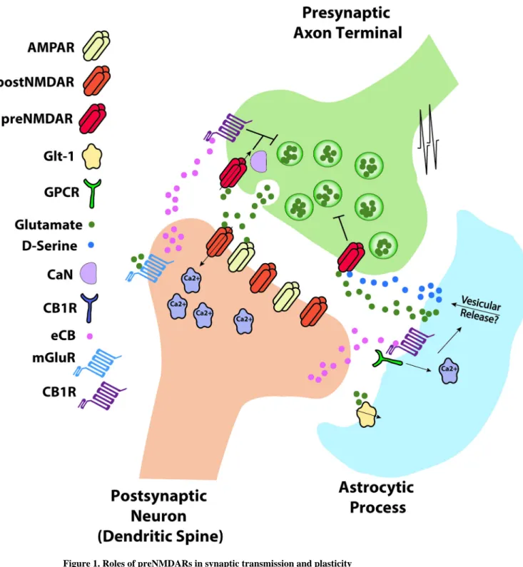
Figure 1. Roles of preNMDARs in synaptic transmission and plasticity
Action potential (AP) arrival at the presynaptic bouton triggers presynaptic release of glutamate that binds to postsynaptic AMPA receptors (AMPAR), NMDA receptors (postNMDARs) and metabotropic glutamate receptors (mGluRs), as well as presynaptic NMDA receptors (preNMDARs). During timing-dependent LTD induction, presynaptically released glutamate activates mGluRs and postsynaptic action potentials enhance Ca2+ influx through voltage-gated calcium channels (VGCCs), leading to synthesis of endocannabinoids (eCBs) which diffuse in a retrograde manner and bind to presynaptic and/or astrocytic CB1
Author Manuscript
Author Manuscript
Author Manuscript
receptors. Co-activation of presynaptic CB1 receptors and presynaptic NMDA receptors causes synaptic depression. Alternatively, activation of astrocytic CB1Rs results in
astrocytic release of glutamate or D-serine, which activates preNMDARs (see [54]), although
in some instances CB1 receptor activation might not be required. Ca2+ influx through preNMDAR activates presynaptic calcineurin (CaN), which may play a role in regulating signalling pathways involved in synaptic depression. PreNMDARs can also be activated by glutamate released from the presynaptic terminal causing synaptic self-depression.
Glutamate transporters (Glt-1) in astrocytes regulate basal levels of glutamate and may therefore influence tonic preNMDAR activation.
Author Manuscript
Author Manuscript
Author Manuscript
Figure 2. Evidence for the axonal expression of preNMDARs
(a) PreNMDARs are localized to presynaptic boutons as demonstrated by immunogold labelling of the obligatory NMDAR subunit GluN1 in L2/3 of the visual cortex (adapted from [42], scale bar indicates 200 nm). (b) In cultured cortical neurons, the NMDAR subunit GluN1 (NR1) colocalizes with the axonal marker tau-1 (adapted from [67]). (c) In
recordings from GABAergic terminals of cultured cerebellar neurons, application of NMDA elicits inward currents that are blocked by the NMDAR antagonist CPP, suggesting these terminals express preNMDARs (adapted from [34]). (d) Pairing action potential firing at 30
Author Manuscript
Author Manuscript
Author Manuscript
Hz with the uncaging of MNI-NMDA near presynaptic boutons produces supralinear calcium responses in a subset of boutons of L5 visual cortical pyramidal neurons (adapted from [23]). This result suggests that a subset of presynaptic boutons express preNMDARs that influence axonal calcium levels and presumably neurotransmitter release. (e) t-LTD at L4-L2/3 synapses requires axonal preNMDARs. Uncaging of the caged NMDAR antagonist cMK-801 over presynaptic axons (but not presynaptic soma or dendrites) prior to the t-LTD induction protocol blocks this form of plasticity (adapted from [78]).
Author Manuscript
Author Manuscript
Author Manuscript
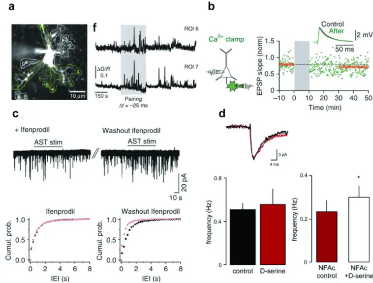
Figure 3. Astrocytic involvement in preNMDAR signalling
(a) During t-LTD induction (pairing), astrocytic calcium transients increase. (b) Blocking this increase in astrocytic calcium signalling by “clamping” astrocyte (green cell) calcium levels during the induction protocol blocks t-LTD induction at neighboring L4-L2/3 cortical synapses (a-b adapted from [54]). (c) In the absence of the GluN2B-selective antagonist ifenprodil, electrical stimulation of astrocytes (AST) increases spontaneous glutamate release onto hippocampal dentate granule cells (washout, right panel). Ifenprodil blocks this increase, suggesting astrocytes influence excitatory neurotransmission at these synapses through interaction with GluN2B subunit-containing preNMDARs (adapted from [68]). (d) In recordings from L5 neurons in the entorhinal cortex, application of the NMDAR co-agonist D-serine does not increase the mEPSC frequency, suggesting preNMDARs in this
brain area are saturated with this co-agonist. However, pre-incubation with the glia-specific metabolic inhibitor NFAc reduces baseline mEPSC frequency and allows the application of
D-serine to increase mEPSC frequency. Indirectly, this suggests that glial release of the
endogenous NMDAR co-agonist D-serine results in the saturation of the preNMDAR
co-agonist binding site at L5 entorhinal cortical synapses (adapted from [86]).