IDENTIFICATION OF HOST AND BACTERIAL FACTORS THAT CONTRIBUTE TO LUNG LESION STRUCTURE DURING PNEUMONIC PLAGUE
Nikolas M. Stasulli
A dissertation submitted to the faculty at the University of North Carolina at Chapel Hill in partial fulfillment of the requirements for the degree of Doctor of Philosophy in
the Department of Microbiology and Immunology in the School of Medicine.
Chapel Hill 2015
Approved by:
William E. Goldman
Peggy A. Cotter
Virginia L. Miller
Thomas H. Kawula
ABSTRACT
Nikolas M. Stasulli: Identification of Host and Bacterial Factors that Contribute to Lung Lesion Structure During Pneumonic Plague
(Under the direction of William E. Goldman)
Yersiniapestis, the causative agent of plague, is a high-priority pathogen that
continues to cause outbreaks worldwide. The ability of Y. pestis to be transmitted via
respiratory droplets and its history of weaponization has led to its classification as a
Tier 1 Select Agent most likely to be used as a biological weapon. The most deadly
form of disease caused by Y. pestis, pneumonic plague, results from the deposition
of bacteria into the lungs and has mortality rates approaching 100% in the absence
of treatment within 24 hours of the onset of symptoms. The Goldman lab has
previously characterized pneumonic plague progression as biphasic, presenting with
two distinct disease phases. Rapid bacterial growth during an initial pre-inflammatory
phase transitions into the second pro-inflammatory phase where disease symptoms
present and lead to death of the host. Using in vivo analyses and focusing on
relevant cell types during pneumonic plague infection host pathways can be
identified that may be manipulated to extend the 24 hour window for treatment of
pneumonic plague.
During pneumonic plague, the bacterium Yersinia pestis elicits the
eventually consolidating entire lobes of the lung during infection. This lesion
development and persistence is poorly understood. In this dissertation I examine
spatially distinct regions of lung lesions using laser capture microdissection and
RNAseq to identify transcriptional differences between lesion microenvironments. I
provide evidence that cellular pathways involved in leukocyte migration and
apoptosis are down-regulated in the center of lung lesions compared to the
periphery. Probing for the bacterial factor(s) important for the alteration in neutrophil
survival, I provide evidence that Y. pestis increases neutrophil survival through a
mechanism that is dependent on the type III secretion system effector YopM.
Additionally, I investigate the roles of reactive oxygen and nitrogen species that are
typically used as neutrophil defense mechanisms, and provide evidence that these
molecules are important for controlling early establishment of Y. pestis in the lungs.
This research explores the complexity of spatially distinct host-microbe interactions
in vivo and emphasizes the importance of cell-relevant assays in understanding Y.
I dedicate this dissertation to Dr. Rebecca Roberts at Ursinus College who gave me my first opportunity in academic research and inspired me through both her teaching
ACKNOWLEDGEMENTS
I want to thank Dr. Bill Goldman for his mentorship throughout my
graduate career. He has taught me how to be thorough and take calculated risks as
a researcher. His mentorship has gone well past the bench and he has shown me
that balancing work and life makes for a fulfilling career. I want to thank him for
encouraging my scientific creativity and supporting my research ideas even when
those ideas seemed crazy… like proposing to dissect out regions of lung lesions 200
microns apart and convincing him there would be transcriptional differences between
the regions.
I want to thank my dissertation committee, Drs. Peggy Cotter, Virginia Miller,
Tom Kawula, and Tony Richardson, for their time and scientific input during my
committee meetings that helped shape the direction of this work. I also want to thank
the Miller laboratory, especially Dr. Eric Weening. His leadership in the BSL 3
laboratory has been invaluable to keeping everything running smoothly. I want to
thank Dr. Joel Parker for his assistance in analyzing the RNAseq datasets and Dr.
Stephanie Montgomery for interpreting lung pathology of mice. I want to thank Bob
Bagnell, Alicia Brandt, and Nicole Maponga for helping me learn new techniques and
providing me with samples for my research. Dixie Flannery also deserves all my
students would be lost without her and I am very grateful for all her work that kept
me on track throughout my graduate career.
My graduate school experience would not have been the same without the
members of the Goldman lab. Their sometimes abrasive and sophomoric antics
have brought a lot of humor to many discouraging days when experiments just would
not work. I especially want to thank Dr. Roger Pechous for mentoring me throughout
my time in the lab and training me to work in the unique BSL 3 setting that Y. pestis
warrants… also of all of the debauchery and “deep thoughts” that kept me focused. I
want to thank Kara Eichelberger for being a superb mentee as I finish up my time in
the lab. It has been great to share all the secrets of Yersinia that I have acquired
over the years and I wish her the best of luck in graduate school. I also want to thank
Vicky Sepulveda for keeping me grounded and giving me sage advice over the
years… and for putting up with all the Colombian jokes and not actually ever setting
me on fire.
My friends and family deserve a special level of thanks for their support over
the years. I want to especially thank my parents, Mike and Karen, and my brother,
Tim, for all their love and support as I’ve been on this journey. Thank you Mom and
Dad for always being there with encouraging words while sharing in my excitements
and my disappointments. Thank you for always reminding me where I come from
and for making so many sacrifices for me to become the man I am today. I can never
thank you enough for all the experiences you afforded me over the years. I hope that
love and support she has provided as we shared this graduate school experience
together. You are one of the first people I met here at UNC and I am glad every day
for having you in my life. Thank you for listening to my science complaints, for
cooking delicious meals, for putting up with my quirks, and for constantly helping me
grow and become a better person. I can’t wait to continue spending our lives
together after this chapter comes to a close.
TABLE OF CONTENTS
LIST OF TABLES ... xi
LIST OF FIGURES ... xii
LIST OF ABBREVIATIONS ... xiii
CHAPTER 1: INTRODUCTION ... 1
1.1 Overview ... 1
1.2 Manifestations of plague ... 2
1.3 History and epidemiology of plague ... 5
1.4 Yersinia pestis as a biological weapon ... 10
1.5 Pulmonary immune defense and neutrophils ... 12
1.6 Pathogenesis and innate immune response during pneumonic plague ... 15
1.7 Significance ... 17
REFERENCES ... 19
CHAPTER 2: CHARACTERIZATION OF THE LUNG LESION MICROENVIRONTMENT BY LASER CAPTURE MICRODISSECTION AND RNAseq ANALYSIS ... 25
2.1 Overview ... 25
2.2 Introduction ... 27
2.3 Methods ... 28
2.4 Results ... 33
2.5 Discussion ... 37
REFERENCES ... 54
CHAPTER 3: INDUCTION OF NETUROPHIL SURVIVAL BY Yersinia pestis THROUGH THE TYPE III SECRETION SYSTEM EFFECTOR YopM ... 59
3.1 Overview ... 59
3.2 Introduction ... 60
3.3 Methods ... 63
3.4 Results ... 67
3.5 Discussion ... 72
3.6 Figures ... 76
REFERENCES ... 83
CHAPTER 4: CHARACTERIZING THE IMPACT OF REACTIVE OXYGEN/NITROGEN SPECIES IN VIVO DURING PNEUMONIC PLAGUE ... 87
4.1 Overview ... 87
4.2 Introduction ... 88
4.3 Methods ... 91
4.4 Results ... 93
4.5 Discussion ... 96
4.6 Figures ... 101
REFERENCES ... 104
CHAPTER 5: DISCUSSION AND FUTURE EXPERIMENTS ... 108
5.1 Summary of results ... 108
5.2 Filling gaps in plague research ... 111
5.3 Implications for the study of plague and pathogen-associated tissue damage ... 114
5.4 Future Experiments ... 117
LIST OF TABLES
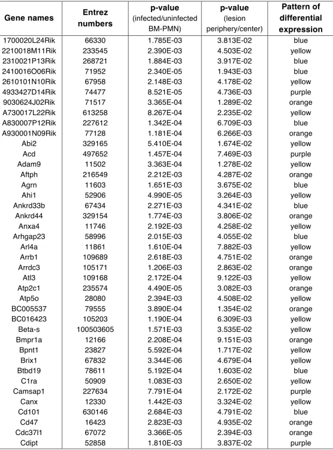
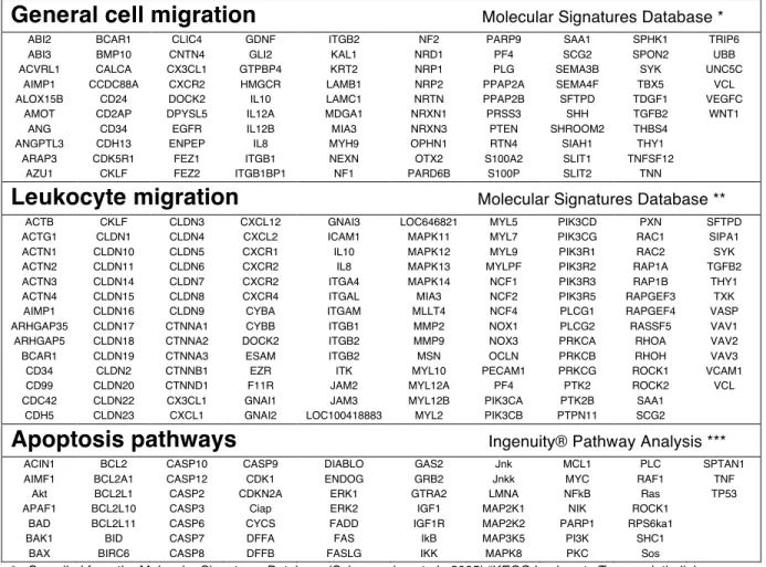
Table 2.1: List of 224 differentially regulated in both the infected versus
uninfected BM-PMN and lesion periphery versus center comparisons ... 48
LIST OF FIGURES
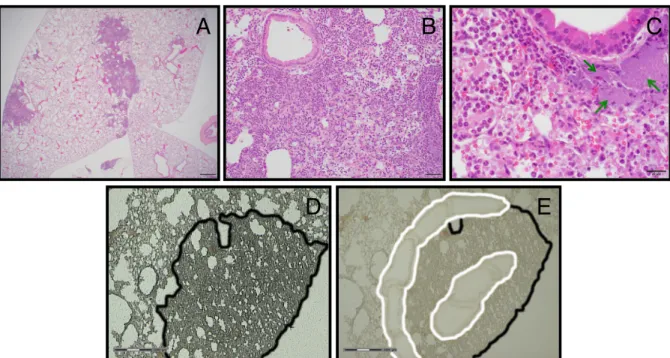
Figure 2.1: Lung lesion histology and laser capture microdissection of
lesions ... 42
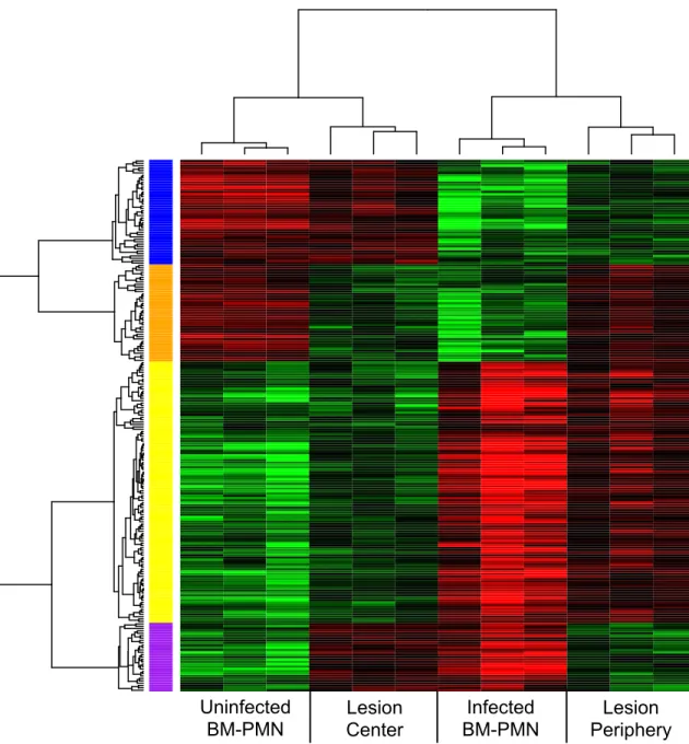
Figure 2.2: Clustered genes showing differential transcription ... 43
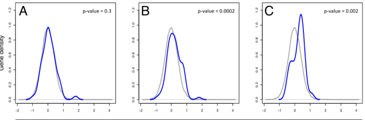
Figure 2.3: Density curves of defined gene sets compared to the entire
transcriptome ... 45
Figure 2.4: Ingenuity® Pathway Analysis “Apoptosis Signaling” pathway
overlaid with relevant genes from density curve analysis ... 46
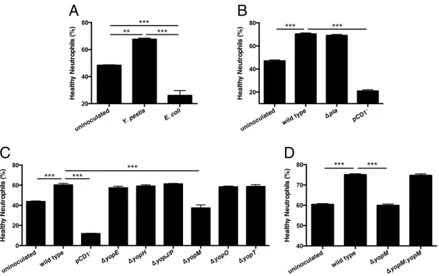
Figure 3.1: The type III secretion system effector YopM is necessary for
enhanced neutrophil survival ... 76
Figure 3.2: Inhibiting known functions of YopM does not alter neutrophil
survival ... 78
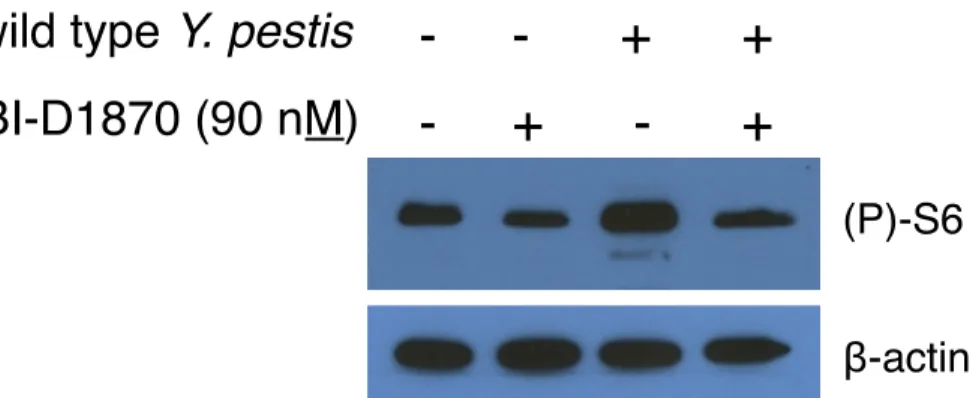
Figure 3.3: BI-D1870 continues to inhibit RSK activity after 24 hours in
isolated human neutrophils ... 79
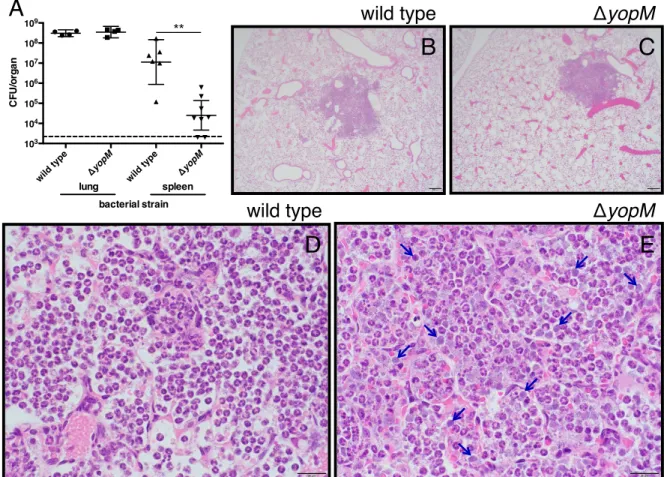
Figure 3.4: YopM-dependent effects on bacterial burden and histopathology
during pulmonary infection with Y. pestis ... 80
Figure 3.5: TUNEL staining of infected mouse lung sections ... 82
Figure 4.1: Comparative histopathology of wild type, iNOS-/-, and
gp91phox-/- mice during intranasal Y. pestis infection ... 101
Figure 4.2: Bacterial burden of both the lung and spleen of wild type, iNOS-/-, and gp91phox-/- mice during intranasal Y. pestis infection ... 102
Figure 4.3: Survival graph of wild type, iNOS-/-, and gp91phox-/- mice during
LIST OF ABBREVIATIONS
AEC – airway epithelial cell
BHI – brain/heart infusion
BM-PMN – bone marrow isolated neutrophil
BSL – biosafety level
BWC – Biological and Toxin Weapons Convention
CFU – colony-forming units
CO92 – Colorado 92 – fully virulent strain of Y. pestis
DEPC – diethylpyrocarbonate
DNA – deoxyribonucleic acid
FBS – fetal bovine serum
FFPE – formalin fixed paraffin embeded
H2O2 – hydrogen peroxide
HOCl – hypochlorous acid
IRB – Institutional Review Board
LCM – laser capture microdissection
MPO – myeloperoxidase
NADPH – nicotinamide adenine dinucleotide phosphate
NCBI – National Center for Biotechnology Information
NO – nitric oxide
NOS (e, i, n) – nitric oxide synthase (epithelial, inducible, neuronal)
O2- – superoxide ion
O.C.T. – optimal cutting temperature
ONOO- – peroxynitrite
PBS – phosphate buffered saline
pgm- – a deletion of the pigmentation locus of Yersinia pestis
PHS – Public Health Service
RNA – ribonucleic acid
RNAseq – RNA deep sequencing
RNS – reactive nitrogen species
ROS – reactive oxygen species
RSK – ribosomal S6 kinase
RSNO – S-nitrosothiol
T3SS – type III secretion system
USDA – United States Department of Agriculture
WHO – World Health Organization
CHAPTER 1: INTRODUCTION
1.1 Overview
The causative agent of the disease known as “plague” was identified
independently in August of 1894 by both Alexandre Yersin and Shibasaburo
Kitasato. Yersin, however, is primarily credited with the official identification because
of his more accurate and descriptive paper describing the gram-negative
coccobacillus, which he named Bacterium pestis. He continued his research and by
1900 had used antiserum to cure a plague patient, and ultimately made the link
between rats and plague outbreaks (Hirst, 1953; Zietz and Dunkelberg, 2004). The
name of the bacterium identified by Yersin has changed several times since its
discovery: first in 1900 to Bacillus pestis, then in 1923 to Pasteurella pestis (after
Louis Pasteur, Yersin’s mentor), then finally in 1970 to Yersinia pestis (after its
identifier) (Zietz and Dunkelberg, 2004).
Y.pestis is a high-priority pathogen that poses a severe threat to both human
and animal health, and continues to cause modern day plague outbreaks worldwide
(Inglesby et al., 2000; Stenseth et al., 2008). The ability of Y. pestis to be transmitted
via respiratory droplets and its past weaponization has led to its classification as a
Tier-1 select agent that must be handled under strict biosafety level 3 (BSL3)
also of concern since the identification of the first antibiotic resistant strains of Y.
pestis in 1995 (Galimand et al., 2006).
The most deadly form of plague disease known as primary pneumonic plague
results from the inhalation of Y. pestis. The Goldman lab has previously
characterized pneumonic plague in a murine model as a biphasic disease with an
initial anti-inflammatory phase followed by rapid activation of the innate immune
system (Lathem et al., 2005). If antibiotic treatment is not administered within 24
hours after the onset of symptoms, the disease approaches 100% mortality
(Inglesby et al., 2000). While not the most common form of plague disease,
pneumonic plague is of the highest concern when considering transmission and
disease treatment. The rapid bacterial growth in the lung during the initial
anti-inflammatory phase primes the patient to be infectious to others once disease
symptoms emerge. Additionally, the rapid disease progression can lead to death
before Y. pestis infection can be diagnosed and confirmed.
1.2 Manifestations of plague
The identification of small mammals as the reservoir of endemic plague
infections was established in 1927. The natural transmission cycle of Y. pestis
between small mammals and fleas was termed sylvatic plague (Zietz and
Dunkelberg, 2004). As humans unintentionally enter this sylvatic cycle (or as Y.
pestis is intentionally dispersed through nefarious means) there are three
manifestations of disease that can result: bubonic, pneumonic, and septicemic
1.2.1 Bubonic plague
Bubonic plague is the most common disease caused by Y. pestis. Bubonic
plague can be caused by contamination of open wounds or direct inoculation
through the bite of an infected flea (Riedel, 2005). After establishing in the dermis, Y.
pestis infiltrates the cutaneous lymphatics and migrates to the nearest draining
lymph node (Gonzalez et al., 2015; Riedel, 2005). After an incubation period of 2-6
days after inoculation, there is a sudden onset of flu-like symptoms followed by a
swelling of the draining lymph nodes that forms the namesake “buboes” (Riedel,
2005). In the pre-antibiotic era the mortality rate of bubonic plague could reach as
high as 70% during epidemics (Stenseth et al., 2008). Today, with early detection
and treatment, mortality has dropped to as low as 5% (Inglesby et al., 2000). Even if
a patient recovers form bubonic plague, buboes may remain for several weeks after
symptoms have dissipated (Riedel, 2005).
1.2.2 Pneumonic plague
When Y. pestis colonizes the lower respiratory system either secondary or
primary pneumonic plague will result. Secondary pneumonic plague occurs after
bubonic or septicemic plague disseminates into the lung compartment, allowing it to
be spread by aerosol from person-to-person. Primary pneumonic plague occurs after
inhalation of Y. pestis directly into the lungs. This can occur by person-to-person
during the intentional release of an aerosol as a biological weapon (Inglesby et al.,
2000). It is believed that person-to-person transmission via respiratory droplets
occurs within approximately one meter of an infected individual (Kool, 2005). Further
discussion of pneumonic plague epidemiology throughout modern times can be
found in Section 1.3.3 of Chapter 1.
There is a short quiescent time of 1-3 days after inhalation of bacteria before
flu-like symptoms emerge. Unfortunately, pneumonic plague must be treated within
24 hours after symptoms begin or mortality approaches 100% only three to four days
after inhalation (Inglesby et al., 2000). Disease progression and symptoms are
similar in the murine model of infection. Based on our animal model, the progression
of pneumonic plague is biphasic: an initial anti-inflammatory phase precedes a
pro-inflammatory phase of disease. In the first phase, bacteria are present in the lungs
and freely replicate with little to no detectable immune responses. Disease
symptoms, increased cytokine levels, and the infiltration of neutrophils into the
airways signal entry into the pro-inflammatory phase of infection. Pneumonic plague
can be characterized by the formation of lung lesions followed by destruction of
surrounding alveolar architecture. Condensed pockets of Y. pestis surrounded by
innate immune cells, primarily neutrophils, form these pulmonary lesions (Lathem et
al., 2005). Death from pneumonic plague can be attributed to pneumonia due to
alveolar destruction and the subsequent septicemia late in disease resulting from
1.2.3 Septicemic plague
If Y. pestis manages to infiltrate the bloodstream of the host the disease will
manifest as septicemic plague. Primary septicemic plague from direct inoculation of
Y. pestis into the blood stream is fairly rare. However, secondary septicemic plague
often results during the late stages of bubonic and pneumonic plague. Once bacteria
have disseminated from initially localized sites, septicemia can induce shock, blood
clots leading to gangrenous extremities, multiple organ failure, and respiratory
distress (Riedel, 2005).
1.3 History and epidemiology of plague
The disease caused by Yersinia pestis known as “plague” has been
described for thousands of years. It is generally agreed upon by scholars that the
oldest written mention of plague is recounted in the Bible (1 Samuel 5&6) referring to
a time between 1320 – 1000 BC (Griffin, 2000; Ligon, 2006). This bacterium has
also been responsible for 3 major pandemics throughout recorded history that were
so devastating to the population that it completely altered the makeup of society.
1.3.1 The Justinian pandemic
The first major plague pandemic is now referred to as the “plague of
Justinian”, after the presiding emperor of Rome. The pandemic may have started as
early as 532 BC, but definitively took hold in 541/42 BC with outbreaks in
that ~40% of Constantinople’s population died during this outbreak. After disease
spread through trade routes, up to 100 million Europeans are believed to have
succumbed to plague over the next 250 – 300 years as pockets of disease
continually reemerged. This devastating death toll depleted trade occupations, the
ability to form armies, and the ability to staff many of the religious monasteries
throughout Europe and Western Asia (Ligon, 2006).
1.3.2 The Black Death
The second and most well known plague pandemic is commonly referred to as “The
Black Death”. The initial outbreak is thought to have started in China in the 1330’s.
The disease then spread west through trade routes and began establishing itself in
Europe. From 1346 – 1352 the spread of Y. pestis resulted in the death of over 25
million people, over one-third of the world’s population at the time. Another 20 million
were believed to have died by the end of the century. This pandemic lasted through
the 1720’s, decimating large cities with resurgences throughout Europe (Ligon,
2006; Zietz and Dunkelberg, 2004).
With over one-third of the world’s population deceased within 6 years, this
pandemic had wide rippling effects throughout society. Commerce, trade, and
government came to a near standstill, and the economy of Europe collapsed from
the large and sudden loss of life. The population and economy only began to
rebound in the late 1600’s. Interestingly, in Western Europe, the sudden decrease in
valuable, laborers. This competition is believed to have started an early form of
capitalism and led to the rise of a “middle class” that was not present prior to the
pandemic. Culture throughout Europe also began to morph and took on a macabre
tone, with the influence of death permeating artwork, literature, and music. Many
people at the time became more religious, as evidenced by the canonization of new
“plague saints” by the Catholic Church. Unfortunately, this new-found focus on
religion also caused societal rifts as Christians, Muslims, and Jews all blamed each
other for incurring “God’s wrath” and causing the horrible disease (Ligon, 2006; Zietz
and Dunkelberg, 2004).
1.3.3 The Modern pandemic: global spread, and tracking of pneumonic plague
The “modern pandemic” of plague is believe to have started in China around
1855 in the Yunnan province, and marked a critical point in the dissemination of
plague into a global issue. By 1894 plague had reached Hong Kong and, with the
growth of the trans-oceanic shipping industry, lead to global spread of Y. pestis
(Caten and Kartman, 1968; Ligon, 2006; Zietz and Dunkelberg, 2004). Stowaway
rats on shipping vessels would transport Y. pestis, and the flea vectors that transmit
it, across oceans. The first reported incidence of plague in the Western Hemisphere
came in Santos, Brazil in October of 1899 (Furman and Williams, 1973), and marked
the beginning of three decades of large epidemics throughout the world. Between
(Ligon, 2006). Even Australia, which has not had a case of plague since 1925, was
struck with over 1,300 cases of plague in the first part of the 1900’s (Garrett, 1991).
In the United States, this first quarter of the 20th century also saw several
epidemics of plague in California. The first epidemic occurred from 1900-1904 in a
Chinese immigrant-populated district of San Francisco. This was followed by a
second outbreak in San Francisco in 1907. These two outbreaks included both
bubonic and pneumonic plague cases, infecting over 280 people. A smaller epidemic
occurred in 1919 in Oakland, CA, where a man developed secondary pneumonic
plague and became the source of 13 primary pneumonic plague cases, 12 of whom
died (Anderson, 1978; Caten and Kartman, 1968; Kool, 2005). This was also
documented as the first instance of a primary pneumonic plague epidemic in the
Western Hemisphere (Kellogg, 1920). The fourth US outbreak occurred in Los
Angeles in late 1924, and was also caused by pneumonic plague, resulting in 39
documented cases with 33 deaths (Anderson, 1978; Kool, 2005). A fifth outbreak
occurred around the Gulf Coast of the United States in 1924, causing over 70 cases
of plague (Anderson, 1978).
While there was a spike of plague cases in the United States between
1900-1908 and again between 1918-1926, after the mid-1920’s there were relatively few
yearly cases of plague. However, unlike Australia that was seemingly able to
eradicate plague disease after 1925 (Garrett, 1991), endemic sylvatic plague
established itself in small-mammal populations in the U.S. west of the Mississippi
information from the Centers for Disease Control and Prevention and other
published sources, beginning in the mid-1960’s through the 1970’s and into the early
1980’s, there was a resurgence of plague cases in the U.S. (Anderson, 1978). These
cases were located throughout the Southwestern U.S., mainly New Mexico, Arizona,
and California (Anderson, 1978; Kaufmann et al., 1980). Between 1900 and 2010,
there were over 1,400 reported cases of plague in the contiguous U.S. and Hawaii
(Kaufmann et al., 1980). Of these U.S. cases, as many as 20% have been due to
pneumonic plague.
In addition to the two pneumonic plague outbreaks in California in the early
1900’s, there have been several global instances of pneumonic plague outbreaks
during the “modern plague” era. The largest of these epidemics took place in the
Manchuria region in China. This region had large pneumonic plague epidemics in
1910-1911 and again in 1920-1921 that left ~60,000 and ~9,300 people dead,
respectively (Wu et al., 1936). Interestingly, the larger of the two epidemics in
1910-1911 was caused by a dramatic increase in the price of marmot fur. Thousands of
poor laborers flocked to this region of China to hunt marmots, whose population was
unknowingly endemically infected with Y. pestis. The butchering and eating of these
animals by hunters unwittingly aerosolized the bacteria and caused a major
epidemic (Summers, 2012). China continues to have relatively large numbers of
plague cases yearly. Between 1994-2003 there were 11 countries that reported
more than 100 confirmed cases of plague (China being number 11). The country
3% of cases being pneumonic plague. For this 10 year time period, that equates to
over 350 cases of pneumonic plague (Butler, 2009). In the first full decade of the 21st
century, however, the WHO reported that the Congo became the number one
reporting country with over 10,500 cases of plague. This increase in cases also
coincided with a large pneumonic plague outbreak in 2005-2006 in the Oriental
Province that left over 130 people dead from pneumonic plague alone (Butler, 2013).
There should continue to be a heightened awareness of outbreaks of pneumonic
plague to ensure that larger epidemics do not continue to occur, especially with the
identification of naturally antibiotic-resistant Y. pestis strains (Galimand et al., 2006).
1.4 Yersinia pestis as a biological weapon
The first known use of Y. pestis as a biological weapon dates back to 1346
during the war between the Tartar army and Genoese sailors at the port city of
Caffa. The Tartars were losing many of their soldiers to plague, and out of
desperation placed the corpses of people who had died of plague onto catapults and
flung them over the city walls to infect enemy troops. Similar corpse-catapulting
strategies were documented in 1422 by the Lithuanians and in 1710 by the Russians
(Ligon, 2006; Riedel, 2004).
During World War II, the Japanese army established a research program
(Unit 731) that secretly studied biological-warfare agents. This unit extensively
studied Yersinia pestis, and infected fleas were released over the Chinese
population several times in the early 1940’s. “Bombs” were built and tested that were
It is believed that tens-of-thousands of people died from plague epidemics
throughout China during the 1940’s due to Unit 731’s biological-weapon
experiments. Unit 731’s field tests of Y. pestis on the Chinese people were ceased
after Japanese troops died of plague due to the unpredictability of the fleas and
spread of disease (Ligon, 2006; Riedel, 2004; Williams and Wallace, 1989).
After WWII, biological weapons programs were slowly dismantled, starting
with the United Kingdom shutting down its program in 1950. The United States
followed suit in 1969, but only after developing at least seven type-classified
microorganisms into biological weapons. The shut down of the US biological
weapons program preceded the drafting of the Biological and Toxin Weapons
Convention (BWC) in 1972. The BWC called for multilateral disarmament of all
biological weapons. The Soviet Union, however, continued with their biological
weapons program until 1992 despite signing the BWC (Ligon, 2006; Riedel, 2004).
In 1970 just prior to the drafting of the BWC, the WHO convened a group of
experts to determine the casualties that would be expected from an aerosolized
biological attack from several select agents. The WHO committee determined that,
under their defined parameters, the release of 50 kg of aerosolized Y. pestis over an
economically developed city of 5,000,000 people would result in the incapacitation of
150,000 and death of 36,000 people, assuming prompt action is taken following the
attack. Out of all biological weapon agents surveyed by this committee, only B.
anthracis would cause more casualties, most likely due to its ability to travel farther
can be spread from person to person, and any extended delay in initiating treatment
could cause casualty numbers to rise. A sylvatic reservoir of Y. pestis could also be
established if small mammals become infected during an attack. Along with the
presence of susceptible flea populations, this new rodent reservoir could lead to
more long-term effects of recurring bubonic plague due to the establishment of a
sylvatic cycle. This assessment of biological and chemical weapons has been
updated as recently as 2004 (World Health Organization, 2004). Despite the unlikely
use of Y. pestis as a biological weapon there has also been an assessment of
antibiotic treatment courses recommended in the case of both contained or mass
casualty outbreaks (Inglesby et al., 2000).
Modeling of well-documented pneumonic plague outbreaks in the 20th century
has found that R0=~1.3 (the ability of an infection to sustain itself in a population). An
R0<1 indicates that an infection cannot sustain it self in a population. An R0>1
indicates that pneumonic plague has the potential to sustain itself and spread in a
population if action is not taken to control the infection. However, an R0 value so
close to 1 signifies the need for close proximity to infect and a small window of
transmission before symptoms begin. The disease could be stopped through
isolation of infected individuals and antibiotic treatment for those who have come into
contact with the infected (Gani and Leach, 2004).
1.5 Pulmonary immune defense and neutrophils
The respiratory tract is continuously in contact with a diverse repertoire of
lungs must maintain an immunological homeostasis, balancing between potentially
harmful and non-harmful antigens. Recognition of antigens begins in the mucosal
tissue of the conducting airways. The airway epithelial cells (AECs) are the first line
of defense. The AECs act as a physical barrier, but also secret proteins and
peptides into the lung mucosa to defend against invading pathogens (Holt et al.,
2008). Secreted proteins and molecules such as lysozyme, peroxidase, lactoferrin,
defensins, collections, ficolins, and complement factors can all directly act on
invading microbes to promote killing and help ensure lung homeostasis (Tsai and
Grayson, 2008). From the conducting airways, the bronchi of the lungs continue to
branch and form the parenchymal lung, consisting of bronchioles and alveolar ducts,
and terminates in alveolar sacs that are necessary for oxygen exchange (Holt et al.,
2008). Besides the AECs there are various resident dendritic cells and macrophages
that patrol and sample the mucosa and alveolar spaces to ensure proper
homeostasis and recruit additional immune cells if necessary (Guilliams et al., 2013;
Holt et al., 2008).
In addition to the resident immune cells, neutrophils are an important and
necessary component of the innate immune system. Neutrophils are short-lived cells
that are constantly being generated in the bone marrow, circulating in the blood
stream, and then returning to the bone marrow for cellular turnover. During their
circulation, neutrophils also monitor the lung environment for invading pathogens.
During a bacterial infection neutrophils are rapidly called to the site of insult and can
pathogens (Craig et al., 2009; Segal, 2005). This rapid antimicrobial response is
facilitated by neutrophil priming that results from signals initiated by the innate
immune system upon detection of a pathogen. This priming retains neutrophils in the
lungs as they “survey” during normal circulation (Singh et al., 2012; Summers et al.,
2010). Upon direct interaction with a secondary stimulus, such as invading microbes,
there is enhancement of the respiratory burst, cytoskeletal rearrangements that
retain neutrophils in the lung and facilitate phagocytosis, regulation of neutrophil
surface antigens (Singh et al., 2012), and release of antimicrobial peptides/proteins
from granules (Kobayashi et al., 2003). After mounting an antimicrobial response,
neutrophils ordinarily undergo necrosis, apoptosis, or NETosis, all of which ultimately
lead to macrophage infiltration. The newly infiltrated macrophages facilitate the
clearance of dead or dying neutrophils as well as any remaining bacteria (Brinkmann
et al., 2004; Kebir and Filep, 2013; Luo and Loison, 2008; Silva, 2011). This double
priming, however, can lead to acute lung injury if the neutrophils are not efficiently
released from the pulmonary compartment back into circulation, or appropriate
neutrophil turnover and removal does not occur (Singh et al., 2012; Summers et al.,
2010). There is also data to suggest that there is cross talk between neutrophils and
various other cell types at sites of inflammation that can promote a continued innate
immune response and even bridge the gap between innate and adaptive immunity
1.6 Pathogenesis and innate immune response during pneumonic plague
Pathogenic microbes have evolved ways to evade the immune system of the
hosts that they infect. The work presented in this dissertation is focused on the
ability of bacteria to manipulate activated neutrophils (Urban et al., 2006), resulting in
increased disease severity and mortality (Balamayooran et al., 2010). Pathogens
have been shown to utilize various mechanisms to alter neutrophil function including
inhibiting neutrophil chemotaxis (Bignold et al., 1991; de Haas et al., 2004; Van Dyke
et al., 1982), preventing phagocytosis (Grosdent et al., 2002; Spinner et al., 2013;
2008; Visser et al., 1995), altering degranulation (Arnett et al., 2014; Bertram et al.,
1986), and, importantly for this work, inhibiting neutrophil apoptosis (Choi et al.,
2005; Erttmann et al., 2014; Ge and Rikihisa, 2006; Schwartz et al., 2013; 2012).
Despite the recruitment of neutrophils to control Y. pestis in the lungs,
bacterial numbers continue to increase throughout the duration of infection (Lathem
et al., 2005). The increase in bacterial burden coincides with continued neutrophil
recruitment, accumulation in the lungs, and subsequent targeting of neutrophils by
the Y. pestis type III secretion system (T3SS) (Marketon et al., 2005; Pechous et al.,
2013). The massive neutrophilic infiltration during pneumonic plague results in the
formation of histopathologically distinct lung lesions that arise during the later
(pro-inflammatory) phase of disease. These lesions expand in the lungs as neutrophils
continue to accumulate until death of the host, with no evidence of bacterial or
neutrophil clearance (Lathem et al., 2005). The continued influx of neutrophils and
also been observed in neutrophils of cystic fibrosis patients (Tirouvanziam et al.,
2008). The continuous neutrophil influx during pneumonic plague results in alveolar
destruction within lung lesions and culminates in a severe and deadly necrotizing
pneumonia (Finegold et al., 1968).
Yersinia pestis is known to inhibit the host immune response via various
virulence mechanisms (Ben-Gurion and Shafferman, 1981; Du et al., 2002; Jackson
and Burrows, 1956; Lathem et al., 2007; Trosky et al., 2008). However, It has just
recently been appreciated that the functions of specific T3SS effectors may vary
depending on the cell type being targeted (Spinner et al., 2010). Neutrophils are the
most prominent immune infiltrate during pneumonic plague, are selectively targeted
by Y. pestis, and are necessary for development of lung lesions (Pechous et al.,
2013). Early recruitment of neutrophils is also linked to decreased bacterial burden
and increased survival of Y. pestis infected mice (Vagima et al., 2015).
In the Yersinia pestis field, pneumonic plague is fairly understudied as a
separate disease from bubonic plague despite the dissimilar disease progression.
Most research is done using bubonic plague models or in in vitro assays. These
results are usually inferred, but not directly tested, to be relevant during pneumonic
plague. Relatively few laboratories use fully virulent Y. pestis to specifically study the
effects of virulence factors on pneumonic plague (Cantwell et al., 2010; Lathem et
al., 2007; van Lier et al., 2014). Most other groups working on pneumonic plague
use attenuated strains missing either the pigmentation locus or the pCD1 plasmid
dissertation and other studies from the Goldman lab continue to show the necessity
of using fully virulent Y. pestis and in vivo infections by presenting data that could not
be elucidated in less pathogenic models (Lathem et al., 2005; Pechous et al., 2013;
2015; Price, 2011).
1.7 Significance
Endemic maintenance of Yersinia pestis in sylvatic cycles continues to cause
spikes of plague disease around the world. Despite the signing of the BWC there is
still fear of Y. pestis being used as a biological weapon during acts of terrorism. With
the vague symptoms and rapid progression of pneumonic plague, it remains
important to be vigilant and continue to characterize this disease, particularly
focusing on more effective late-stage treatments. With the development of
pneumonia being caused by the continued replication of Y. pestis and infiltration of
neutrophils into the pleural cavity, it is important to move away from in vitro studies
and to study the disease in a more relevant in vivo setting.
I hypothesized that spatially distinct transcriptional modulation of the
important neutrophil population would be observable in Y. pestis lung lesions. This
line of in vivo questioning, which could not be undertaken in vitro, specifically looked
at the most relevant cell type important during pneumonic plague disease. In this
body of work, fully virulent Y. pestis is employed along with the novel use of laser
capture microdissection (LCM) and RNAseq technology to evaluate host
transcription within the expanding neutrophil-rich lung lesions that arise during
vivo for disease progression, and to characterize both bacterial and host factors
responsible for the distinctive pneumonic plague lung lesions that form late during
infection. It is necessary to interrogate this inflammatory environment in the context
of neutrophils to understand how interactions with Y. pestis determine pneumonic
plague presentation and progression. By determining how interaction of Y. pestis
and neutrophils induce responses uniquely within sites of infection, the scientific
community will better understand the mechanisms by which neutrophils typically
work in a broad range of diseases in which neutrophil infiltration becomes harmful to
REFERENCES
Anderson, E.T. (1978). Plague in the continental United States, 1900-76. Public Health Rep 93, 297–301.
Arnett, E., Vadia, S., Nackerman, C.C., Oghumu, S., Satoskar, A.R., McLeish, K.R., Uriarte, S.M., and Seveau, S. (2014). The pore-forming toxin listeriolysin O is
degraded by neutrophil metalloproteinase-8 and fails to mediate Listeria
monocytogenes intracellular survival in neutrophils. Journal of Immunology 192,
234–244.
Balamayooran, G., Batra, S., Fessler, M.B., Happel, K.I., and Jeyaseelan, S. (2010). Mechanisms of neutrophil accumulation in the lungs against bacteria. American Journal of Respiratory Cell and Molecular Biology 43, 5–16.
Ben-Gurion, R., and Shafferman, A. (1981). Essential virulence determinants of different Yersinia species are carried on a common plasmid. Plasmid 5, 183–187.
Bertram, T.A., Canning, P.C., and Roth, J.A. (1986). Preferential inhibition of primary granule release from bovine neutrophils by a Brucella abortus extract. Infection and Immunity 52, 285–292.
Bignold, L.P., Rogers, S.D., SIAW, T.M., and Bahnisch, J. (1991). Inhibition of chemotaxis of neutrophil leukocytes to interleukin-8 by endotoxins of various bacteria. Infection and Immunity 59, 4255–4258.
Brinkmann, V., Reichard, U., Goosmann, C., Fauler, B., Uhlemann, Y., Weiss, D.S., Weinrauch, Y., and Zychlinsky, A. (2004). Neutrophil extracellular traps kill bacteria. Science 303, 1532–1535.
Butler, T. (2009). Plague into the 21st century. Clinical Infectious Diseases 49, 736– 742.
Butler, T. (2013). Plague gives surprises in the first decade of the 21st century in the United States and worldwide. American Journal of Tropical Medicine and Hygiene
89, 788–793.
Cantwell, A.M., Bubeck, S.S., and Dube, P.H. (2010). YopH inhibits early pro-inflammatory cytokine responses during plague pneumonia. BMC Immunology 11, 29.
Choi, K.-S., Park, J.T., and Dumler, J.S. (2005). Anaplasma phagocytophilum delay of neutrophil apoptosis through the p38 mitogen-activated protein kinase signal pathway. Infection and Immunity 73, 8209–8218.
Craig, A., Mai, J., Cai, S., and Jeyaseelan, S. (2009). Neutrophil recruitment to the lungs during bacterial pneumonia. Infection and Immunity 77, 568–575.
de Haas, C.J., Veldkamp, K.E., Peschel, A., Weerkamp, F., Van Wamel, W.J., Heezius, E.C., Poppelier, M.J., Van Kessel, K.P., and van Strijp, J.A. (2004).
Chemotaxis inhibitory protein of Staphylococcus aureus, a bacterial antiinflammatory agent. The Journal of Experimental Medicine 199, 687–695.
Du, Y., Rosqvist, R., and Forsberg, A. (2002). Role of fraction 1 antigen of Yersinia
pestis in inhibition of phagocytosis. Infection and Immunity 70, 1453–1460.
Erttmann, S.F., Gekara, N.O., and Fällman, M. (2014). Bacteria induce prolonged PMN survival via a phosphatidylcholine-specific phospholipase C- and protein kinase C-dependent mechanism. PLoS One 9, 0087859.
Fetherston, J.D., Kirillina, O., Bobrov, A.G., Paulley, J.T., and Perry, R.D. (2010). The yersiniabactin transport system is critical for the pathogenesis of bubonic and pneumonic plague. Infection and Immunity 78, 2045–2052.
Finegold, M.J., Petery, J.J., Berendt, R.F., and Adams, H.R. (1968). Studies on pathogenesis of plague - blood coagulation and tissue responses of Macaca mulatta
following exposure to aerosols of Pasteurella pestis. The American Journal of Pathology 53, 99.
Furman, B., and Williams, R.C. (1973). A profile of the United States Public Health Service, 1798-1948 (National Institutes of Health).
Galimand, M., Carniel, E., and Courvalin, P. (2006). Resistance of Yersinia pestis to antimicrobial agents. Antimicrobial Agents and Chemotherapy 50, 3233–3236.
Galvan, E.M., Nair, M.K.M., Chen, H., Del Piero, F., and Schifferli, D.M. (2010). Biosafety level 2 model of pneumonic plague and protection studies with F1 and Psa. Infection and Immunity 78, 3443–3453.
Gani, R., and Leach, S. (2004). Epidemiologic determinants for modeling pneumonic plague outbreaks. Emerg Infect Dis 10, 608–614.
Ge, Y., and Rikihisa, Y. (2006). Anaplasma phagocytophilum delays spontaneous human neutrophil apoptosis by modulation of multiple apoptotic pathways. Cellular Microbiology 8, 1406–1416.
Gonzalez, R.J., Lane, C.M., Wagner, N.J., Weening, E.H., and Miller, V.L. (2015). Dissemination of a highly virulent pathogen: tracking the early events that define infection. PLoS Pathogens 11.
Griffin, J.P. (2000). Bubonic plague in biblical times. Journal of the Royal Society of Medicine 93, 449.
Grosdent, N., Maridonneau-Parini, I., Sory, M.P., and Cornelis, G.R. (2002). Role of Yops and Adhesins in Resistance of Yersinia enterocolitica to Phagocytosis.
Infection and Immunity 70, 4165–4176.
Guilliams, M., Lambrecht, B.N., and Hammad, H. (2013). Division of labor between lung dendritic cells and macrophages in the defense against pulmonary infections. Mucosal Immunol 6, 464–473.
Hirst, L.F. (1953). The Conquest of Plague (Oxford : Clarendon Press).
Holt, P.G., Strickland, D.H., Wikstrom, M.E., and Jahnsen, F.L. (2008). Regulation of immunological homeostasis in the respiratory tract. Nature Reviews Immunology 8, 142–152.
Inglesby, T.V., Dennis, D.T., Henderson, D.A., Bartlett, J.G., Ascher, M.S., Eitzen, E., Fine, A.D., Friedlander, A.M., Hauer, J., Koerner, J.F., et al. (2000). Plague as a biological weapon - Medical and public health management. The Journal of the American Medical Association 283, 2281–2290.
Jackson, S., and Burrows, T.W. (1956). The Pigmentation of Pasteurella pestis on a Defined Medium Containing Haemin. British Journal of Experimental Pathology 37, 570–576.
Kaufmann, A.F., Boyce, J.M., and Martone, W.J. (1980). From the Center for Disease Control. Trends in human plague in the United States. J. Infect. Dis. 141, 522–524.
Kebir, El, D.D., and Filep, J.G.J. (2013). Modulation of Neutrophil Apoptosis and the Resolution of Inflammation through β2 Integrins. Frontiers in Immunology 4, 60.
Kellogg, W.H. (1920). An epidemic of pneumonic plague. Am J Public Health (N Y)
10, 599–605.
Kool, J.L. (2005). Risk of person-to-person transmission of pneumonic plague. Clinical Infectious Diseases 40, 1166–1172.
Lathem, W.W., Price, P.A., Miller, V.L., and Goldman, W.E. (2007). A plasminogen-activating protease specifically controls the development of primary pneumonic plague. Science 315, 509–513.
Lathem, W.W., Crosby, S.D., Miller, V.L., and Goldman, W.E. (2005). Progression of primary pneumonic plague: A mouse model of infection, pathology, and bacterial transcriptional activity. Proceedings of the National Academy of Sciences 102, 17786–17791.
Lee-Lewis, H., and Anderson, D.M. (2009). Absence of inflammation and pneumonia during infection with nonpigmented Yersinia pestis reveals a new role for the pgm locus in pathogenesis. Infection and Immunity 78, 220–230.
Ligon, B.L. (2006). Plague: a review of its history and potential as a biological weapon. Seminars in Pediatric Infectious Diseases.
Luo, H.R., and Loison, F. (2008). Constitutive neutrophil apoptosis: mechanisms and regulation. American Journal of Hematology 83, 288–295.
Mantovani, A., Cassatella, M.A., Costantini, C., and Jaillon, S. (2011). Neutrophils in the activation and regulation of innate and adaptive immunity. Nature Reviews Immunology 11, 519–531.
Marketon, M.M., DePaolo, R.W., DeBord, K.L., Jabri, B., and Schneewind, O. (2005). Plague bacteria target immune cells during infection. Science 309, 1739– 1741.
Pechous, R.D., Broberg, C.A., Stasulli, N.M., Miller, V.L., and Goldman, W.E. (2015).
In vivo transcriptional profiling of Yersinia pestis reveals a novel bacterial mediator of
pulmonary inflammation. mBio 6, e02302–e02314.
Pechous, R.D., Sivaraman, V., Price, P.A., Stasulli, N.M., and Goldman, W.E. (2013). Early host cell targets of Yersinia pestis during primary pneumonic plague. PLoS Pathogens 9, e1003679.
Price, P.A. (2011). Dominant suppression of early innate immune mechanisms by
Yersinia pestis. All Theses and Dissertations (ETDs).
Riedel, S. (2004). Biological warfare and bioterrorism: a historical review. Proceedings (Baylor University. Medical Center) 17, 400–406.
Schwartz, J.T., Bandyopadhyay, S., Kobayashi, S.D., McCracken, J., Whitney, A.R., DeLeo, F.R., and Allen, L.-A.H. (2013). Francisella tularensis alters human
neutrophil gene expression: insights into the molecular basis of delayed neutrophil apoptosis. Journal of Innate Immunity 5, 124–136.
Schwartz, J.T., Barker, J.H., Kaufman, J., Fayram, D.C., McCracken, J.M., and Allen, L.-A.H. (2012). Francisella tularensis inhibits the intrinsic and extrinsic pathways to delay constitutive apoptosis and prolong human neutrophil lifespan. Journal of Immunology (Baltimore, Md. : 1950) 188, 3351–3363.
Segal, A.W. (2005). How neutrophils kill microbes. Immunology 23, 197–223.
Silva, M.T. (2011). Macrophage phagocytosis of neutrophils at
inflammatory/infectious foci: a cooperative mechanism in the control of infection and infectious inflammation. Journal of Leukocyte Biology 89, 675–683.
Singh, N.R.P., Johnson, A., Peters, A.M., Babar, J., Chilvers, E.R., and Summers, C. (2012). Acute lung injury results from failure of neutrophil de-priming: a new
hypothesis. European Journal of Clinical Investigation 42, 1342–1349.
Spinner, J.L., Carmody, A.B., Jarrett, C.O., and Hinnebusch, B.J. (2013). Role of
Yersinia pestis toxin complex family proteins in resistance to phagocytosis by
polymorphonuclear leukocytes. Infection and Immunity 1, 4041–4052.
Spinner, J.L., Cundiff, J.A., and Kobayashi, S.D. (2008). Yersinia pestis type III secretion system-dependent inhibition of human polymorphonuclear leukocyte function. Infection and Immunity 76, 3754–3760.
Spinner, J.L., Seo, K.S., O'Loughlin, J.L., Cundiff, J.A., Minnich, S.A., Bohach, G.A., and Kobayashi, S.D. (2010). Neutrophils are resistant to Yersinia YopJ/P-induced apoptosis and are protected from ROS-mediated cell death by the type III secretion system. PLoS One 5, e9279–e9279.
Stenseth, N.C., Atshabar, B.B., Begon, M., Belmain, S.R., Bertherat, E., Carniel, E., Gage, K.L., Leirs, H., and Rahalison, L. (2008). Plague: past, present, and future. PLoS Medicine 5, e3–e3.
Summers, C., Rankin, S.M., Condliffe, A.M., Singh, N., Peters, A.M., and Chilvers, E.R. (2010). Neutrophil kinetics in health and disease. Trends Immunol 31, 318–324.
Tirouvanziam, R., Gernez, Y., Conrad, C.K., Moss, R.B., Schrijver, I., Dunn, C.E., Davies, Z.A., Herzenberg, L.A., and Herzenberg, L.A. (2008). Profound functional and signaling changes in viable inflammatory neutrophils homing to cystic fibrosis airways. Proceedings of the National Academy of Sciences of the USA 105, 4335– 4339.
Trosky, J.E., Liverman, A.D.B., and Orth, K. (2008). Yersinia outer proteins: Yops. Cellular Microbiology 10, 557–565.
Tsai, K.S., and Grayson, M.H. (2008). Pulmonary defense mechanisms against pneumonia and sepsis. Current Opinion in Pulmonary Medicine 14, 260–265.
Urban, C.F., Lourido, S., and Zychlinsky, A. (2006). How do microbes evade neutrophil killing? Cellular Microbiology 8, 1687–1696.
Vagima, Y., Zauberman, A., Levy, Y., Gur, D., Tidhar, A., Aftalion, M., Shafferman, A., and Mamroud, E. (2015). Circumventing Y. pestis virulence by early recruitment of neutrophils to the lungs during pneumonic plague. PLoS Pathogens 11,
e1004893.
Van Dyke, T.E., Bartholomew, E., Genco, R.J., Slots, J., and Levine, M.J. (1982). Inhibition of neutrophil chemotaxis by soluble bacterial products. Journal of
Periodontology 53, 502–508.
van Lier, C.J., Sha, J., Kirtley, M.L., Cao, A., Tiner, B.L., Erova, T.E., Cong, Y., Kozlova, E.V., Popov, V.L., Baze, W.B., et al. (2014). Deletion of braun lipoprotein and plasminogen-activating protease-encoding genes attenuates Yersinia pestis in mouse models of bubonic and pneumonic plague. Infection and Immunity 82, 2485.
Visser, L.G., Annema, A., and van Furth, R. (1995). Role of Yops in inhibition of phagocytosis and killing of opsonized Yersinia enterocolitica by human granulocytes. Infection and Immunity 63, 2570–2575.
Williams, P., and Wallace, D. (1989). Unit 731 (New York : Free Press).
World Health Organization (2004). Public health response to biological and chemical weapons: WHO guidance.
Wu, L.-T., Chun, J.W.H., Pollitzer, R., and Wu, C.Y. (1936). Plague. A Manual for Medical and Public Health Workers (Shanghai Station: National Quarantine Service).
CHAPTER 2: CHARACTERIZATION OF THE LUNG LESION
MICROENVIRONTMENT BY LASER CAPTURE MICRODISSECTION AND RNAseq ANALYSIS*
2.1 Overview
This chapter describes the use of fully virulent Y. pestis, laser capture
microdissection (LCM), and RNAseq technology to evaluate host transcription within
the expanding neutrophil-rich lung lesions that arise during the pro-inflammatory
disease phase of pneumonic plague. This work provides evidence that lung lesions
begin as small foci that expand outward throughout infection, and that Y. pestis
inhibits apoptosis of neutrophils in the center of lung lesions compared with those
found around the lesion periphery. This work is the first application of LCM to
evaluate the effects of a bacterial pathogen on a microenvironment within its host
during infection. This novel use of LCM allowed us to identify spatially distinct
transcriptional changes in vivo within sites of tissue injury that cannot be
characterized in vitro. This work evaluates how interactions with Y. pestis induce
unique transcriptional responses in neutrophils within spatially distinct sites of
* This data has been published in: Stasulli NM, Eichelberger KR, Price PA, Pechous
infection. Research of this nature will better help expound on mechanisms by which
neutrophils typically work in a broad range of diseases where neutrophil infiltration
2.2 Introduction
Large dataset analyses have become invaluable for new and innovative
research in the bacterial pathogenesis field. Several studies have investigated the
regulation of the Yersinia transcriptome by both microarrays (Du et al., 2009; Han et
al., 2007; Liu et al., 2009; Pechous et al., 2015; Sebbane et al., 2006; Zhou et al.,
2006) and deep sequencing (Koo et al., 2011; Schiano et al., 2014; Yan et al., 2013)
under varying environmental conditions in vitro and during disease in vivo. Very few
studies, on the other hand, have focused on the transcriptional changes of the host
during Y. pestis infection. One microarray study by Liu et al. evaluated both bacterial
and host transcription in multiple organs by qRT-PCR and microarray analysis after
pulmonary challenge with Y. pestis. This host analysis focused on transcriptional
changes in cytokines (Liu et al., 2009). A less high-throughput study using Northern
blot analysis also observed transcriptional changes in human neutrophils after
interaction with Y. pestis (Subrahmanyam et al.).
Despite previous research on interactions between neutrophils and Y. pestis
in vivo (Laws et al., 2010; Lukaszewski et al., 2005; Marketon et al., 2005; Pechous
et al., 2013; Shannon et al., 2013), there is little information to explain the
architecture and rapid expansion of neutrophil-rich lesions that are the pathological
hallmark of pneumonic plague. Previous research has indicated that Y.
pseudotuberculosis regulates its genes spatially within infected tissue (Davis et al.,
2015). Combined with the information that bacterial pathogens can alter neutrophil
gene expression (Schwartz et al., 2013; Subrahmanyam et al.), the research in this
neutrophils is observable in lung lesions that develop during pneumonic plague.
Laser capture microdissection (LCM) was first described in 1996
(Emmert-Buck et al., 1996), and is a technique that can be used to investigate spatial gene
regulation in tissue samples. The work in this dissertation was performed using a UV
laser Zeiss P.A.L.M. microscope. With this system, lung tissue slices are adhered
onto a slide containing a polyethylene napthalate membrane for use with the UV
laser. The laser is focused and cuts around the desired section of tissue. The
defocused laser is then pulsed in the center of the cut membrane where it is
catapulted off of the slide and into an RNA preserving resin in the top of an
Eppendorf tube, where the RNA can be isolated for downstream applications
(Espina et al., 2007; Schütze et al., 2007). This technique is sophisticated enough to
allow for DNA, RNA, and protein extraction at the single-cell level (Suarez-Quian et
al., 1999). Its use in the field of microbial pathogenesis, however, has rarely been
exploited and has mainly been limited to plant pathogens (Balestrini et al., 2007;
Berruti et al., 2013; Klink et al., 2005; Klink and Matthews, 2008; Klink et al., 2007)
or in vitro studies (Schulte et al., 2011).
2.3 Methods
All reagents were obtained form Sigma-Aldrich™ unless otherwise noted.
2.3.1 Ethics statement
The use of live vertebrate animals was performed in accordance with the
the Amended Animal Welfare Act of 1985, and the regulations of the United States
Department of Agriculture (USDA). All animal studies were approved by the
University of North Carolina at Chapel Hill Office of Animal Care and Use, protocols
#12-028.0 and #15-022.0.
2.3.2 Bacterial strains and culture conditions
The fully virulent Yersinia pestis strain CO92 was obtained from the U.S.
Army, Ft. Detrick, MD. Y. pestis was grown on brain-heart infusion (BHI) agar (Difco
Laboratories) at 26°C for two days. For infections, liquid cultures of Y. pestis CO92
were grown in BHI broth for 6–12 h at 26°C. The cultures were then diluted to an
OD620 of 0.05–0.1 in BHI supplemented with 2.5 mM CaCl2 and grown 12–16h at
37°C with constant shaking.
2.3.3 Animals and infections
Six- to eight-week old female C57BL/6J mice were obtained from Jackson
Laboratories. Mice were provided with food and water ad libitum and maintained at
25°C and 15% humidity with alternating 12 h periods of light and dark. For animal
infections, groups of three to ten mice were lightly anesthetized with
ketamine/xylazine and inoculated intranasally with a lethal dose of 104
colony-forming units (CFUs) suspended in 20μL PBS. Actual CFUs inoculated was
2.3.4 Tissue preparation, Laser Capture Microdissection, and RNA isolation
All solutions are either RNase free or have been treated with
diethylpyrocarbonate (DEPC). Lungs were inflated via tracheal cannulation with
RNAlater® (Ambion®) and incubated for 30 min. Lungs were then fully inflated with
4% fresh paraformaldehyde and incubated for 2 h for fixation and removal from the
BSL 3 laboratory. Lungs were placed in phosphate buffer (pH 7.4) with 30% Sucrose
and 20% O.C.T. compound (Tissue-Tek®) for 3 h with intermittent inverting. Lungs
were removed, covered in O.C.T. for 10 min, frozen on dry ice, and stored at -80°C.
Lungs were sectioned into 20-μm slices using a Leica®-1850 cryostat and
adhered to 1.0 mm PEN-MembraneSlides (Zeiss™). To remove O.C.T. media from
sections, slides were dipped in 100% ethanol and allowed to dry, incubated in
DEPC-treated water for 3-5 min, then dipped into increasing concentrations of
ethanol (70%, 95%, 100%) and allowed to fully dry.
For LCM, slides were then loaded onto a Zeiss PALM LCM microscope and a
4X objective was used to observe lung lesions. Opaque AdhesiveCap Eppendorf
tubes were loaded and positioned directly over the slides. The desired sections of
each lung lesion were traced, cut by a laser, and laser pulse-catapulted into the resin
preservative in the top of the AdhesiveCap tubes. The tubes containing isolated
lesion pieces were stored at -80°C prior to RNA isolation. Total RNA was isolated
from the LCM-extracted lesion pieces using an RNeasy FFPE kit (Qiagen™) as per
the manufacturers instructions with the exception of a 3 h incubation at 56°C after
For bone marrow neutrophil isolation, femurs of C57BL/6J mice were
collected and flushed with cold PBS to extract marrow. Marrow cells were
disaggregated by repetitive passes through an 18 gauge needle. Bone marrow
neutrophils (BM-PMNs) were then isolated using a MACS Neutrophil isolation kit
(Miltenyi Biotec) per the manufacturer’s instructions. The final volume of BM-PMNs
was resuspended in TRIzol® Reagent (Ambion®) and total RNA was isolated per
the manufacturer’s instructions.
2.3.5 RNAseq library prep and analysis
Total RNA was sent for library preparation and sequencing at the High
Throughput Sequencing Facility at The University of North Carolina at Chapel Hill.
Briefly, a Qubit™ RNA Assay Kit for use with a Qubit® Fluorometer was used to
quantify the total RNA concentration of each sample. Both eukaryotic and
prokaryotic ribosomal-RNA was then removed from the samples using an
Epidemiology Ribo-Zero™ Magnetic Gold Kit (Epicentre®). Complementary DNA
was generated using a SMARTer® Universal Low Input RNA Kit (Clontech®)
followed by RNAseq library sequencing template preparation using a Low Input
Library Prep Kit (Clontech®). The library was prepared and run on a MiSeq®
sequencer (Illumina®) to test the quality of the library. After library verification,
samples were diluted and barcoded for paired end amplification and clustered
(TruSeq® SBS Kit v2 (200cycles) and TruSeq® PE Cluster Kit v2 (Illumina®)) using
control steps throughout the RNAseq library preparation were performed using a
DNA 12K Analysis Kit (Experion®) or a LabChipGX HT DNA LabChip® Kit
(Caliper™). All MiSeq and HiSeq kits used the v2 chemistry.
Purity filtered reads were aligned to the Mus musculus reference genome
(mm9) using MapSplice (Davis et al., 2015; Wang et al., 2010). The alignment profile
was summarized by Picard Tools (Broad Institute) v1.64. Aligned reads were sorted
and indexed using SAMtools (Li et al., 2009; Schwartz et al., 2013; Subrahmanyam
et al.), translated to transcriptome coordinates, and filtered for indels, large inserts,
and zero mapping quality using UBU v1.0. Transcript abundance estimates for each
sample were performed using an Expectation-Maximization algorithm implemented
in RSEM (Li and Dewey, 2011) based on the UCSC knownGene definitions. Raw
counts were normalized to the upper quartile (Bullard et al., 2010). Log transformed
normalized gene expression estimates were assessed for differential expression
using Student’s t-test, and these results were filtered to highlight genes achieving p <
0.05 in both comparisons (BM-PMN and lung lesions). Cluster analysis was
performed to assess the patterns of differential expression (hierarchical clustering
based on complete linkage and a Pearson correlation) for several defined gene sets
testing for differential activity between the conditions. The gene set test performed a
Wilcoxon rank-sum test of the log fold change estimates between genes in a set and
those in the entire transcriptome where there were greater than three gene
reads. Gene set results were visualized using density plots in R (version 3.1.1). Raw
2.4 Results
2.4.1 Characterizing host gene regulation in spatially defined regions of pneumonic plague lung lesions
Pulmonary infection of mice with Y. pestis results in the formation of distinct
inflammatory lesions in the lung during the pro-inflammatory phase of disease. The
lesions consist primarily of Y. pestis bacteria and densely packed pockets of
neutrophils, and these inflamed sites expand throughout the duration of infection,
ultimately covering entire lobes of the lung (Lathem et al., 2005). At 48 hours
post-inoculation (hpi) with Y. pestis strain CO92, we observed that a typical H&E-stained
lung section contained up to 5 distinct foci of inflammation per lobe. These sites
were as large as 3 mm in diameter, with inflammation occupying 5-10% of the total
examined tissue plane (Figure 2.1A). The inflammatory infiltrate displayed very little
cellular necrosis and was composed predominantly of neutrophils, with fewer
dispersed macrophages. Interspersed amid inflammatory infiltrate were abundant
extracellular organisms and variable amounts of fibrin, hemorrhage, edema, and
cellular debris. The majority of large and small bronchi remained unaffected, even
those located immediately adjacent to inflammatory foci. Occasional dense
aggregates of extracellular bacteria formed small colonies, particularly at the
periphery of lesions (Figure 2.1C). These observations suggested that I could learn
more quantitative information from these lesions by spatially dissecting them to
To further characterize these inflammatory lung lesions, I focused on
analyzing transcriptional responses in neutrophils to understand the lack of turnover
and clearance of this key cell type. The T3SS of Y. pestis has potent
anti-inflammatory effects on targeted mammalian cells (Aepfelbacher et al., 2007). As a
result of their direct contact with Y. pestis, we predicted that those neutrophils found
within the initiating focus of the lung lesions, the lesion “center,” would have a
different transcriptional profile than those found in the lesion periphery, where I
assumed the most recent wave of migrating neutrophils would be found. To test this,
I devised an approach to identify transcriptionally regulated host pathways in
neutrophils found within these spatially distinct regions. Briefly, I infected mice with
104 CFU of Y. pestis CO92, a fully virulent strain previously isolated from a
pneumonic plague patient. At 48 hpi I treated lungs with an RNA preservative, then
used LCM on fixed and frozen sections to collect tissue from the center and
periphery of the lesions (Figure 2.1D, E).
Total RNA was isolated from these captured microenvironments, RNAseq
libraries were prepared, and samples were sequenced to define host transcriptional
responses. Additionally, I harvested total RNA from MACS-isolated neutrophils
residing in the bone marrow (BM-PMNs) of Y. pestis-infected (and mock-infected)
mice at 48 hpi for comparison. Using Student’s t-test, 976 genes were identified that
were differentially expressed between the periphery and center of the lung lesions
(p-value < 0.05). This analysis was simultaneously performed on the infected- and
that were differentially expressed in BM-PMNs from infected mice compared to
mock-infected mice. Importantly, transcriptionally altered BM-PMN genes represent
neutrophil-specific responses to Y. pestis infection at 48 hpi, and are a heterologous
tissue match to the lung lesion samples collected at the same time point during
infection. Matching genes in the lesion periphery/center comparison to those genes
found in the BM-PMN comparison allowed me to narrow the lung lesion gene set to
those genes regulated specifically in neutrophils rather than other cell types in the
lung. The initial list of 976 genes was reduced to 224 genes that were differentially
expressed in both the BM-PMN and lung lesion comparisons, with a significant
enrichment value of p = 0.02 (Figure 2.2, Table 2.1). This confirms that there are
transcriptional alterations in neutrophil genes between the lesion periphery and
center, and establishes that Y. pestis causes a sustained change in neutrophil
transcription at distinct locations in lung lesions. Interestingly, when the four
conditions (infected BM-PMN, mock-infected BM-PMN, lesion periphery, lesion
center) were clustered across samples, the differential expression patterns of the
224 genes showed that (i) the BM-PMN from mock-infected mice and lesion center
conditions clustered together, and (ii) the BM-PMN from Y. pestis-infected mice and
lesion periphery conditions clustered together (Figure 2.2). This suggests that the
neutrophils in the lesion center had a transcriptional pattern more similar to
“unstimulated” bone marrow neutrophils, despite residing in a highly inflammatory