GENX INHIBITS P-GLYCOPROTEIN AND BREAST CANCER RESISTANCE PROTEIN TRANSPORT ACTIVITY AND EXPRESSION AT THE BLOOD-BRAIN BARRIER IN
SPRAGUE-DAWLEY RATS
Alicia Carolyn Richards
A thesis submitted to the faculty at the University of North Carolina at Chapel Hill in partial fulfillment of the requirements for the degree of Master of Science in the Department of Environmental Science and Engineering in the Gillings School of Global Public Health.
Chapel Hill 2018
Approved by: Linda S. Birnbaum Louise Ball
© 2018
ABSTRACT
Alicia Carolyn Richards: GenX Inhibits P-glycoprotein and Breast Cancer Resistance Protein Transport Activity and Expression at the Blood-Brain Barrier in Sprague-Dawley Rats
(Under the direction of Linda S. Birnbaum)
ACKNOWLEDGEMENTS
I would like to express my gratitude to the Laboratory of Toxicology and Toxicokinetics at the National Cancer Institute at the National Institute of Environmental Health Sciences. Thank you to Dr. Linda Birnbaum, Dr. Ron Cannon, Dr. Gabriel Knudsen, Andrew Trexler, Sam Hall, and Sherry Coulter. I greatly appreciate the assistance provided by the Fluorescence and Microscopy Core at the NIEHS, especially Dr. Erika Scappini, Dr. Jeff Tucker, and Dr. Agnes Janoshazi. Thank you to Dr. Birandra Sinha from the NIEHS for providing the human cell line work. Thank you to Dr. J. Earl Grey for providing us with rat pups. In addition, I would like to thank my advisor Dr. Louise Ball and the Department of Environmental Sciences and
TABLE OF CONTENTS
LIST OF TABLES ... vi
LIST OF FIGURES ... vii
LIST OF ABBREVIATIONS ... ix
CHAPTER 1. INTRODUCTION ... 1
CHAPTER 2. METHODS ... 7
Chemicals ... 7
Animals ... 8
Capillary Isolation in Adult Rats ... 9
Capillary Isolation in Pups ... 12
Ex vivo Transport Assay ... 12
In vivo Transport Assay ... 13
Western Blotting ... 13
Statistical Analysis ... 17
CHAPTER 3. RESULTS ... 19
P-glycoprotein Transport Activity and Expression ... 19
Breast Cancer Resistance Protein Transport Activity ... 24
Multidrug Resistance Protein 2 Transport Activity ... 28
CHAPTER 4. DISCUSSION ... 32
LIST OF TABLES
LIST OF FIGURES
Figure 1. GenX Structure ... 1 Figure 2. Specific P-gp transport activity versus concentration of GenX treatment
(3h, ex vivo). GenX-mediated decreases in P-gp activity were
concentration-dependent in both (A) males and (B) females. ... 19 Figure 3. Specific P-gp transport activity versus duration GenX treatment (100
nM, ex vivo). GenX-mediated decreases in P-gp transport activity were
time-dependent in both (A) males and (B) females. ... 20 Figure 4. Specific P-gp transport activity versus time. GenX-mediated decreases
in P-gp transport activity are reversible in males (1h ex vivo 100 nM treatment). ... 21 Figure 5. P-gp expression in male and female at brain capillary membranes,
following ex vivo 3-hour exposure to 100 nM GenX. GenX decreased P-gp
expression in (A) male and (B female capillaries. ... 21 Figure 6. Percent P-gp expressing human ovarian cancer cell survival versus
concentration of Adriamycin. Co-treatment of GenX and Adriamycin increases
cytotoxicity compared to Adriamycin alone. ... 22 Figure 7. Specific P-gp transport activity versus concentration of GenX (3h in
vivo treatment with 0.03 nmol/kg male or 0.3 nmol/kg female). (A) P-gp transport activity does not change in the presence of GenX in males, but (B) increases in
females. ... 23 Figure 8. Specific P-gp transport activity versus concentration of GenX treatment
(GD8-PND4, in vivo, 28 µmol/kg or 86 µmol/kg). P-gp activity decreases in
female PND4 pups in the presence of the highest dose of GenX. ... 24 Figure 9. Specific BCRP transport activity versus concentration of GenX
treatment. GenX-mediated decreases in BCRP transport activity are
concentration-dependent in (A) males and (B) females. ... 25 Figure 10. Specific BCRP transport activity versus duration of treatment GenX
(100 nM, ex vivo). GenX-mediated decreases in BCRP transport activity are
time-dependent in (A) males and (B) females. ... 25 Figure 11. Specific P-gp transport activity versus time. GenX-mediated decreases
in P-gp transport activity are irreversible in males (2h ex vivo 100 nM treatment). ... 26 Figure 12. Specific BCRP transport activity versus concentration of GenX (3h in
Figure 13. Specific BCRP transport activity versus concentration of GenX treatment (GD8-PND4, in vivo, 28 µmol/kg or 86 µmol/kg). BCRP activity
increases in female PND4 pups in the presence of the highest dose of GenX. ... 28 Figure 14. MRP2 transport activity versus concentration of GenX treatment (3h
ex vivo). MRP2 transport activity decrease significantly at (A) 1000 nM in males
and (B) 1 nM in females. ... 29 Figure 15. MRP2 transport activity versus duration of GenX treatment (100 nM,
ex vivo). MRP2 transport activity increases in (A) males and (B) females after 4
hours. ... 29 Figure 16. MRP2 transport activity versus concentration of GenX (3h in vivo
treatment with 0.03 nmol/kg male or 0.3 nmol/kg female). MRP2 transport
activity does not change in the presence of GenX in (A) males or (B) females. ... 30 Figure 17. MRP2 transport activity versus concentration of GenX treatment
(GD8-PND4, in vivo, 28 µmol/kg or 86 µmol/kg). MRP2 transport activity does
LIST OF ABBREVIATIONS
ABC ATP-Binding Cassette
ACUC Animal Care and Use Committee BBB Blood-brain Barrier
BCRP Breast Cancer Resistance Protein DEQ Department of Environmental Quality DMSO Dimethyl Sulfoxide
EU European Union
HPLC High Performance Liquid Chromatography
IV Intravenous
KO Knock-out
MRP2 Multidrug Resistance Protein 2
NBD-CSA [N-ε(4-nitrobenzofurazan-7-yl)-d-Lys8]-cyclosporin A
NC North Carolina
NIEHS National Institute of Environmental Health Sciences PBS Phosphate-buffered Saline
PFAS Poly and per fluoroalkyl substances PFNA Perfluorononanoic acid
PFOA Perfluorooctanoic Acid PFOS Perfluorooctane sulfonic acid PFTE, Teflon Polytetrafluoroethylene
P-gp P-glycoprotein
ppt Parts per trillion
REACH Regulation for the Registration, Evaluation, Authorization and Restriction of Chemicals Act
SD Sprague-Dawley
CHAPTER 1. INTRODUCTION
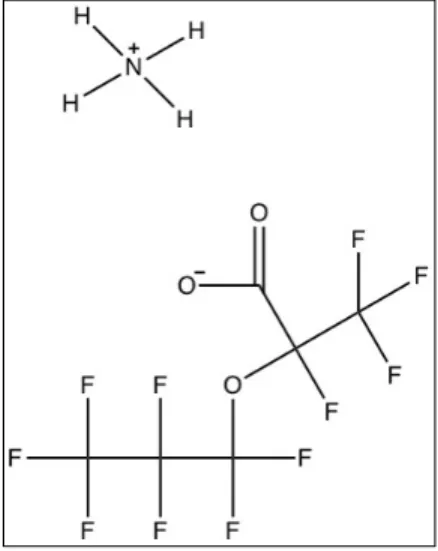
Ammonium 2,3,3,3-tetrafluoro-2-(heptafluoropropoxy) propanoate (CAS number: 62037-80-3, GenX) is the ammonium salt of the hexafluoropropylene dimer acid. It is a member of a class of compounds called per- and polyfluorinated alkyl substances (PFAS) (PubChem). Because of its surfactant properties, GenX and related substances are useful as a polymerization processing aids in the production of fluoropolymers. Under the Regulation for the Registration, Evaluation, Authorization and Restriction of Chemicals (REACH) Act, the European Union (EU) regulated and began the phase-out of perfluorooctanoic acid (PFOA) due to its human and environmental exposure in addition to increasing reports of
toxicity in laboratory animals. Fluoropolymer manufacturers introduced GenX as a PFOA replacement in the production of polytetrafluoroethylene (PTFE, Teflon), which creates nonstick coatings on cookware. GenX is also a constituent in food
packaging and firefighting foams. GenX and other short-chain or ether linked PFAS were chosen to minimize concerns about persistence and toxicity.
GenX is structurally related to PFOA, but it is distinguished by an ether linkage and a carboxylic acid group at the second carbon (Figure 1). GenX is amphipathic, due to its lipophilic hydrocarbon chain tail and hydrophilic head. Because of its strong carbon-fluorine bonds and steric hindrance, this structure is both stable and unlikely to be metabolized. GenX is a
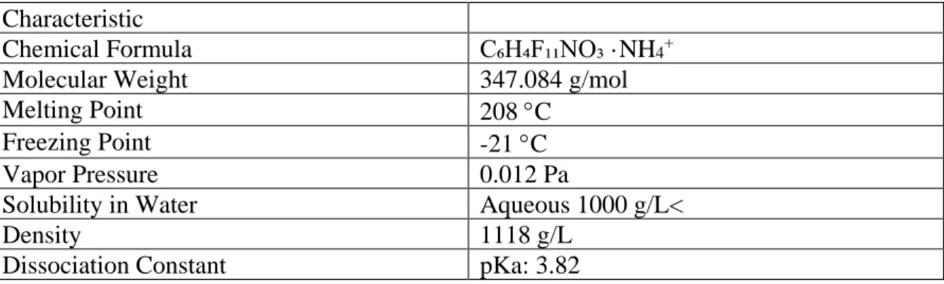
g/mol, is highly soluble in water, and is a strong acid with a pKa of 3.82 ("<Chemours Dordrecht Evaluation of substances used in genx tech.pdf>,").
Table 1. GenX chemical characteristics
Characteristic
Chemical Formula C₆H₄F₁₁NO₃ ·NH4+
Molecular Weight 347.084 g/mol
Melting Point 208 °C
Freezing Point -21 °C
Vapor Pressure 0.012 Pa
Solubility in Water Aqueous 1000 g/L<
Density 1118 g/L
Dissociation Constant pKa: 3.82
GenX has a production volume of 10-100 tons per year in the EU, but its worldwide production volume is unknown ("<Chemours Dordrecht Evaluation of substances used in genx tech.pdf>,"). It enters the environment through the manufacturing process in liquid and
aerosolized effluent. GenX contaminates rivers in the Netherlands, Germany, and China at concentrations of 0.210 nM, 0.248 nM, and 8.9 nM, respectively (Heydebreck, Tang, Xie, & Ebinghaus, 2015). In a study on acute and chronic aquatic toxicity of GenX, researchers
determined the 96-hour LC50 for rainbow trout was >96.9 mg/mL. At 120 mg/L, GenX did not produce immobility or sublethal effects in Daphnia magna, growth inhibition in algae, or changes in Daphnia reproduction or rainbow trout hatching (Hoke, Ferrell, Sloman, Buck, & Buxton, 2016). In addition, GenX did not bioaccumulate in any of the test species (Daphnia, trout, carp, and algae). These results suggest that GenX is unlikely to pose significant aquatic hazard, but it will be persistent in environmental media.
in finished drinking water at concentrations from 340-1100 ppt (approximately 1-3 nM) (NCDEQ, 2018). NC DEQ reported that the Fayetteville, NC facility releases approximately 2,700 pounds of GenX into the air per year (NCDEQ, 2018). The chemical is also present in private well water and some local food products, such as oysters and honey. Following state direction, the manufacturer ceased further discharge of GenX, and levels of GenX in drinking water began to decline. There are no mandatory federal or state regulatory requirements concerning GenX at the time of this publication, but NC set a health guideline of 140 ppt (0.40 nM) GenX in drinking water. This guideline is intended to be protective for bottle-feeding infants, children, nursing and pregnant women, and adults (NCDEQ, 2018).
Human exposure to GenX most likely occurs through contaminated drinking water, air, and potentially in food. There are no blood or urine concentration data available in the U.S. population. In an occupational biomonitoring study in the Netherlands, serum concentrations of GenX ranged from below the limit of detection to 486 nM with a mean concentration in serum of 47 nM (Vandenberg, 2018). Estimations of this population’s environmental exposure were not provided.
In the manufacturer’s study of the toxicokinetics of GenX, Gannon et al. described oral and intravenous routes of exposure in rats, mice, and cynomolgus monkeys. In the oral kinetic studies, they administered a single dose of 10 or 30 mg/kg GenX (water vehicle, PO) to Sprague-Dawley (SD) rats (3/sex/dose) and to Crl:CD1 mice (3/sex/dose) (Gannon et al., 2016). GenX was rapidly absorbed (absorption half-life of 0.21 h in males and 0.46 h in females), and the Cmax
hours for male and female rats, respectively, indicating a moderate sex difference in the rate of elimination. In the intravenous (IV) studies, they dosed Crl:CD SD rats (6/sex/dose) with 10 or 50 mg/kg GenX (PBS, single tail vein injection), as well as cynomolgus monkeys (3/sex, 10 mg/kg, PBS, peripheral vein injection). All test species showed similar blood concentration curves and elimination of the parent compound, suggesting that humans will behave similarly.
In a 2-year oral dosing study of GenX in SD rats, Caverly Rae et al. investigated the carcinogenicity and toxicity potential of GenX, using doses of 0, 0.1, 1, or 50 mg/kg in male rats and 0, 1, 50, or 500 mg/kg in female rats (Caverly Rae et al., 2015). They observed rapid
absorption and elimination of the unmetabolized compound in urine. In the 50 mg/kg males and the 500 mg/kg females, GenX decreased red cell mass and increased some liver enzymes indicative of liver injury. The high dose (500 mg/kg) females displayed kidney, tongue, and stomach changes, in addition to hepatocellular adenoma and carcinoma. At the high doses (50 and 500 mg/kg) GenX exposure resulted in neoplastic lesions of the liver, pancreas and testes in the male SD rats and in livers of female SD rats. Liver damage was attributed to peroxisome proliferator alpha (PPARα) agonism (Caverly Rae et al., 2015).
Rushing et al. investigated markers of liver peroxisome proliferation and T
cell-dependent antibody responses (TDAR) in C57Bl/6 mice in response to 28-day GenX exposure (Rushing et al., 2017). Females exposed to 100 mg/kg and males exposed to 10 mg/kg of the test compound exhibited increased hepatic acyl-CoA oxidase activity, indicative of peroxisome proliferation (Rushing et al., 2017). Liver weights were increased in both sexes at 10 and 100 mg/kg, and TDAR was significantly decreased in females at 100 mg/kg. PFOA had more potent effects on PPARα and TDAR than GenX. It is unlikely that the PFOA-mediated TDAR
PFOA decreases TDAR in PPARα knockout (KO) animals (DeWitt, Williams, Creech, & Luebke, 2016). Additional studies are needed to investigate if GenX’s immunotoxicity is also independent of PPARα.
Although the potential for GenX to cause central nervous system (CNS) toxicity is unknown, previous research showed that other PFAS (PFOS and PFNA) modulate ATP-binding cassette (ABC) transporter activity and expression at the Blood-Brain Barrier (BBB) (More et al., 2017). The BBB regulates the movement of endogenous and exogenous compounds into and out of the brain through a network of brain microvessels. Tight junction protein complexes connect endothelial cells and prevent the paracellular movement of compounds. Pericytes, astrocytes, neurons, and a basal membrane surround the abluminal side of the capillary, helping to form a physical barrier. Enzymes on the luminal-facing membrane of endothelial cells add an additional layer of chemical protection. Selective channels and carrier proteins located on both sides of the capillary membrane control transcellular transport (Fricker, 2016).
transporter-inhibiting drugs can increase the efficacy of chemotherapeutics and other drugs that normally cannot enter the target organ.
Table 2. Examples of endogenous and exogenous substrates for ABC transporters
Transporter Endogenous Substrates Exogenous Substrates P-glycoprotein Phospholipids Cyclosporine A, digoxin,
doxorubicin, tamoxifen Breast Cancer Resistance
Protein
Dietary flavonoids, porphyrins, estrones
Doxorubicin, irinotecan,
methotrexate, prazosin, 2-amino-1- methyl-6-phenylimidazo[4,5-b]pyridine (PhIP), statins, topotecan
Multidrug Resistance Protein 2
Glucuronide and glutathione conjugates, bilirubin
Cisplatin, doxorubicin, irinotecan,
methylmercury-N-acetyl-L-cysteine, methotrexate, ochratoxin A, 2-amino-1-
methyl-6-phenylimidazo[4,5-b]pyridine (PhIP), statins (Jonker et al., 2005), (Jemnitz et al., 2010), (Biotechnology)
In a previous study, 10 nM perfluorooctane sulfonic acid (PFOS) and 10 nM
perfluorononanoic acid (PFNA), two longer chain perfluoroalkyl surfactants, induce the activity of P-gp, BCRP, and MRP2 (More et al., 2017). However, shorter chain PFAS (10 nM
CHAPTER 2. METHODS
Chemicals
The fluorescent P-glycoprotein substrate N-" (4-nitrobenzofurazan-7-yl)-D-Lys cyclosporine A (NBD-CSA) was custom synthesized. We purchased the fluorescent Breast Cancer Resistance Protein substrate BODIPY-Prazosin from Life Technologies, and the
fluorescent MRP2 substrate sulforhodamine 101 free acid (Texas Red) from Sigma-Aldrich. The specific P-glycoprotein inhibitor, PSC-833
(3S,6S,9S,12R,15S,18S,21S,24S,30S,33S)-1,4,7,10,12,15,19,25,28-nonamethyl-33-[(E,2R)-2-methylhex-4-enoyl]-6,9,18,24-tetrakis (2-methylpropyl) 3,21,30-tri(propan-2-yl)-1,4,7,10,13,16,19,22,25,28,31
undecazacyclotritriacontane-2,5,8,11,14,17,20,23,26,29,32-undecone) was kindly provided by Novartis. Specific BCRP inhibitor KS-176
(N-[4-(2-Hydroxyethyl)phenyl]-2-[(4-nitrobenzoyl)amino]-benzamide) and specific MRP2 inhibitor MK-571,
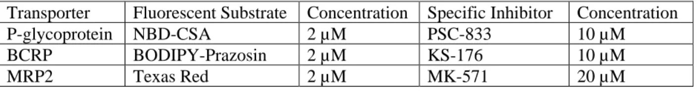
5-(3-(2-(7-Chloroquinolin-2-yl)ethenyl)phenyl)-8-dimethylcarbamyl-4,6-dithiaoctanoic acid sodium salt were purchased from Sigma-Aldrich . Solutions of specific fluorescent substrates and inhibitors were prepared at the concentrations 1000 times stronger DMSO (Table 3), then diluted in isolation buffer.
Table 3. Transporter-specific fluorescent substrates and inhibitors
Transporter Fluorescent Substrate Concentration Specific Inhibitor Concentration
P-glycoprotein NBD-CSA 2 µM PSC-833 10 µM
BCRP BODIPY-Prazosin 2 µM KS-176 10 µM
Isolation buffers were prepared 4-48 hours prior to the experiment and are stored in a 4 ℃ refrigerator. Isolation buffer was prepared using the following recipe: 500 mL phosphate-buffered saline (PBS) containing calcium and magnesium, 0.055 g sodium pyruvate (Sigma-Aldrich, St. Louis, MO), and 0.45 g glucose (Sigma-(Sigma-Aldrich, St. Louis, MO). Isolation buffer containing 1% BSA solution was prepared using the following recipe: 500 mL PBS containing Calcium and Magnesium, 0.055 g sodium pyruvate (Sigma-Aldrich, St. Louis, MO), 0.45 g glucose, (Sigma-Aldrich, St. Louis, MO), and 5.0 g of Bovine Serum Albumin (Sigma-Aldrich, St. Louis, MO). The two bottles of isolation buffer were placed on a shaking platform to mix them. A 30% Ficoll solution was prepared by combining 40 mL of Isolation Buffer (not
containing BSA) and 15 g of Ficoll (Sigma-Aldrich, St. Louis, MO) and mixed using a magnetic stir plate for 30 minutes. During the experiment, isolation buffers and the 30% Ficoll solution are stored on ice.
Ammonium 2,3,3,3-tetrafluoro-2-(heptafluoropropoxy)propanoate (CAS No. 62037-80-3, GenX) was purchased from Synquest (Alachua, FL) in powder form. The concentrations of GenX tested include 0.01 nM, 0.1 nM, 1 nM, 10 nM, 100 nM, and 1 µM. 1000X stock solutions were prepared in DMSO in 4 mL glass vials for immediate use. We vortexed for 10 seconds immediately after mixing and prior to formation of the next solution. A 1:1000 dilution was then used in the treatment of GenX dissolved in PBS. After each addition of GenX, the vial was vortexed for 5 seconds.
Animals
under a 12-h light/dark cycle in a temperature controlled facility and allowed access to food and water ad libitum. Animals were euthanized using carbon dioxide inhalation and decapitation with guillotine. We isolated brain capillaries using the protocol described below for immediate use in transport assays, or they were flash frozen in liquid nitrogen and stored in a -80 ℃ freezer for use in Western Blotting.
Capillary Isolation in Adult Rats
Procedures for capillary isolation for use in ex vivo or in vivo transport assays have been outlined previously (Chan & Cannon, 2017). Some modifications have been made to the method to optimize the number of capillaries, minimize contamination, and improve repeatability. Four rats per treatment group were used in in vivo assays; 2-6 rats were used in ex vivo assays; and 5 rats were used for Western Blotting. After euthanizing the rats via CO2 inhalation, we severed
the head at the base of the skull with a guillotine. Using a scalpel, we scored the skull from between the eyes to the base of the neck. We spit the skull with a Littauer bone cutter (Fine Science Tools, cat. no. 16152-15) along the score mark, and removed the brain with a spatula. We stored the brains in 50-ml Falcon polypropylene conical tubes (Becton, Dickinson and Company) containing 20 mL cold isolation buffer.
grinding vessel (prechilled on ice with the pestle) using a 7-ml bulb polyethylene transfer pipette (USA Scientific, cat. no. 1020-2500) and ice-cold isolation buffer. We rinsed the small petri dish and sides of the grinding vessel with additional isolation buffer.
We placed the grinding vessel in a 250 mL beaker containing ice. Using a pestle (Size C serrated, Thomas Scientific, cat. no. 3431F25) (clearance: 150 to 230 µm) to a drill, we
homogenized up and down slowly (approximately 5-10 seconds per up and down motion) for a total of 25 strokes at a speed of 20 RPM. We used 1 mL of isolation buffer to rinse remaining homogenized tissue on the pestle into the grinding vessel. We poured the homogenate into a pre-chilled 15 ml KONTES dounce tissue grinder (VWR, cat. no. 885300-0015), and we dounced 8-10 times using the pestle (size B; clearance: 165 to 889 µm). Then, we poured the mixture into a 50-ml polycarbonate centrifuge tube with polyethylene cap (Beckman Coulter, cat. no. 363664) and added 22mL of 30% Ficoll PM400 solution to the tube, inverting at least 6 times.
Then, we centrifuged at 7000 rpm, 4℃, for 24 minutes using a Sorvall RC-5B centrifuge using an SS-34 rotor. Removing the tubes carefully, we placed the tubes back on ice. We poured off the top layer of fat and supernatant, leaving behind a red pellet. Then, we wiped away any remaining fat on the sides of the tube with a Kimwipe, being careful to not disturb the pellet. Immediately we added 1-3 mL of ice cold isolation buffer containing 1% BSA and resuspended the pellet using a transfer pipette. Once it was fully dissolved into the BSA solution, we added another 15 mL of BSA solution and store on ice.
tension, then transferred the capillary-rich solution from the Beckman centrifuge tube through the mesh using a transfer pipette. We removed the mesh filter. Then, we washed the
pluriStrainers with the 1% BSA solution before pouring the filtered capillary-rich solution through them.
Once all the capillary-rich solution was poured through the filter, we removed the filter and flipped it over on top of a clean, open 50-mL Falcon tube. We used a transfer pipette to rinse the back side of the filter with one pipette-full 1% BSA solution, lightly touching filter with the tip of the pipette. We repeated this action two more times. Then, we rinsed the top side of the filter into the tube, using another pipette-full of 1% BSA solution. We repeated this action two times. We then poured the remaining solution in the tubes with the pluriStrainers through an additional pluristrainer to catch any capillaries that may have passed through the other filters. We then rinsed the additional pluriStrainer into the “clean capillaries” tube using the same process described above. We then centrifugde the filtered capillary-rich solution for 7 minutes at 1300 RPM in a ThermoFisher Sorvall RC 6+ Centrifuge.
the chamber slides and waited 30 minutes before adding 1 mL of room temperature isolation buffer.
Capillary Isolation in Pups
To isolate capillaries from rat pups (8h – 14 days old), the process is similar to the process in adult rats, but the differences are noted below. Four to five brains per treatment group were used in our in vivo assay. The skin and skull are much thinner, so only a scalpel or razor blade are necessary to open the skull and slide out the brain. We removed meninges if present. We skipped the step using the homogenizing drill, since the brain tissue is much more fragile. We reduced the number of douncing motions from 10 to 5. We then poured the dounced mixture into a Beckman centrifuge tube and added 1:2 volume of 30% Ficoll solution. After the 7000 RPM centrifugation, we removed the supernatant and add approximately 10 mL of 1% BSA solution. We omitted the nylon filter step and just filtered using one pluriStrainer per 5 brains. Only one 1300 RPM centrifugation for 7 minutes was necessary prior to applying the capillaries to the slides.
Ex vivo Transport Assay
After adding 1 mL of room temperature isolation buffer to each slide, we began adding the treatments as soon as possible. Each experiment included a control slide, which was treated with 2 mL of isolation buffer with 0.1% DMSO, and an inhibited slide, which was pre-treated with a transporter-specific inhibitor for 30 minutes before adding the fluorescent substrate with the inhibitor. The treated slides received 2 mL of isolation buffer prepared with the desired concentration of GenX (solutions were prepared as described previously). Fluorescent substrates were added 45 minutes prior to imaging on a 710 Zeiss confocal microscope.
solution was removed from the chamber slide. One mL of isolation buffer was added then
removed, then this process was repeated to rinse any remaining GenX from the slide. 2 milliliters of GenX were then added until it was time to add the fluorescent substrate. Control and inhibited slides were also rinsed twice to simulate the rinse on the treated capillaries
In vivo Transport Assay
Four age-matched male Sprague-Dawley rats (13 weeks, Taconic) were dosed with 0.03 µmol/kg of GenX dissolved in water by oral gavage, and four rats were treated with vehicle. Four age-matched female Sprague-Dawley rats (13 weeks, Taconic) were dosed with 0.3 µmol/kg of GenX dissolved in water by oral gavage, and four rats were treated with vehicle. After three hours, all rats were euthanized by CO2 euthanasia, and blood was collected by
cardiac puncture. They were then decapitated and the capillaries from each treatment group were isolated separately as described previously. All tools used in decapitation were cleaned in
deionized water prior to being used on the next treatments group or were single-use. After capillaries were isolated, they were immediately added to the slides. For each treatment group, three slides received the fluorescent substrate for P-gp, BCRP, and MRP2, and three slides received a 30-minute pretreatment with a specific inhibitor before being treated with the fluorescent substrate.
Western Blotting
3 hours, we centrifuged both treatment tubes at 1300 RPM for 7 minutes (ThermoFisher, Sorvall RC 6+ Centrifuge), poured off the isolation buffer solution, and submerged them in liquid nitrogen. We stored the tubes in a -80 ℃ freezer until they were needed.
We prepared the lysis buffer by combining 6 ml of Cellytic MT Lysis buffer (Sigma-Aldrich, St. Louis, MO) and 1 ml of 7X protease inhibitor [one pellet (Roche Diagnostics, Mannheim, Germany)/1.5 ml of 1X PBS]. We then directly added 150 µL of lysis buffer to resuspend the brain capillary pellet. We transferred this volume to a microcentrifuge tube (Beckman) and repeated this process for a total volume of 300 µL of lysis buffer and capillaries in the microcentrifuge tube. We vortexed the tubes for 10 seconds every five minutes and stored them on ice. After one hour, we centrifuged the tubes at 10,000 g for 30 minutes (Eppendorf centrifuge, 5430 R). Then, we pipetted off the denucleated supernatant into a new
microcentrifuge tube, and we centrifuged that supernatant at 100,000 g for 90 mins (Beckman Coulter, Optima MAX-XP Ultracentrifuge). Then, we pipetted off the cytoplasmic supernatant into a new microcentrifuge tube. The pellet left behind is the isolated membrane protein fraction. We resuspended the nuclear pellet and the membrane pellet in 30 µL of lysis buffer, and protein concentrations were determined by Bradford Assay.
measurement for each BSA standard versus its concentration in ug/mL. We used the standard curve to determine the protein concentration of each unknown sample.
After the protein concentrations have been determined by Bradford assay, we determined the volumes necessary to generate 25 µL total volume of sample, NuPAGE LDS Sample Buffer at 4X concentration, NuPAGE Reducing Agent at 10X concentration, and the remaining of deionized water. We denatured the samples at 80 ℃ in a GeneMate Digital Dry Bath for 10 minutes. We prepared 1000 mL of Running Buffer by adding 50 mL of 20X NuPAGE SDS Running Buffer and 950 mL of deionized water in a graduated cylinder and mixing on a stir plate. We removed 200 mL of running buffer and added 500 µL of NuPAGE Antioxidant. We prepared 1000 mL of transfer buffer by adding 50 mL of NuPAGE Transfer Buffer (20X), 1 mL of NuPAGE Antioxidant, 100 mL of methanol, and 849 mL of Deionized water to a graduated cylinder and mixing on a stir plate.
We loaded the samples and two molecular weight markers into each well using gel-loading pipette tips. Then, we filled the lower buffer chamber with 600 mL of the appropriate 1X running buffer. We ran the electrophoresis under constant 200 V for 35 minutes. After complete, we shut off power, disconnected the electrodes and removed gel from the XCell SureLock Mini-Cell. Then, we separated each of the three bonded sides of the cassette by inserting the gel knife into the gap between the two plastic plates that make up the cassette. We cut off the foot of the gel. The notched “well” side of the cassette should face up. We pushed down gently on the knife handle to separate the plates and repeated on each side.
We soaked the PVDF membrane for 30 seconds in methanol, and briefly rinsed in
deionized water, then placed it in a shallow dish with 50 mL of 1X NuPAGE Transfer Buffer for several minutes. We placed a piece of pre-soaked filter paper on top of the gel, with the edge above the slot in the bottom of the cassette. We wet the surface of the gel with transfer buffer and placed the pre-soaked transfer membrane on the protein side of the gel. Then, we placed another pre-soaked filter paper on top of the membrane and rolled out any bubbles with a roller. We placed three soaked blotting pads into the cathode core of the blot module and oriented the sandwich such that the gel is closest to the cathode core. Then, we placed three additional soaked blotting pads on the other side of the sandwich. Finally, we held the blot module together firmly and slid it into the guide rails on the lower buffer chamber. We inserted the gel tension wedge so that its vertical face is against the blot module and locked it into place by pulling the lever forward.
Then, we filled the blot module with 1X NuPAGE Transfer Buffer until the
25 V constant (began at 330 mA, ended at 130 mA) for 90 minutes. After the transfer was
complete, we removed the membrane from the box and rinsed with 1X PBS three times. We then applied a 50/50 Licor blocking buffer and 1X PBS (25 mL) for 30 minutes on a plate shaker.
We removed the blocking buffer and rinsed the membrane with 1X PBS. We then applied an 4 mL solution of primary antibodies. In the 4 mL solution was 20 µL (1:200) rabbit P-gp primary (Ab170904), 20 µL (1:200) rat BCRP primary (ab24115), 0.8 µL (1:5000) mouse Actin (Sigma) and 4 µL Tween. The membrane was vacuum sealed with this solution in a plastic envelope, and it was placed on a plate shaker in a 4 °C refrigerator overnight.
The next day, we removed the membrane from the envelope and poured off the primary solution. We rinsed the filter with 1X PBS then placed it in a container with 1X PBS and rocked on a plate shaker for 30 minutes. This was repeated three times. Then we poured off the PBS and added 20 mL secondary treatment, which included 2 µL AlexaFluor 647 goat anti-mouse Actin secondary antibody, 2 µL AlexaFluor 647 goat anti-rabbit P-gp secondary antibody, and 2 µL Licor 800 goat anti-rat BCRP secondary antibody. After 90 minutes, we removed the secondary treatment and imaged the membrane in a digital imager.
Statistical Analysis
Adult in vivo experiments used 4 age-matched control and 4 treated animals, pooled. The pup in vivo experiment containted 4-5 pup brains pooled per treatment group. Ex vivo
Average luminal fluorescence intensity of each in focus and intact capillary was quantified using ImageJ software. Approximately 10-15 images were taken for each treatment group and controls, and 15 ≤ capillaries per group were analyzed. We adjusted the means of the treated and control groups for the average fluorescence intensity of the group treated with a specific inhibitor. In all cases, the capillaries treated with MK-571 were not significantly different from controls, so these results were reported unadjusted. The adjusted and unadjusted means of fluorescence intensity were analyzed using GraphPad Prism (GraphPad Software, Inc, La Jolla, CA). The data were subjected to one-way ANOVA and multiple comparisons.
Statistical difference compared to control are reported as follows: ****, significantly different than control, p ≤ 0.0001; ***, significantly different than control, p ≤ 0.001; **, significantly different than control, p ≤ 0.01; *, significantly different than control, p ≤ 0.05; ns, not
CHAPTER 3. RESULTS
P-glycoprotein Transport Activity and Expression
We first examined the effects of the concentration of GenX on P-glycoprotein using an ex vivo steady-state luminal fluorescence assay in male and female Sprague-Dawley rats. In males,
all concentrations of GenX tested significant decreased P-gp transport activity, from 1 nM to 100 nM, following a 3-hour ex vivo exposure (Figure 2A). In females, a 3-hour ex vivo exposure to GenX decreased P-gp transport activity from 10 nM to 1000 nM (Figure 2B).
(A) 0 1 10 10 0 10 00 - 2 0
0 2 0 4 0
C o n c e n t r a t i o n ( n M )
S p eci f i c P -g l yco p r o t ei n T r an sp o r t A ct i vi t y * * * * * * * * * * * * * * * (B) 0
0. 0 1
0. 1 1 10 10 0 10 00 0 1 0 2 0 3 0 4 0 5 0
C o n c e n t r a t i o n ( n M )
S p eci f i c P -g l yco p r o t ei n T r an sp o r t A ct i vi t y * * * * * * * * * * *
Figure 2. Specific P-gp transport activity versus concentration of GenX treatment (3h, ex vivo).
GenX-mediated decreases in P-gp activity were concentration-dependent in both (A) males and (B) females.
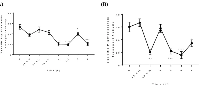
full inhibition occurring after 2 hours. GenX elicits a nonlinear decrease in P-gp transport activity in both males and females.
(A) 0 15 m i n 30 m i n 45 m
i n 1
1. 5
2 3 0 2 0 4 0 6 0 8 0
T i m e ( h )
S p eci f i c P -g l yco p r o t ei n T r an sp o r t A ct i vi t y * * * * * * * * * * * * * * (B) 0 15 m i n 30 m
i n 1 2 3 4
0 1 0 2 0 3 0 4 0
T i m e ( h )
S p eci f i c P -g l yco p r o t ei n T r an sp o r t A ct i vi t y * * * * * * * * * * *
Figure 3. Specific P-gp transport activity versus duration GenX treatment (100 nM, ex vivo).
GenX-mediated decreases in P-gp transport activity were time-dependent in both (A) males and (B) females.
P-gp (170 kDa) - GLUT1 (55 kDa) -
Control
0 1 2 3 4
0 10 20 30 40 50 Time (h) S p e c if ic P -g ly c o p ro te in T ra n s p o rt A c ti v it y GenX Removed ****
Figure 4. Specific P-gp transport activity versus time. GenX-mediated decreases in P-gp
transport activity are reversible in males (1h ex vivo 100 nM treatment).
Changes in transport activity are not necessarily indicative of changes in amount of protein present in the membrane. To quantify changes in protein expression, we treated male and female SD rat brain capillaries ex vivo with 100 nM GenX for 3 hours prior to performing
Western Blotting. When normalized for GLUT1 or Actin, P-gp expression in males and in females was significantly decreased in the GenX-treated groups (Figure 5).
(A) (B)
P-gp (170 kDa) - β-Actin (42 kDa) -
GenX Control GenX
Figure 5. P-gp expression in male and female at brain capillary membranes, following ex
We then wanted to know whether the decreased expression and activity of P-gp in rat cells treated ex vivo would also occur in human ovarian cancer cells. This cell line expresses only P-gp, which protects the cell under normal conditions from Adriamycin, a toxic
chemotherapeutic P-gp substrate. When we exposed the cells to 100 nM GenX and increasing concentrations of Adriamycin, the percent cell survival significantly declined compared to Adriamycin-only treatment (Figure 6). GenX-only treatment had little to no effect on cell survival.
E -8
E -7
E -6
E -5
4 0 6 0 8 0 1 0 0
C o n c e n t r a t i o n o f A d r i a m y c i n ( M )
P
er
cen
t
C
e
l
l
S
u
r
v
i
va
l
A d r i a m y c i n
A d r i a m y c i n + G e n X
Figure 6. Percent P-gp expressing human ovarian cancer cell survival versus concentration of
Adriamycin. Co-treatment of GenX and Adriamycin increases cytotoxicity compared to Adriamycin alone.
Ve hi c
l e , M
al e
0. 0 3 n
m ol /
kg , M
al e
Ve hi c
l e , F
em al e
0. 3 n
m ol /
kg , F
em al e
0 1 0 2 0 3 0 4 0 S p eci f i c P -g l yco p r o t ei n T r an sp o r t A ct i vi t y n s *
Figure 7. Specific P-gp transport activity versus concentration of GenX (3h in vivo treatment
with 0.03 nmol/kg male or 0.3 nmol/kg female). (A) P-gp transport activity does not change in the presence of GenX in males, but (B) increases in females.
0 2 8 8 6 0
5 0 1 0 0 1 5 0
C o n c e n t r a t i o n ( µ m o l / k g b w )
N
o
n
-sp
eci
f
i
c P
-g
l
yco
p
r
o
t
ei
n
T
r
a
n
sp
o
r
t
A
ct
i
vi
t
y
*
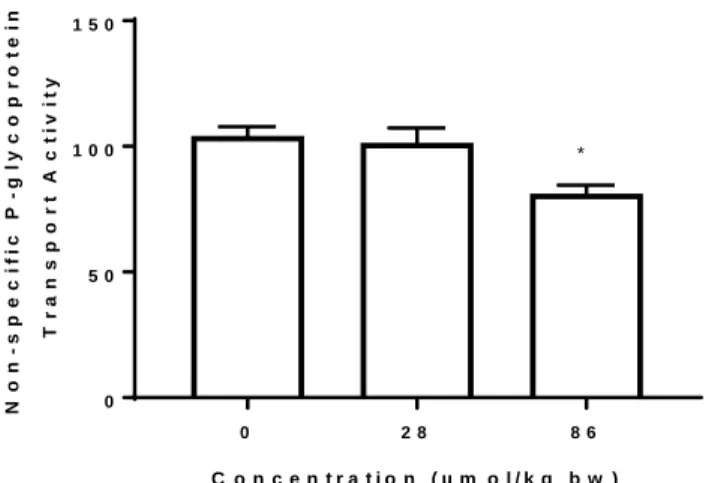
Figure 8. Specific P-gp transport activity versus concentration of GenX treatment (GD8-PND4,
in vivo, 28 µmol/kg or 86 µmol/kg). P-gp activity decreases in female PND4 pups in the presence of the highest dose of GenX.
Breast Cancer Resistance Protein Transport Activity
(A) 0 1 10 10 0 10 00 0 2 0 4 0 6 0
C o n c e n t r a t i o n ( n M )
S p eci f i c B C R P T r an sp o r t A ct i vi t y * * * * * * * * * * * * * * (B) 0 0. 1
1 10 10 0 10 00
- 4 0 - 2 0 0 2 0 4 0 6 0
C o n c e n t r a t i o n ( n M )
S p eci f i c B C R P T r an p so r t A ct i vi t y * * * * * * * * * * * * * * * * * * * *
Figure 9. Specific BCRP transport activity versus concentration of GenX treatment.
GenX-mediated decreases in BCRP transport activity are concentration-dependent in (A) males and (B) females.
We then looked at the effects of 100 nM GenX ex vivo exposure over time. In the presence of 100 nM GenX, ex vivo BCRP transport activity decreases beginning at one hour in both sexes (Figure 10). BCRP activity remains significantly inhibited until the latest time points we investigated in males (6h) and females (4h).
(A)
0 . 5 1
1. 5 2
2. 5 3
3. 5
4 5 6
- 2 0 0 2 0 4 0 6 0
T i m e ( h )
S p eci f i c B C R P T r an sp o r t A ct i vi t y * * * * * * * * * * * * * * * * * * * * * * * * * * * * * * * (B)
0 1 2 3 4
0 2 0 4 0 6 0
T i m e ( h )
S p e c i f ic BCRP T r a n sp o r t A ct i vi t y * * * * * * * * * * * * * * * *
Figure 10. Specific BCRP transport activity versus duration of treatment GenX (100 nM, ex
To test the reversibility of GenX-mediated inhibition of BCRP, we exposed the capillaries to a 2-hour ex vivo treatment with 100 nM GenX. A treatment of 100 nM of GenX significantly inhibits BCRP transport activity after two hours. Up to 4 hours following removal, BCRP transport activity remains inhibited (Figure 11).
0 2 3 4 5 6
- 2 0 0 2 0 4 0 6 0
T o t a l T i m e ( h )
S
p
eci
f
i
c
B
C
R
P
T
r
an
sp
o
r
t
A
ct
i
vi
t
y
* * * *
* * * *
G e n X R e m o v e d
* * * * * * * * * * * *
Figure 11. Specific P-gp transport activity versus time. GenX-mediated decreases in P-gp
transport activity are irreversible in males (2h ex vivo 100 nM treatment).
We then looked at the effects of an in vivo exposure to 0.03 nmol/kg GenX or water vehicle (4 rats/treatment group) in males and 0.3 nmol/kg GenX or water vehicle (4
Ve hi c
l e , M
al e
0. 0 3 n
m ol /
kg , M
al e
Ve hi c
l e , F
em al e
0. 3 n
m ol /
kg , F
em al e
0 2 0 4 0 6 0 8 0 S p eci f i c B C R P T r an sp o r t A ct i vi t y n s *
Figure 12. Specific BCRP transport activity versus concentration of GenX (3h in vivo treatment
with 0.03 nmol/kg male or 0.3 nmol/kg female). (A) BCRP transport activity decreases in the presence of GenX in males, but (B) does not change in females.
0
28 86
0 5 0 1 0 0 1 5 0
C o n c e n t r a t i o n ( µ m o l / k g b w )
N
on-s
pe
c
i
f
i
c
B
C
R
P
T
r
an
sp
o
r
t
A
ct
i
vi
t
y
*
Figure 13. Specific BCRP transport activity versus concentration of GenX treatment
(GD8-PND4, in vivo, 28 µmol/kg or 86 µmol/kg). BCRP activity increases in female PND4 pups in the presence of the highest dose of GenX.
Multidrug Resistance Protein 2 Transport Activity
Next, we investigated the effects of ex vivo GenX exposure on MRP2 transport activity. A 3-hour exposure to GenX did not change MRP2 transport activity significantly at
(A) 0 1 10 10 0 10 00 0 5 0 1 0 0 1 5 0
C o n c e n t r a t i o n ( n M )
N o n -sp eci f i c M R P 2 T r an sp o r t A ct i vi t y * * * (B) 0 0. 0
1 0. 1
1 10 10 0 10 00 0 5 0 1 0 0 1 5 0
C o n c e n t r a t i o n ( n M )
N o n -sp eci f i c M R P 2 T r an sp o r t A ct i vi t y * *
Figure 14. MRP2 transport activity versus concentration of GenX treatment (3h ex vivo). MRP2
transport activity decrease significantly at (A) 1000 nM in males and (B) 1 nM in females.
Then, we investigated the effects of a single concentration of GenX over a period of 4 hours on MRP2 transport activity. GenX significantly increases MRP2 transport activity following a 4-hour ex vivo exposure to 100 nM of GenX in males and females (Figure 15).
(A)
0 1 2 3 4
9 0 1 0 0 1 1 0 1 2 0 1 3 0 1 4 0
T i m e ( h )
N o n -sp eci f i c M R P 2 T r an sp o r t A ct i vi t y * * (B)
0 1 2 3 4
9 0 1 0 0 1 1 0 1 2 0 1 3 0
T i m e ( h )
N o n -sp eci f i c M R P 2 T r an sp o r t A ct i vi t y *
Figure 15. MRP2 transport activity versus duration of GenX treatment (100 nM, ex vivo). MRP2
Then, we looked at the effects of a 3-hour in vivo exposure to GenX in adult males and females. In both sexes, we observed no significant change in MRP2 transport activity (Figure 16).
Ve hi c
l e , M
al e
0. 0 3 n
m ol /
kg , M
al e
Ve hi c
l e, Fe
m al e
0. 3 n
m ol /
kg , F
em al e
0 5 0 1 0 0 1 5 0
N o n -s p eci f i c M R P 2 T r an sp o r t A ct i vi t y n s n s
Figure 16. MRP2 transport activity versus concentration of GenX (3h in vivo treatment with
0.03 nmol/kg male or 0.3 nmol/kg female). MRP2 transport activity does not change in the presence of GenX in (A) males or (B) females.
Finally, we investigated the effects of GenX in vivo on MRP2 transport activity in female PND4 pups exposed developmentally. We observed no significant changes at either
0 2 8 8 6 0
5 0 1 0 0 1 5 0
C o n c e n t r a t i o n ( µ m o l / k g b w )
N
o
n
-s
p
eci
f
i
c M
R
P
2
T
r
an
sp
o
r
t
A
ct
i
vi
t
y
Figure 17. MRP2 transport activity versus concentration of GenX treatment (GD8-PND4, in
CHAPTER 4. DISCUSSION
The aim of this study was to investigate the potential effects of GenX on the function and expression of ABC transporters at the blood-brain barrier in Sprague-Dawley rats. GenX is a known global contaminant in drinking water and air (Heydebreck et al., 2015; Sun et al., 2016). It shares structural similarities to PFOA and PFNA, which cause toxicity and carcinogenicity. These two PFAS induce P-gp and BCRP activity and expression at the BBB, likely through PPARα agonism (More et al., 2017). GenX is also a PPARα agonist (Caverly Rae et al., 2015), but is has a shorter carbon chain compared to PFOA and PFNA. No previous study has
investigated the toxicity of GenX at low concentrations or its potential for CNS toxicity. We hypothesized that GenX would modulate P-gp and BCRP activity and expression at the blood-brain barrier. Changes in P-gp and BCRP activity and expression have implications for the pharmacokinetics of the substrates of these efflux transporters.
P-glycoprotein is the most well-characterized ABC efflux transporter we studied. It has broad substrate specificity. P-gp is expressed in many tissues, such as brain, liver, and
GenX (100 nM) rapidly inhibits P-gp in male and female rat capillaries ex vivo by one hour. For other molecules we studied, rapid inhibition occurred via direct signaling in less than 30 minutes (Cannon, Peart, Hawkins, Campos, & Miller, 2012), but inhibition occurring after approximately one hour coincided with a complex indirect signaling event (Mesev, Miller, & Cannon, 2017). Due to the nonlinear and rapid effects we observed, it is possible that GenX has a dynamic impact on indirect cellular signaling pathways. To understand if the GenX-mediated inhibition is reversible, we administered a 1-hour exposure to 100 nM GenX, then removed the treatment and quantified P-gp transport activity 1, 2, 3, and 4 hours following removal. In males, P-gp transport activity returns to baseline levels when GenX is removed. These data also indicate that a 1-hour exposure to 100 nM GenX does not damage or affect the integrity of the capillary membrane, since activity is able to return to baseline when GenX is removed. These experiments have not been completed in females.
We observed no change in P-gp transport activity following in vivo exposure to 0.03 nmol/kg in male rats. We extrapolated this dose based on the maximum concentration in blood reported in Gannon et al. (50,000 ng/mL, 10 mg/kg rat, oral), in order to create a maximum concentration of 100 nM in blood. The process of isolating capillaries following the in vivo exposure requires approximately 3 hours. Since our ex vivo studies demonstrated that P-gp transport activity is rapidly reversible, and since the half-life of GenX is approximately 3 hours, it is possible that the animals fully eliminated the test compound before euthanasia or that P-gp activity returned to baseline during the capillary isolation process. The kinetics of GenX at lower doses, such as the ones used in our experiments, are unknown. In female rats, we increased the dose tenfold to approximate 1000 nM Cmax in blood, due to lower sensitivity to GenX ex vivo and
female rat P-gp transport activity increased significantly. This response is different from what we observed ex vivo. We do not know if GenX has a reversible effect in females or how the estrous cycle affects transport activity.
We performed Western Blotting using capillaries treated ex vivo to assess the effects of 100 nM GenX on the expression of P-gp. The capillary protein purification methods we used were effective, and we were unable to use β-actin as a loading control since it was not expressed
in males. For the males, we used GLUT1, a glucose transporter associated with endothelial cells in the BBB, as a loading control. β-actin was present in females, so we used it as a loading control in females. In both sexes, a 3-hour ex vivo treatment of 100 nM GenX significantly decreased P-gp expression. This finding suggests that the decreased P-gp activity could be due to the decreased expression of P-gp.
The second efflux transporter we investigated was Breast Cancer Resistance Protein. This ABC protein is “half-transporter,” so it requires two units to function. More than half of
In our in vivo experiments, we observed that a 3-hour exposure to 0.03 nmol/kg GenX in male rats caused significant inhibition of BCRP transport activity. These effects could be due to the irreversibility or the potency of the effects of GenX on this transporter. In females, a 3-hour exposure to 0.3 nmol/kg GenX caused no change in BCRP transport activity. We do not know whether GenX has an irreversible effect on BCRP activity in females, nor do we know the potential for the estrous cycle to impact BCRP expression or activity.
At 100 nM, preliminary data suggest that a 3-hour exposure to GenX decreases the expression of BCRP in isolated male brain capillaries treated ex vivo. These data support our finding that the same concentration of GenX lowers BCRP transport activity. We did not detect BCRP in our female capillaries. The literature indicates that BCRP expression is up to eight times lower than P-gp expression. In addition, 17β-Estradiol, which is present at significantly higher levels in females, inhibits BCRP (Mahringer & Fricker, 2010). So, a higher concentration of protein is likely necessary to detect BCRP in capillaries isolated from female rats in a Western Blotting.
The final ABC transporter we investigated was Multidrug Resistance Protein 2. MRP2 has the smallest active site of the three transporters we studied. In males, 1000 nM GenX (3h, ex vivo) and 100 nM GenX (4h, ex vivo) significantly decreased MRP2 transport activity. In
females, 1 nM GenX (3h, ex vivo) slightly decreased MRP2 transport activity, however these experiments have not been repeated and no other concentration showed any change. Since effects were either not repeated, at high concentrations, or after a long incubation with the test
compound, it is likely that the effects on MRP2 are due to an abnormal toxic response.
that the inhibition of P-gp and BCRP are specific to those transporters rather than a total inhibition of efflux transport.
We then used Adriamycin, a P-gp substrate chemotherapeutic, and a human ovarian cancer cell line containing P-gp (no BCRP or MRP2) to investigate the effects of GenX in human cells. In the absence of GenX, the efflux activity of P-gp protects the cells from Adriamycin toxicity. However when we exposed the cells to 100 nM GenX and increasing concentrations of Adriamycin, we observed increased cytotoxicity. These findings indicate that GenX inhibits human P-gp transport activity in vitro, allowing for a higher concentration of Adriamycin within the cell leading to cell death.
Our study was not without limitations. Firstly, this study did not investigate the mechanism underlying the GenX-induced P-gp and BCRP inhibition. Understanding GenX’s mechanism of action could allow us to predict which PFAS will cause similar effects on transporter activity and expression at the BBB. We were also limited by the lack of knowledge surrounding the sex differences in transporter expression and the effects of estrous cycle on both activity and expression of efflux transporters. Since the estrous cycle includes known fluctuation of hormones that affect at least BCRP expression, future studies in female animals should track the animals stage in estrous for at least a week prior to the experiment and group animals by their stage. Preliminary data show that GenX significantly increases BCRP activity and decreases P-gp activity in female PND4 pups, whose mothers were dosed with 30 mg/kg GenX from GD8 to PND4. Method development is in progress to create a viable assay to study young animal
The studies presented here are the first to demonstrate the effects of GenX on efflux transport at the Blood-Brain Barrier. At biologically and environmentally relevant doses, GenX inhibits P-gp and BCRP transport activity and expression. P-gp and BCRP are common
REFERENCES
Biotechnology, S. Knowledge Center: Transporters A-Z. Retrieved 7/3/2018, from Solvo Biotechnology https://www.solvobiotech.com/knowledge-center/transporters-a-z
Cannon, R. E., Peart, J. C., Hawkins, B. T., Campos, C. R., & Miller, D. S. (2012). Targeting blood-brain barrier sphingolipid signaling reduces basal P-glycoprotein activity and improves drug delivery to the brain. Proc Natl Acad Sci U S A, 109(39), 15930-15935. doi:10.1073/pnas.1203534109
Caverly Rae, J. M., Craig, L., Slone, T. W., Frame, S. R., Buxton, L. W., & Kennedy, G. L. (2015). Evaluation of chronic toxicity and carcinogenicity of ammonium
2,3,3,3-tetrafluoro-2-(heptafluoropropoxy)-propanoate in Sprague-Dawley rats. Toxicol Rep, 2, 939-949. doi:10.1016/j.toxrep.2015.06.001
Chan, G. N., & Cannon, R. E. (2017). Assessment of Ex Vivo Transport Function in Isolated Rodent Brain Capillaries. Curr Protoc Pharmacol, 76, 7 16 11-17 16 16.
doi:10.1002/cpph.21
<Chemours Dordrecht Evaluation of substances used in genx tech.pdf>.
DeWitt, J. C., Williams, W. C., Creech, N. J., & Luebke, R. W. (2016). Suppression of antigen-specific antibody responses in mice exposed to perfluorooctanoic acid: Role of
PPARalpha and T- and B-cell targeting. J Immunotoxicol, 13(1), 38-45. doi:10.3109/1547691X.2014.996682
Fricker, G. O., Melanie; Mahringer, Anne. (2016). The Blood Brain Barrier: Springer Verlag. Gannon, S. A., Fasano, W. J., Mawn, M. P., Nabb, D. L., Buck, R. C., Buxton, L. W., . . . Frame,
S. R. (2016). Absorption, distribution, metabolism, excretion, and kinetics of 2,3,3,3-tetrafluoro-2-(heptafluoropropoxy)propanoic acid ammonium salt following a single dose in rat, mouse, and cynomolgus monkey. Toxicology, 340, 1-9.
doi:10.1016/j.tox.2015.12.006
Heydebreck, F., Tang, J., Xie, Z., & Ebinghaus, R. (2015). Alternative and Legacy
Perfluoroalkyl Substances: Differences between European and Chinese River/Estuary Systems. Environ Sci Technol, 49(14), 8386-8395. doi:10.1021/acs.est.5b01648
Hoke, R. A., Ferrell, B. D., Sloman, T. L., Buck, R. C., & Buxton, L. W. (2016). Aquatic hazard, bioaccumulation and screening risk assessment for ammonium 2,3,3,3-tetrafluoro-2-(heptafluoropropoxy)-propanoate. Chemosphere, 149, 336-342.
doi:10.1016/j.chemosphere.2016.01.009
Jonker, J. W., Merino, G., Musters, S., van Herwaarden, A. E., Bolscher, E., Wagenaar, E., . . . Schinkel, A. H. (2005). The breast cancer resistance protein BCRP (ABCG2)
concentrates drugs and carcinogenic xenotoxins into milk. Nat Med, 11(2), 127-129. doi:10.1038/nm1186
Mahringer, A., & Fricker, G. (2010). BCRP at the blood-brain barrier: genomic regulation by 17beta-estradiol. Mol Pharm, 7(5), 1835-1847. doi:10.1021/mp1001729
Mesev, E. V., Miller, D. S., & Cannon, R. E. (2017). Ceramide 1-Phosphate Increases P-Glycoprotein Transport Activity at the Blood-Brain Barrier via Prostaglandin E2 Signaling. Mol Pharmacol, 91(4), 373-382. doi:10.1124/mol.116.107169
More, V. R., Campos, C. R., Evans, R. A., Oliver, K. D., Chan, G. N., Miller, D. S., & Cannon, R. E. (2017). PPAR-alpha, a lipid-sensing transcription factor, regulates blood-brain barrier efflux transporter expression. J Cereb Blood Flow Metab, 37(4), 1199-1212. doi:10.1177/0271678X16650216
NCDEQ. (2018). GenX Investigation: North Carolina Department of Environmental Quality. Rushing, B. R., Hu, Q., Franklin, J. N., McMahen, R., Dagnino, S., Higgins, C. P., . . . DeWitt, J.
C. (2017). Evaluation of the immunomodulatory effects of 2,3,3,3-tetrafluoro-2-(heptafluoropropoxy)-propanoate in C57BL/6 mice. Toxicol Sci.
doi:10.1093/toxsci/kfw251
Sun, M., Arevalo, E., Strynar, M., Lindstrom, A., Richardson, M., Kearns, B., . . . Knappe, D. R. U. (2016). Legacy and Emerging Perfluoroalkyl Substances Are Important Drinking Water Contaminants in the Cape Fear River Watershed of North Carolina. Environmental Science & Technology Letters, 3(12), 415-419. doi:10.1021/acs.estlett.6b00398