Clinical Factors Associated with Long-Term
Complete Remission versus Poor Response to
Chemotherapy in HIV-Infected Children and
Adolescents with Kaposi Sarcoma Receiving
Bleomycin and Vincristine: A Retrospective
Observational Study
Nader Kim El-Mallawany1,2*, William Kamiyango3, Jeremy S. Slone2,4, Jimmy Villiera3, Carrie L. Kovarik5, Carrie M. Cox6, Dirk P. Dittmer7, Saeed Ahmed3,8, Gordon E. Schutze8,
Michael E. Scheurer2,4, Peter N. Kazembe3, Parth S. Mehta2,4
1Department of Pediatrics, Division of Hematology, Oncology, and Hematopoietic Stem Cell
Transplantation, New York Medical College, Valhalla, New York, United States of America,2Department of Pediatrics, Section of Hematology and Oncology, Baylor College of Medicine, Houston, Texas, United States of America,3Baylor College of Medicine Children’s Foundation Malawi, Lilongwe, Malawi,4Texas Children’s Cancer and Hematology Centers, Houston, Texas, United States of America,5Department of Dermatology, University of Pennsylvania, Philadelphia, Pennsylvania, United States of America,
6Department of Pediatrics, Loyola University Chicago, Stritch School of Medicine, Maywood, Illinois, United States of America,7Department of Microbiology and Immunology, Lineberger Comprehensive Cancer Center, University of North Carolina, Chapel Hill, North Carolina, United States of America,8Department of Pediatrics, Baylor College of Medicine, Houston, Texas, United States of America
Abstract
Kaposi sarcoma (KS) is the most common HIV-associated malignancy in children and adolescents in Africa. Pediatric KS is distinct from adult disease. We evaluated the clinical characteristics associated with long-term outcomes. We performed a retrospective observa-tional analysis of 70 HIV-infected children and adolescents with KS less than 18 years of age diagnosed between 8/2010 and 6/2013 in Lilongwe, Malawi. Local first-line treatment included bleomycin and vincristine plus nevirapine-based highly active anti-retroviral therapy
(HAART). Median age was 8.6 years (range 1.7–17.9); there were 35 females (50%). Most
common sites of presentation were: lymph node (74%), skin (59%), subcutaneous nodules (33%), oral (27%), woody edema (24%), and visceral (16%). Eighteen (26%) presented with lymphadenopathy only. Severe CD4 suppression occurred in 28%. At time of KS diagnosis,
49% were already on HAART. Overall, 28% presented with a platelet count<100 x 109/L and
37% with hemoglobin<8 g/dL. The 2-year event-free (EFS) and overall survival (OS) were
46% and 58% respectively (median follow-up 29 months, range 15–50). Multivariable
analy-sis of risk of death and failure to achieve EFS demonstrated that visceral disease (odds ratios
[OR] 19.08 and 11.61, 95% CI 2.22–163.90 and 1.60–83.95 respectively) and presenting with
more than 20 skin/oral lesions (OR 9.57 and 22.90, 95% CI 1.01–90.99 and 1.00–524.13
respectively) were independent risk factors for both. Woody edema was associated with
a11111
OPEN ACCESS
Citation:El-Mallawany NK, Kamiyango W, Slone JS, Villiera J, Kovarik CL, Cox CM, et al. (2016) Clinical Factors Associated with Long-Term Complete Remission versus Poor Response to Chemotherapy in HIV-Infected Children and Adolescents with Kaposi Sarcoma Receiving Bleomycin and Vincristine: A Retrospective Observational Study. PLoS ONE 11(4): e0153335. doi:10.1371/journal.pone.0153335
Editor:Shou-Jiang Gao, University of Southern California Keck School of Medicine, UNITED STATES
Received:December 28, 2015
Accepted:March 28, 2016
Published:April 15, 2016
Copyright:© 2016 El-Mallawany et al. This is an open access article distributed under the terms of the
Creative Commons Attribution License, which permits unrestricted use, distribution, and reproduction in any medium, provided the original author and source are credited.
Data Availability Statement:The dataset has been uploaded to Dryad at the following DOI: doi:10.5061/ dryad.75tn6.
failure to achieve EFS (OR 7.80, 95% CI 1.84–33.08) but not death. Univariable analysis
revealed that lymph node involvement was favorable for EFS (OR 0.28, 95% CI 0.08–0.99),
while T1TIS staging criteria, presence of cytopenias, and severe immune suppression were
not associated with increased mortality. Long-term complete remission is achievable in pedi-atric KS, however outcomes vary according to clinical presentation. Based on clinical hetero-geneity, treatment according to risk-stratification is necessary to improve overall outcomes.
Introduction
Kaposi sarcoma (KS) is the most common human immunodeficiency virus (HIV)-associated malignancy in children and adolescents in sub-Saharan Africa.[1] KS is caused by human her-pesvirus-8 (HHV-8) infection, a virus with prevalence rates that vary geographically.[2] East-ern and central Africa in particular have the highest prevalence of HHV-8 infection in the
world.[3–9] Consequently, KS has become an important and widespread complication of the
HIV epidemic in sub-Saharan Africa, affecting not only adults, but children and adolescents as well. While clinical descriptions and treatment paradigms for adult KS have been well
estab-lished, there exist only a few clinical descriptions of pediatric KS cohorts in Africa.[10–14]
Standardized therapeutic strategies and risk factors associated with survival outcomes for chil-dren have yet to be established. Furthermore, since the largest published series on HIV-associ-ated malignancies from the United States or Europe reported only eight pediatric KS patients
from 1978–1996 in the AIDS-Cancer Match Registry Study Group, the pre-highly active
anti-retroviral therapy (HAART) era in the western world fails to serve as a guide.[15–20]
HHV-8, or Kaposi sarcoma associated herpesvirus (KSHV), is endemic in eastern and
cen-tral Africa with reported prevalence rates in children of 25–60% in Malawi, Uganda, and
Tan-zania; the HHV-8 seroprevalence in adults ranges between 60–90% in the same region.[3–5,8,
21–24] HHV-8 transmission often occurs during childhood from mother to child or between
siblings through exposure to oral secretions in endemic regions of the world.[9,25–27]
There-fore, with the onset of HHV-8 infection occurring at any point during childhood, KS can
develop at any time in the life of an HIV-infected child—either as a consequence of primary
HHV-8 infection or a secondary viral re-activation.
Pediatric KS is distinct from adult disease in many ways. Some of the unique features of pediatric KS in sub-Saharan Africa include the high rates of lymph node involvement, inade-quate response to HAART alone, and lack of prognostic significance in the AIDS Clinical Trial
Group (ACTG) TIS staging classification.[10,11,13,28,29] One-year overall survival (OS) for
most pediatric cohorts is approximately 40%, and risk factors predicting failure to respond to
chemotherapy plus HAART have yet to be determined.[10–14,29]
Despite high mortality rates in pediatric KS patients, long-term (i.e.>3 years) complete
remission (CR) has been achieved in HIV-infected children with KS in Malawi through the combination of chemotherapy and long-term HAART.[10] We evaluated the clinical charac-teristics of HIV-infected children and adolescents with KS in Lilongwe, Malawi and identified specific clinical factors that influence survival outcomes in patients receiving bleomycin and vincristine (BV) in combination with HAART.
Methods
Study Setting
We retrospectively analyzed the medical records of 70 consecutive HIV-infected children and adolescents with KS between August 2010 and June 2013 at the Baylor College of Medicine
purchase chemotherapy from ConocoPhillips. The Kamuzu Central Hospital pathology laboratory and Kaposi sarcoma research at the University of North Carolina Project-Malawi are supported by public health service grants CA019014, CA190152, and CA192744. The funders had no role in study design, data collection and analysis, decision to publish, or preparation of the manuscript.
Children’s Foundation Malawi Clinical Centre of Excellence (MW-COE) on the campus of Kamuzu Central Hospital (KCH) in Lilongwe, Malawi, a tertiary care facility that serves a
catchment area of approximately 5–8 million people. The primary clinical team included one
pediatric hematologist-oncologist, two specialized clinical officers, two staff nurses and addi-tional support staff comprised of social workers, pharmacy technicians, and clinic volunteers. Data were extracted from an electronic medical record system and standardized treatment flow sheets; patient health information was protected for confidentiality.
Diagnostic and Therapeutic Strategies
Standard clinical evaluation included a physical exam, full blood count, CD4 count analysis, and chest x-ray (CXR). Standardized KS diagnosis/treatment forms were used to maintain con-sistency in evaluation and documentation. Due to resource constraints, there were occasions when standard diagnostic studies were not available. There was very limited availability of HIV viral load analysis throughout the study period.
Biopsies were only performed in patients where a clinical diagnosis was uncertain as the availability of pathology resources was limited until 2013. Biopsy specimens were initially pro-cessed for histological evaluation with hematoxylin and eosin (H&E) stains at the University of Pennsylvania in Philadelphia, USA. Starting in 2013, through support from the University of
North Carolina Project–Malawi, specimens were processed and evaluated at the local KCH
pathology laboratory.[30] Morphology of the malignant cells was determined on H&E and confirmed with the HHV-8 latency-associated nuclear antigen (LANA) immunohistochemical stain.
The up-front chemotherapy regimen included bleomycin 15 units/m2via intramuscular
(IM) or intravenous (IV) injection and vincristine 1.4 mg/m2via IV injection. For naïve
patients, HAART was initiated within 2 weeks of chemotherapy. This standard practice is based on extrapolation from the standard approach provided by the Malawi National HAART Guidelines to initiate anti-tuberculosis treatment within 2 weeks of HAART initiation in patients with newly diagnosed HIV and tuberculosis co-infection.[31] BV was given for a total of 8 cycles in two phases: induction phase (4 cycles given every 2 weeks), followed by a consoli-dation phase (4 additional cycles given every 4 weeks). In 2012 doxorubicin became available, and the addition of doxorubicin to BV (ABV) served as a salvage regimen (doxorubicin 35 mg/
m2IV, bleomycin 10 units/m2IM, and vincristine 1.4 mg/m2IV, administered every 3 weeks).
Cumulative dosing of bleomycin and doxorubicin was tallied for each patient; the total lifetime
limit of bleomycin was established as 250 units/m2and for doxorubicin as 300 mg/m2. The
third line chemotherapeutic option was paclitaxel 75 mg/m2IV infusion every 3 weeks based
upon a modified protocol from our experience in Botswana.[32]
General guidelines for chemotherapy included delaying and/or dose-adjusting
chemother-apy for absolute neutrophil count (ANC)<1,000 cells/mm3and/or platelet count<100 x 109/
L. However, because a significant percentage of patients presented with cytopenias as part of the original clinical picture of their KS, the initial two cycles of chemotherapy were given at full-dose regardless of blood counts. For patients experiencing multiple occurrences of chemo-therapy-induced cytopenias, adjustments were made to the co-trimoxazole regimen and/or patients were switched off of zidovudine (AZT) containing-HAART.
co-infection were treated with efavirenz-based regimens instead of nevirapine. Additional modifi-cations of the standard HAART regimen were available for patients with specific contra-indica-tions and/or clinical scenarios. Daily co-trimoxazole was given for prophylaxis against
pneumocystis jiroveci according to World Health Organization (WHO) guidelines. Financial support for transportation was provided to patients requiring financial assistance.
Resources for supportive care were limited but consistently available. Moderate-severe mal-nutrition was treated according to WHO guidelines with peanut butter-based supplements and/or milk-based formulas. Anti-bacterial medicines included parenteral ceftriaxone and the following oral antibiotics: ciprofloxacin, amoxicillin, metronidazole, azithromycin, and co-tri-moxazole. Tuberculosis was treated according to WHO guidelines utilizing a 4-drug RHZE (rifampicin, isoniazid, pyrazinamide, and ethambutol) initial phase, followed by a 2-drug RH (rifampicin and isoniazid) continuation phase. Streptomycin was available for patients requir-ing re-treatment. The only anti-fungal medicine available was oral fluconazole; there were no medicines available for the treatment of invasive mold infections. There were no anti-viral agents at our disposal other than HAART. Anti-parasitic agents included artesunate-based oral medicines and parenteral quinine for malaria, albendazole and/or mebendazole (and metroni-dazole) for intestinal parasite infections, and praziquantel for schistosomiasis. Oral co-trimoxa-zole was given for treatment of pneumocystis jiroveci pneumonia as well as toxoplasma infections. Cultures of body fluids and advanced diagnostic methods of isolating infectious organisms were not available, therefore opportunistic infections were clinically diagnosed and treated based upon WHO clinical guidelines. We generally maintained a low threshold to empirically treat opportunistic infections with the available supportive care medicines. Whole blood transfusion was usually available from the Malawi Blood Bank, however platelets for transfusion were extremely difficult to obtain.
Clinical Definitions
The ACTG staging criteria were applied to all patients classifying tumor extent (T), immune status (I) and systemic illness (S) based upon the guidelines established for adults with KS.[33] However, because in our experience the ACTG staging criteria did not consistently provide prognostic relevance for pediatric patients, we also carefully documented the extent of KS involvement in each patient using standardized data collection forms.[10]
We differentiated sites of KS involvement based upon the following definitions:
• Skin: hyperpigmented macules/patches or papules
• Oral: hyperpigmented macules/patches or papular/nodular lesions of the oral mucosa
• Woody Edema: firm, non-pitting edema that typically occurs in the extremities and/or pubic
region, its texture resembling that of tree bark
• Lymphadenopathy: bulging, firm, non-tender, enlarged lymph nodes measuring>2 cm in
diameter, and not attributable to another cause of lymphadenopathy
• Subcutaneous Nodules: non-hyperpigmented nodules that are distinctly subcutaneous and
may occur as isolated nodules or as clusters of knotty lesions in the subcutaneous tissue, the latter often associated with underlying woody edema
• Facial Edema: non-woody, soft edema of the face, often in the peri-orbital region
• Other: includes exophytic masses and conjunctival lesions
• Pulmonary: in those patients with KS and a CXR demonstrating reticulo-nodular infiltrates and/or large pleural effusions not attributable to other pulmonary pathology (including dem-onstration of a failure to improve on empiric anti-tuberculosis and other anti-bacterial treat-ment). Serosanguineous effusions without evidence of infectious etiology were considered confirmatory for pulmonary KS. Additionally, a clinical diagnosis of upper airway pulmo-nary KS is defined in patients with KS and clinical signs of upper airway obstruction that improve with chemotherapy.
• Abdominal Cavity: in those patients with KS and persistent bloody stools not attributable to
other causes of hemorrhagic colitis (ie demonstration of failure to improve on empiric anti-bacterial and anti-parasitic treatment). Also, in patients with KS and severe ascites not attrib-utable to another disease process.
Definitions for disease response to chemotherapy were based upon previously established criteria from the ACTG.[34] The ACTG criteria define CR as the absence of any detectable residual lesions. CR was always clinically determined, as patients did not have confirmatory biopsies performed. However, CR was uniformly validated by the lack of progression in this cohort with consistent long-term follow-up. Relapse was defined as the recurrence of KS after having previously achieved CR. We have defined an additional response group called very good partial response (VGPR), in which patients achieve a subjective measure of at least 90% reduction in the size of all lesions. Induction failure was defined as failing to achieve a VGPR or CR after induction phase BV. For the survival analyses, event was defined as the occurrence of relapse, disease progression, failure to achieve CR, or death from any cause.
Immunological definitions from WHO guidelines were used, establishing severe and advanced immunosuppression as follows for the following age groups: infants younger than 12
months, a CD4% of<20% and<25% respectively. For children aged 1–5 years, a CD4%
of<15% and<20% respectively, and for children 5 years and older, an absolute CD4
count<200 and<350 respectively. Immune reconstitution inflammatory syndrome (IRIS)
was defined as occurrence of new-onset or worsening KS within 6 months of the initiation of HAART in patients who have not received chemotherapy. Virologic suppression of HIV is an important criteria to monitor in all patients, however due to limitations in resources, informa-tion on HIV viral load was only available in a few select clinical scenarios over the time period of the study, and therefore not discussed. The diagnosis of tuberculosis was clinical and WHO definitions of severe acute malnutrition were used.[35]
Statistical Analysis and Ethical Considerations
Demographic, disease and treatment information were described by standard descriptive
statis-tics. Dichotomous variables were analyzed using theχ2test, and continuous variables were
ana-lyzed using the t-test for two outcome categories and reported as mean and standard deviation. Univariate and multivariable analyses were done to evaluate the risk of death and the risk of failure to achieve event-free survival (EFS). Analyses were conducted utilizing either standard logistic regression or penalized maximum likelihood regression if there were any variables for which a subset of analyzed subjects were not exposed, i.e., zero cells in a contingency table.[36]
The stepwise backward selection procedure (p<0.05) was used to build the most
parsimoni-ous multivariable model for each outcome.
Both OS and EFS are reported using standard Kaplan-Meier curves. All statistical analyses
were done using STATATM, version 14 (StataCorp LP, College Station, TX, USA). Ethics
Review Board for Human Subjects Research. Due to the retrospective nature of this study, informed consent was not obtained for the review of medical records. However, data was anon-ymized and de-identified prior to analysis to maintain strict protection of confidentiality.
Results
Patient Characteristics
During the study period, 70 HIV-infected (presumed vertical transmission) children and
adolescents<18 years of age were diagnosed with KS at the MW-COE and KCH. (Table 1) Using
a denominator of children initiated on HAART at the MW-COE as a proxy for number of new pediatric HIV diagnoses during that time period, KS represented 5.2% (70/1,359) of children and adolescents newly diagnosed with HIV infection. There were 35 females (50%) and the median age
at the time of KS diagnosis was 8.6 years (range 1.7–17.9). The most common sites of KS clinical
involvement were lymph node (74.3%) and skin (58.6%). Visceral disease occurred in 16%. Notably,
18 patients (25.7%) presented withlymphadenopathy only, and no classic KS lesions of the skin, oral
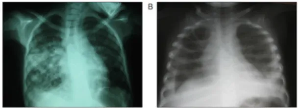
mucosa, or woody edema.Fig 1demonstrates the radiographic presentations of pulmonary KS in
our patients. Seven patients (10%) presented with>20 hyperpigmented skin/oral lesions in a
wide-spread,“disseminated”distribution all over the body. The inguinal region, including the groin/inner
thigh, was involved in 58 patients (82.9%), either in the form of lymphadenopathy, skin lesions, sub-cutaneous nodules, or woody edema. Diagnosis was pathologically confirmed in 14 patients (20%); Fig 2demonstrates a representation of the histologic and immunohistochemical lymph node biopsy findings. Baseline CXR was obtained at the time of KS diagnosis in 51 of 70 patients (72.9%).
Among the 18 patients who presented with bulging lymphadenopathy and none of the pro-totypical KS skin/oral lesions or woody edema, 13 (72%) diagnoses were established by biopsy. Of the five patients who were diagnosed clinically, all of them presented with massive, bulging, firm lymphadenopathy in the neck and inguinal regions plus/minus the axilla, with lymph
node masses measuring>5cm in diameter in each patient.
Baseline CD4 count samples were available for 60 of 70 patients and 17 (28.3%) had severe CD4 suppression. Moderate-severe cytopenias at the time of KS diagnosis was a common find-ing in this cohort; 67 of 70 patients had baseline full blood counts. At the time of KS diagnosis,
28.4% of children had moderate thrombocytopenia (platelet count<100 x 109/L) while 20.9%
had severe thrombocytopenia (platelets<50 x 109/L). Moderate anemia (hemoglobin<8 g/
dL) was noted in 37.3% with severe anemia (hemoglobin<6 g/dL) in 19.4%. Cytopenias
promptly improved after initiation of chemotherapy, with 88% of patients with moderate-severe thrombocytopenia resolving within 2 weeks without transfusion.
Nearly half of this cohort (48.6%) was already on HAART at the time of KS diagnosis. Six-teen patients (22.9%) were on HAART for more than 1 year, an additional ten (14.2%) on
HAART for 2–6 months, and eight (11.4%) were on HAART for less than 2 months at the time
of the KS diagnosis. Focusing on patients with an apparent IRIS phenomenon, among the eight patients diagnosed within 2 months of HAART initiation, 4 were event-free survivors, 4 died (two from KS, three within one month of diagnosis), and none of them had visceral or
dissemi-nated disease. Of the ten patients diagnosed with KS on HAART for 2–6 months, 9 died (six
from KS, two within one month of diagnosis) and only one survived; among the nine deaths, 3
patients had abdominal visceral disease and 2 presented with>20 skin/oral lesions. All of the
Response to Chemotherapy
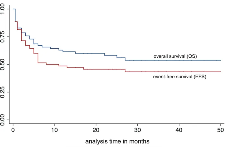
With a median follow-up of 29 months (range 15–50), the 2-year OS was 58.2%. Long-term
(median 31 months, range 15–50) CR was achieved in 33 children (47.1%), with an additional
six patients (8.6%) alive with stable disease. Two-year EFS was 45.7% for the overall cohort
Table 1. Clinical Characteristics of HIV-infected Children and Adolescents with Kaposi Sarcoma.
Number of Patients 70
Gender Female 35 (50%)
Median Age (range) 8.6 years (1.7–17.9)
Pathology Confirmation 14 (20%)
Clinical Site of KS Involvement
Lymph Node 52 (74.3%)
Hyperpigmented Skin Lesions 41 (58.6%)
Non-Hyperpigmented Subcutaneous Nodules 23 (32.9%)
Oral 19 (27.1%)
Woody Edema 17 (24.3%)
Facial Edema 11 (15.7%)
Pulmonary 8 (11.4%)
Abdominal Visceral 3 (4.3%)
Lymph Node ONLY, biopsy-proven in 13 of 18 (72.2%) 18 (25.7%) Inguinal Region Involvement (any type of lesion) 58 (82.9%) More than 20 Hyperpigmented Skin/Oral Lesions 7 (10%) HAART Status at Time of KS Diagnosis
Naïve to HAART 34 (48.6%)
IRIS 18 (25.7%)
On HAART>12 months 16 (22.9%)
Previously Defaulted off HAART 2 (2.9%)
Median Absolute CD4 Count at Time of KS Diagnosis (range) 368 (3–1039) CD4 Suppression at KS Diagnosis (WHO Definitions by Age), n = 60
None-Mild Immune Suppression 27 (45%)
Advanced Immune Suppression 16 (26.7%)
Severe Immune Suppression 17 (28.3%)
Presence of Concurrent Opportunistic Illness 32 (45.7%)
Tuberculosis 25 (35.7%)
Severe Malnutrition 12 (17.1%)
Cytopenias at Time of KS Diagnosis (n = 67)
Median Platelet Count (range) 201 (6–672)
Platelet Count<50 14 (20.9%)
Platelet Count<100 19 (28.4%)
Median Hemoglobin (range) 8.8 (1.8–13.6)
Hemoglobin<6 13 (19.4%)
Hemoglobin<8 25 (37.3%)
Median Absolute Neutrophil Count (range) 1953 (300–5930)
Absolute Neutrophil Count<500 2 (2.9%)
Absolute Neutrophil Count<1000 12 (17.9%)
ACTG TIS Staging Classification
T1 (n = 68) 42 (61.8%)
I1 (n = 60) 17 (28.3%)
S1 (n = 67) 32 (47.8%)
(Fig 3). Relapse due to primary failure of the HAART regimen, with successful re-induction of
CR after switching to 2ndline HAART plus additional doses of chemotherapy, was not
consid-ered an event; this occurred in four patients with lymphadenopathic disease. Median time to
event occurrence was 2 months (range 0.1–27 months). Five patients (7%) abandoned
treat-ment; three were traced by telephone communication and found to have died, while two returned to care.
Fifty patients (71%) completed the full course of BV. The response to chemotherapy was
often prompt, with resolution of KS lesions within 2–4 cycles of BV. Fourteen patients (20%)
received less than the 4 cycles of induction BV; two abandoned therapy early, while 12 experi-enced early death. Of the 56 patients who completed induction BV, 32 (57%) achieved a CR or VGPR after induction. The response of woody edema to chemotherapy was notoriously slow; of the 14 patients with woody edema who completed induction BV, 12 (86%) were induction failures. Some patients with woody edema eventually experienced resolution of the edema, with the only remnant being subtle fibrotic knotty changes in the subcutaneous tissue, espe-cially in the inner thigh. These patients were considered to be in CR if there was no residual edema and no asymmetry in the circumference of the extremity. Eight patients received salvage chemotherapy with ABV and three also received paclitaxel subsequently. Three of the patients
receiving 2nd/3rdline chemotherapy were long-term survivors, one after ABV alone, two after
ABV plus paclitaxel.
BV was well tolerated. Based upon CTCAE criteria, 18.6% of patients experienced grade 4 neutropenia and 35.7% grade 3 neutropenia. There were no occurrences of grade 3 or 4 throm-bocytopenia. Eight patients (11.4%) experienced grade 2 constipation and one patient
Fig 1. Representations of pulmonary KS on chest x-ray in patients with classic hyperpigmented KS skin lesions.(A) Multiple, scattered reticulo-nodular infiltrates in the right lung and (B) large pleural effusions.
doi:10.1371/journal.pone.0153335.g001
Fig 2. Morphology of KS in a lymph node biopsy.Complete effacement of the nodal architecture by a spindle cell infiltrate at (A) low and (B) high power on hematoxylin and eosin stain that are diffusely positive for the HHV-8 latency-associated nuclear antigen immunohistochemical stain shown at (C) low and (D) high power.
experienced grade 2 peripheral neuropathy from vincristine. Pulmonary function testing and echocardiograms were not available to assess end-organ damage from bleomycin and doxoru-bicin respectively. All patients that died from respiratory failure did so in the setting of severe pleural effusions, which is not a characteristic of bleomycin toxicity. No patients developed clinical signs of cardiopulmonary insufficiency throughout the study period and strict guide-lines were in place to avoid surpassing the total lifetime cumulative dosing limits of bleomycin and doxorubicin as outlined above.
Death occurred at a median time of 3 months after KS diagnosis (range 0.1–27 months).
The majority of deaths (23 of 31, 74.2%) occurred less than 6 months from KS diagnosis. Median time to death for patients who died from progressive KS (n = 19) was 3 months (range
0.1–22, interquartile range 1–4 months), while median time to death for patients who died
from other causes (multifactorial in patients that presented with KS and multiple other oppor-tunistic illnesses [OI, n = 5], severe infection [n = 4], complications of severe malnutrition
[n = 2], and unknown [n = 1]) was 3.5 months (range 0.1–27, interquartile range 1–6 months).
Ten patients died within 1–2 weeks of presentation; five had clinically refractory and
pro-gressive KS, however the other five (7.1%) presented with multiple acute severe OI and the cause of death is uncertain and therefore presumed treatment-related mortality (TRM). Six patients died in CR, three of them while on chemotherapy (one from presumed sepsis without neutropenia, one from bacterial meningitis without neutropenia, and one from complications of severe malnutrition). The latter two had poor control of their underlying HIV and it is
difficult to definitively determine if their causes of death were treatment related versus second-ary to uncontrolled HIV infection. Three patients died in CR long after completing
chemother-apy (11, 15, and 27 months from KS diagnosis), all in the setting of poorly controlled HIV—
one each from complications of severe malnutrition, severe pneumonia, and presumed sepsis.
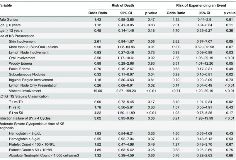
Risk Factors Associated with Death
Univariate logistic regression analysis was performed evaluating the risk of death and the risk of failure to achieve EFS in the cohort (Table 2). The following clinical variables demonstrated the strongest association with risk of death: visceral involvement (odds ratio (OR) 19,
p<0.01) and presenting with>20 skin/oral lesions (OR 9.5, p = 0.01). Notable variables that
were not associated with an increased risk of death included moderate-severe cytopenias,
severe immune suppression, age, and T1TIS tumor stage classification.
The univariate analysis of variables associated with a failure to achieve EFS demonstrated very similar findings (Table 2). Important findings included the association of woody edema with failure to achieve EFS (OR 3.51, p = 0.05), and the favorable association of lymph node
involvement (OR 0.28, p = 0.03). T1staging did prove adversely associated with EFS (OR 3.4,
p = 0.02), along with failure of induction phase BV (OR 6.21, p<0.01). Using the log-rank
test, there were no statistically significant associations between HAART status at the time of KS diagnosis and OS (p = 0.40) or EFS (0.50).
Table 2. Univariate Analysis of Variables Associated with Risk of Death and of Experiencing an Event.
Variable Risk of Death Risk of Experiencing an Event
Odds Ratio 95% CI p value Odds Ratio 95% CI p value
Male Gender 1.42 0.55–3.65 0.47 1.12 0.44–2.9 0.81
Age6 years 1.12 0.41–3.05 0.83 2.31 0.84–6.34 0.11
Age12 years 0.45 0.14–1.46 0.18 1.70 0.55–5.27 0.36
Site of KS Presentation
Skin Involvement 2.61 0.94–1.07 0.06 2.62 0.97–7.07 0.05
More than 20 Skin/Oral Lesions 9.50 1.08–83.86 0.01 15.00 0.82–273.98 0.07
Lymph Node Involvement 0.83 0.27–2.48 0.73 0.28 0.08–0.99 0.03
Oral Involvement 3.50 1.17–10.41 0.02 7.56 1.96–29.19 <0.01
Woody Edema 0.88 0.29–2.68 0.83 3.51 1.01–12.20 0.05
Facial Edema 0.70 0.19–2.67 0.6 0.63 0.17–2.31 0.49
Subcutaneous Nodules 0.32 0.11–0.97 0.04 0.28 0.10–0.81 0.02
Inguinal Region Involvement 1.18 0.30–4.63 0.81 0.79 0.20–3.09 0.73
Lymph Node Only Presentation 0.26 0.08–0.91 0.02 0.14 0.04–0.49 <0.01
Visceral Involvement 19.00 2.27–159.20 <0.01 10.71 1.29–89.19 <0.01
ACTG TIS Staging Classification
T1 vs T0 2.00 0.73–5.45 0.17 3.40 1.24–9.34 0.02
I1 vs I0 1.78 0.56–5.61 0.33 1.57 0.50–4.91 0.43
S1 vs S0 4.22 1.50–11.89 <0.01 1.98 0.75–5.26 0.17
Induction Failure of BV x 4 Cycles 3.02 0.95–9.65 0.06 6.21 1.93–19.99 <0.01
Moderate-Severe Cytopenias at time of KS Diagnosis
Hemoglobin<8 g/dL 1.83 0.54–6.21 0.33 1.50 0.55–4.09 0.43
Hemoglobin<6 g/dL 2.55 0.92–7.04 0.07 1.49 0.43–5.13 0.53
Platelet Count<100 x 10^9/L 1.52 0.47–4.98 0.49 1.27 0.43–3.70 0.67
Platelet Count<50 x 10^9/L 1.85 0.63–5.42 0.26 0.83 0.25–2.69 0.75
Absolute Neutrophil Count<1,000 cells/mm3 1.32 0.38–4.59 0.66 0.76 0.22–2.63 0.66
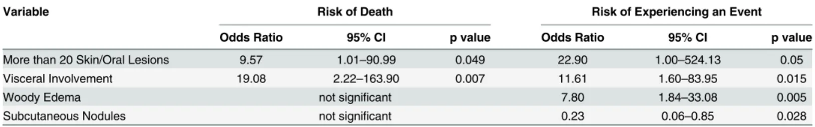
Multivariable analysis (Table 3) revealed that the only independent variables associated with a significant risk of death were visceral involvement and having more than 20 skin/oral lesions (OR 19.08, p = 0.007 and OR 9.57, p = 0.049 respectively). Failure to achieve EFS was independently predicted by visceral involvement, having more than 20 skin/oral lesions, and woody edema (OR 11.61, p = 0.015, OR 22.90, p = 0.05 and OR 7.80, p = 0.005 respectively). Oral involvement was not an independent risk factor.
Discussion
Analysis of detailed records from a retrospective pediatric KS cohort in Lilongwe, Malawi dem-onstrated that while lymphadenopathic KS was associated with improved long-term complete remission, visceral disease and disseminated skin/oral lesions were associated with a poor response to BV chemotherapy. Prior to a heightened awareness of lymphadenopathic KS in HIV-infected children, delays in diagnosis often resulted in patient deaths for lack of initiating proper KS-directed chemotherapy.[37] Instead, patients were usually misdiagnosed and mis-takenly treated for tuberculosis and other infectious etiologies of lymphadenopathy. As dem-onstrated by the univariate risk analyses, children with lymphadenopathic KS demonstrate favorable outcomes when treated with BV; therefore greater identification of lymph node dis-ease in this cohort may explain the superior 12-month OS in comparison to our published his-torical control (61% vs 43%).[10] This study highlights the virtually curative potential of a subset of HIV-infected children and adolescents with KS and suggests pathways towards better implementation of pediatric KS care in sub-Saharan Africa.
Very few studies on pediatric KS cohorts in sub-Saharan Africa exist, with most of them
reporting only 1-year outcome data.[10–13] As a result, our knowledge of the long-term
response to treatment is limited to and often based upon extrapolation from experiences in adult patients in the region. Benefiting from the integration of pediatric oncology and HIV ser-vices, our Pediatric HIV-Malignancy Program at the MW-COE has the advantage of long-term follow-up of HIV-infected patients that consistently return for refills of HAART. Of the 39 survivors in this cohort, 26 were in CR with greater than two years follow-up; the longest duration of follow-up was greater than four years.
Similar to other pediatric KS reports from sub-Saharan Africa, lymphadenopathy was the most common clinical presentation (74.3%) in our cohort as well. It is well established that the immunology of pediatric HIV infection is distinct from that in adults.[38] Early HIV infection and immune suppression of a naïve immune system may lead to unchecked primary HHV-8 lytic-phase infection, potentially explaining this phenomenon in children, which is seldom seen in adults who acquire HIV infection into a presumably competent immune system that has most likely been previously exposed to HHV-8 in regions with endemic prevalence.
Other unique features of pediatric KS demonstrated in our cohort include the low preva-lence of severe CD4 suppression (28%), and the high rates of presentation with moderate-severe cytopenias. Low prevalence of moderate-severe CD4 suppression has also been observed in adult
Table 3. Multivariate Analysis of Variables Associated with Risk of Death and of Experiencing an Event.
Variable Risk of Death Risk of Experiencing an Event
Odds Ratio 95% CI p value Odds Ratio 95% CI p value
More than 20 Skin/Oral Lesions 9.57 1.01–90.99 0.049 22.90 1.00–524.13 0.05
Visceral Involvement 19.08 2.22–163.90 0.007 11.61 1.60–83.95 0.015
Woody Edema not significant 7.80 1.84–33.08 0.005
Subcutaneous Nodules not significant 0.23 0.06–0.85 0.028
patients in Malawi.[39] Meanwhile, although anemia has been described in adult KS cohorts in
Africa, the percentage of patients presenting with hgb<8 g/dL (37%) in this pediatric cohort
far exceeds that of adult descriptions.[40] Additionally, the high rate of presentation with severe thrombocytopenia (21%) in this cohort is a phenomenon never reported in adult KS patients. We theorize once again that lytic-phase HHV-8 infection may contribute to this phe-nomenon in HIV-infected children akin to the clinical patterns of other lytic-phase HHV-8-associated malignancies such as multicentric Castleman disease (MCD) and the KSHV
inflammatory cytokine syndrome (KICS).[41–43] Bone marrow descriptions in patients with
MCD reveal normocellular marrows with reactive plasmacytosis consistent with excess inter-leukin-6 (IL-6) or its HHV-8-encoded homolog, viral IL-6.[44] Although none of the patients in this cohort had a bone marrow evaluation, the near-uniform normalization of the cytopenias within 2 weeks of starting BV chemotherapy may lend credence to the theory of a cytokine-mediated pathophysiology. Ultimately, severe cytopenias were not associated with adverse outcomes.
Regarding KS staging, our data suggest that the adult KS T1versus T0staging classification
is not significantly associated with higher mortality rates in children. Although presentation
with visceral disease was a significant risk factor for death, other features of the T1staging
defi-nitions such as woody edema were not. Conversely, although patients with widespread skin
disease are classified as T0, those presenting with>20 skin/oral lesions demonstrated high
mortality rates in our cohort. Only 2 of the 7 patients with>20 skin/oral lesions also had
vis-ceral disease, and the multivariable analysis revealed that both visvis-ceral disease and having>20
skin/oral lesions are independent risk factors for EFS and OS. T1staging though, did have a
sig-nificant predictive value for the failure to achieve EFS in children. S1staging demonstrated a
higher risk for death though, and certainly the presence of another OI—especially severe
mal-nutrition—renders these patients vulnerable to infectious, cardiovascular, and metabolic
complications.
Selecting a threshold of 20 hyperpigmented skin/oral lesions was an arbitrary decision made retrospectively based upon evaluation of our historical experience in Lilongwe. While adult
descriptions of KS often include much higher numbers of skin lesions (ie>50), we found it a
rare occurrence in our pediatric cohort.[34] Observing the patterns of clinical behavior in our patients based upon the descriptions of their original presentation, we decided upon a thresh-old of 20. These criteria will require prospective validation in larger clinical trials to become a
universal definition of“disseminated”skin disease in children and adolescents. As such, we
consider the ultimate decision on the threshold to define disseminated disease a dynamic issue that may be modified as the experience treating pediatric KS in Africa evolves.
Considering the practical limitations and challenges in low-income countries to prospec-tively identify all patients with visceral KS and other OI, it is important to monitor response to chemotherapy as well as empiric anti-microbial treatment. Although it is expected for woody
edema to exhibit a slow response to BV chemotherapy, for all patientswithoutwoody edema,
failure to respond to BV appears to be a poor prognostic sign and perhaps an indication for an alternative therapeutic approach. Patients with induction failure demonstrated an increased
OR (6.21, p value<0.01) for failure to achieve EFS in the univariate analysis, and represents
an important variable to be evaluated in future, prospective clinical trials.
the 18 were confirmed with biopsy, the diagnosis was made clinically for five patients. The clin-ical diagnosis of lymphadenopathic KS in the absence of prototypclin-ical lesions presents a major challenge throughout the region. Although these five patients carrying clinical diagnoses of lymph node KS cannot be considered definitive, there were 13 confirmed lymph node biopsies and 34 other patients presenting with lymph node involvement plus prototypical KS skin/oral/ edematous lesions, strengthening the pattern of observation of this distinct clinical presenta-tion in eastern and central Africa, for which clinical descrippresenta-tions (and images) actually date
back to the 1960–70’s.[45,46]
The five patients with a clinical diagnosis of lymph node KS presented with massive
lymph-adenopathy at the outset. Four presented with severe thrombocytopenia (range 13–24 x 109/L)
and without availability of platelets for transfusion, biopsies could not safely be performed con-sidering the risk of bleeding. The fifth patient did not have severe thrombocytopenia, however he had already been on anti-tuberculosis treatment for more than 1 month and experienced worsening of all of the enlarged lymph nodes. Taking into account the severity of their clinical presentations, our historical observations of high mortality rates in lymphadenopathic KS patients experiencing delayed initiation of chemotherapy, their lack of improvement on anti-tuberculosis therapy (albeit of short duration), and the safety profile of the BV chemotherapy regimen, these patients were empirically treated for KS. We recognize that our threshold for the clinical diagnosis was low, however this stems from our historical experience of witnessing rapid clinical demise in especially those patients presenting with severe cytopenias. Three of the five patients remain in active follow-up in long-term CR. The other two both died; one from sepsis 6 weeks into treatment in a clinical CR, while the other died from severe pneumo-nia one day after starting chemotherapy.
While these five clinical diagnoses remain probable, we gave consideration to the following differential: generalized lymphadenopathy associated with HIV infection rarely presents as massively enlarged nodes, and patients generally are not critically ill. Although the duration of empiric anti-tuberculosis treatment was only 2 weeks or greater (and not sufficient to defini-tively determine failure of therapy), four of the patients clinically worsened despite it. The other most likely diagnoses on the differential would be a mature B-cell non-Hodgkin lym-phoma or Hodgkin lymlym-phoma, both of which cannot be adequately treated by merely bleomy-cin and vincristine alone. Therefore, with a retrospective lens and long-term follow-up, we feel that these five patients represent probable diagnoses of lymphadenopathic KS.
Limitations in pathology resources (i.e. exhaustive panels of immunohistochemical stains, flow cytometry, and HHV-8 virologic assays) also present a significant hurdle to establishing the co-existence of other HHV-8 malignancies such as primary effusion lymphoma, MCD, and KICS. This represents an important limitation in an HHV-8 endemic region with a high preva-lence of pediatric HIV, where it would be logical for these other HHV-8 associated malignan-cies to occur. The retrospective aspect of the current study is another limitation. However, ultimately this manuscript presents a comprehensive and detailed account of the clinical char-acteristics, response to treatment, and long-term outcomes in pediatric KS and the data may serve to guide strategic planning for future clinical trials or the implementation of treatment paradigms.
Conclusions
is associated with good outcomes. Patients with woody edema have increased risk of failure to achieve EFS, but not mortality, ultimately exhibiting a more chronic disease course. However,
patients with visceral disease and/or>20 widespread“disseminated”skin/oral lesions
demon-strated significantly higher rates of death with BV; identifying these patients is critical to deter-mining which patients will require alternative therapeutic strategies.
Acknowledgments
We wish to acknowledge the courage and strength of our patients and their families, fighting a brave battle against both HIV and cancer in the setting of severe limitations in resources. We
would like to thank our colleagues at the Baylor Children’s Foundation Malawi inclusive of the
Tingathe Outreach Program. We additionally appreciate the vision and achievements of Dr. Mark Kline and our colleagues in the Baylor International Pediatric AIDS Initiative at Texas
Children’s Hospital. We express gratitude to Dr. David Poplack and our colleagues at the
Texas Children’s Cancer and Hematology Centers and the Global HOPE Program. We
acknowledge the excellent clinical care provided by our many colleagues at KCH as well as the
valuable collaboration of our colleagues from the University of North Carolina Project–
Malawi, including their expertise and support for the KCH Pathology Laboratory through Pro-fessor George Liomba.
Author Contributions
Conceived and designed the experiments: NKE WK JV CLK CMC SA PNK PSM. Performed the experiments: NKE WK JV CLK. Analyzed the data: NKE JSS MES PSM. Contributed reagents/materials/analysis tools: NKE JSS CLK SA DPD MES. Wrote the paper: NKE JSS DPD GES MES PNK PSM.
References
1. Orem J, Otieno MW, Remick SC. AIDS-associated cancer in developing nations. Current opinion in oncology. 2004; 16(5):468–76. PMID:15314517.
2. Chang Y, Cesarman E, Pessin MS, Lee F, Culpepper J, Knowles DM, et al. Identification of herpesvi-rus-like DNA sequences in AIDS-associated Kaposi's sarcoma. Science. 1994; 266(5192):1865–9. PMID:7997879.
3. Martro E, Bulterys M, Stewart JA, Spira TJ, Cannon MJ, Thacher TD, et al. Comparison of human her-pesvirus 8 and Epstein-Barr virus seropositivity among children in areas endemic and non-endemic for Kaposi's sarcoma. Journal of medical virology. 2004; 72(1):126–31. doi:10.1002/jmv.10548PMID: 14635020.
4. Sarmati L. HHV-8 infection in African children. Herpes: the journal of the IHMF. 2004; 11(2):50–3. PMID:15955270.
5. Chatlynne LG, Ablashi DV. Seroepidemiology of Kaposi's sarcoma-associated herpesvirus (KSHV). Seminars in cancer biology. 1999; 9(3):175–85. doi:10.1006/scbi.1998.0089PMID:10343069.
6. Olsen SJ, Chang Y, Moore PS, Biggar RJ, Melbye M. Increasing Kaposi's sarcoma-associated herpes-virus seroprevalence with age in a highly Kaposi's sarcoma endemic region, Zambia in 1985. Aids. 1998; 12(14):1921–5. PMID:9792393.
7. Rohner E, Wyss N, Heg Z, Faralli Z, Mbulaiteye SM, Novak U, et al. HIV and human herpesvirus 8 co-infection across the globe: Systematic review and meta-analysis. International journal of cancer Journal international du cancer. 2015. Epub 2015/07/16. doi:10.1002/ijc.29687PMID:26175054.
8. Mbulaiteye SM, Pfeiffer RM, Whitby D, Brubaker GR, Shao J, Biggar RJ. Human herpesvirus 8 infection within families in rural Tanzania. The Journal of infectious diseases. 2003; 187(11):1780–5. doi:10. 1086/374973PMID:12751036.
10. Cox CM, El-Mallawany NK, Kabue M, Kovarik C, Schutze GE, Kazembe PN, et al. Clinical characteris-tics and outcomes of HIV-infected children diagnosed with Kaposi sarcoma in Malawi and Botswana. Pediatric blood & cancer. 2013; 60(8):1274–80. doi:10.1002/pbc.24516PMID:23487320.
11. Gantt S, Kakuru A, Wald A, Walusansa V, Corey L, Casper C, et al. Clinical presentation and outcome of epidemic Kaposi sarcoma in Ugandan children. Pediatric blood & cancer. 2010; 54(5):670–4. doi: 10.1002/pbc.22369PMID:20205254; PubMed Central PMCID: PMC2839022.
12. Vaz P, Macassa E, Jani I, Thome B, Mahagaja E, Madede T, et al. Treatment of Kaposi sarcoma in human immunodeficiency virus-1-infected Mozambican children with antiretroviral drugs and chemo-therapy. The Pediatric infectious disease journal. 2011; 30(10):891–3. doi:10.1097/INF.
0b013e318228fb04PMID:21730886.
13. Chagaluka G, Stanley C, Banda K, Depani S, Nijram'madzi J, Katangwe T, et al. Kaposi's sarcoma in children: an open randomised trial of vincristine, oral etoposide and a combination of vincristine and bleomycin. European journal of cancer. 2014; 50(8):1472–81. doi:10.1016/j.ejca.2014.02.019PMID: 24636877.
14. Stefan DC, Stones DK, Wainwright L, Newton R. Kaposi sarcoma in South African children. Pediatric blood & cancer. 2011; 56(3):392–6. doi:10.1002/pbc.22903PMID:21225916.
15. Biggar RJ, Frisch M, Goedert JJ. Risk of cancer in children with AIDS. AIDS-Cancer Match Registry Study Group. Jama. 2000; 284(2):205–9. PMID:10889594.
16. Pollock BH, Jenson HB, Leach CT, McClain KL, Hutchison RE, Garzarella L, et al. Risk factors for pedi-atric human immunodeficiency virus-related malignancy. Jama. 2003; 289(18):2393–9. doi:10.1001/ jama.289.18.2393PMID:12746363.
17. Kest H, Brogly S, McSherry G, Dashefsky B, Oleske J, Seage GR 3rd. Malignancy in perinatally human immunodeficiency virus-infected children in the United States. The Pediatric infectious disease journal. 2005; 24(3):237–42. PMID:15750460.
18. Granovsky MO, Mueller BU, Nicholson HS, Rosenberg PS, Rabkin CS. Cancer in human immunodefi-ciency virus-infected children: a case series from the Children's Cancer Group and the National Cancer Institute. Journal of clinical oncology: official journal of the American Society of Clinical Oncology. 1998; 16(5):1729–35. PMID:9586885.
19. Caselli D, Klersy C, de Martino M, Gabiano C, Galli L, Tovo PA, et al. Human immunodeficiency virus-related cancer in children: incidence and treatment outcome—report of the Italian Register. Journal of clinical oncology: official journal of the American Society of Clinical Oncology. 2000; 18(22):3854–61. PMID:11078499.
20. Evans JA, Gibb DM, Holland FJ, Tookey PA, Pritchard J, Ades AE. Malignancies in UK children with HIV infection acquired from mother to child transmission. Archives of disease in childhood. 1997; 76 (4):330–3. PMID:9166025; PubMed Central PMCID: PMC1717129.
21. DeSantis SM, Pau CP, Archibald LK, Nwanyanwu OC, Kazembe PN, Dobbie H, et al. Demographic and immune correlates of human herpesvirus 8 seropositivity in Malawi, Africa. International journal of infectious diseases: IJID: official publication of the International Society for Infectious Diseases. 2002; 6 (4):266–71. PMID:12718819.
22. Kasolo FC, Spinks J, Bima H, Bates M, Gompels UA. Diverse genotypes of Kaposi's sarcoma associ-ated herpesvirus (KSHV) identified in infant blood infections in African childhood-KS and HIV/AIDS endemic region. Journal of medical virology. 2007; 79(10):1555–61. doi:10.1002/jmv.20952PMID: 17705172; PubMed Central PMCID: PMC2683451.
23. Pfeiffer RM, Wheeler WA, Mbisa G, Whitby D, Goedert JJ, de The G, et al. Geographic heterogeneity of prevalence of the human herpesvirus 8 in sub-Saharan Africa: clues about etiology. Annals of epidemi-ology. 2010; 20(12):958–63. doi:10.1016/j.annepidem.2010.07.098PMID:21074111.
24. Dollard SC, Butler LM, Jones AM, Mermin JH, Chidzonga M, Chipato T, et al. Substantial regional dif-ferences in human herpesvirus 8 seroprevalence in sub-Saharan Africa: insights on the origin of the "Kaposi's sarcoma belt". International journal of cancer Journal international du cancer. 2010; 127 (10):2395–401. doi:10.1002/ijc.25235PMID:20143397; PubMed Central PMCID: PMC2895015.
25. Andreoni M, Sarmati L, Nicastri E, El Sawaf G, El Zalabani M, Uccella I, et al. Primary human herpesvi-rus 8 infection in immunocompetent children. Jama. 2002; 287(10):1295–300. PMID:11886321.
26. Bourboulia D, Whitby D, Boshoff C, Newton R, Beral V, Carrara H, et al. Serologic evidence for mother-to-child transmission of Kaposi sarcoma-associated herpesvirus infection. Jama. 1998; 280(1):31–2. PMID:9660357.
28. Dow DE, Cunningham CK, Buchanan AM. A Review of Human Herpesvirus 8, the Kaposi's Sarcoma-Associated Herpesvirus, in the Pediatric Population. Journal of the Pediatric Infectious Diseases Soci-ety. 2014; 3(1):66–76. doi:10.1093/jpids/pit051PMID:24567845; PubMed Central PMCID:
PMC3933043.
29. Anglemyer A, Agrawal AK, Rutherford GW. Treatment of Kaposi sarcoma in children with HIV-1 infec-tion. The Cochrane database of systematic reviews. 2014; 1:CD009826. doi:10.1002/14651858. CD009826.pub2PMID:24464843.
30. Gopal S, Krysiak R, Liomba NG, Horner MJ, Shores CG, Alide N, et al. Early experience after develop-ing a pathology laboratory in Malawi, with emphasis on cancer diagnoses. PloS one. 2013; 8(8): e70361. doi:10.1371/journal.pone.0070361PMID:23950924; PubMed Central PMCID: PMC3737192.
31. Tweya H, Ben-Smith A, Kalulu M, Jahn A, Ng'ambi W, Mkandawire E, et al. Timing of antiretroviral ther-apy and regimen for HIV-infected patients with tuberculosis: the effect of revised HIV guidelines in Malawi. BMC public health. 2014; 14:183. Epub 2014/02/22. doi:10.1186/1471-2458-14-183PMID: 24555530; PubMed Central PMCID: PMCPMC3943509.
32. Reddy-Holdcraft S, Mehta PS, Agrawal AK. Paclitaxel for relapsed or recurrent HIV-associated pediat-ric Kaposi's sarcoma. Aids. 2014; 28(5):800–2. doi:10.1097/QAD.0000000000000157PMID: 24983546.
33. Krown SE, Testa MA, Huang J. AIDS-related Kaposi's sarcoma: prospective validation of the AIDS Clinical Trials Group staging classification. AIDS Clinical Trials Group Oncology Committee. Journal of clinical oncology: official journal of the American Society of Clinical Oncology. 1997; 15(9):3085–92. PMID:9294471.
34. Krown SE, Metroka C, Wernz JC. Kaposi's sarcoma in the acquired immune deficiency syndrome: a proposal for uniform evaluation, response, and staging criteria. AIDS Clinical Trials Group Oncology Committee. Journal of clinical oncology: official journal of the American Society of Clinical Oncology. 1989; 7(9):1201–7. Epub 1989/09/01. PMID:2671281.
35. WHO, UNICEF. WHO child growth standards and the identification of severe acute malnutrition in infants and children: a Joint Statement by the World Health Organization and the United Nations Chil-dren's Fund. Geneva: 2009.
36. Firth D. Bias Reduction of Maximum Likelihood Estimates. Biometrika. 1993; 80(1):27–38. doi:10. 2307/2336755
37. Arkin LM, Cox CM, Kovarik CL. Kaposi's sarcoma in the pediatric population: the critical need for a tis-sue diagnosis. The Pediatric infectious disease journal. 2009; 28(5):426–8. doi:10.1097/INF. 0b013e318193ee21PMID:19295460.
38. Tobin NH, Aldrovandi GM. Immunology of pediatric HIV infection. Immunological reviews. 2013; 254 (1):143–69. Epub 2013/06/19. doi:10.1111/imr.12074PMID:23772619; PubMed Central PMCID: PMCPmc3737605.
39. Hosseinipour MC, Sweet KM, Xiong J, Namarika D, Mwafongo A, Nyirenda M, et al. Viral profiling iden-tifies multiple subtypes of Kaposi's sarcoma. PMID:mBio. 2014; 5(5):e01633–14. doi:10.1128/mBio. 01633-14PMID:25249280; PubMed Central PMCID: PMC4173763.
40. Herce ME, Kalanga N, Wroe EB, Keck JW, Chingoli F, Tengatenga L, et al. Excellent clinical outcomes and retention in care for adults with HIV-associated Kaposi sarcoma treated with systemic chemother-apy and integrated antiretroviral therchemother-apy in rural Malawi. Journal of the International AIDS Society. 2015; 18:19929. doi:10.7448/IAS.18.1.19929PMID:26028156; PubMed Central PMCID: PMC4450240.
41. Polizzotto MN, Uldrick TS, Wang V, Aleman K, Wyvill KM, Marshall V, et al. Human and viral interleu-kin-6 and other cytokines in Kaposi sarcoma herpesvirus-associated multicentric Castleman disease. Blood. 2013; 122(26):4189–98. doi:10.1182/blood-2013-08-519959PMID:24174627; PubMed Cen-tral PMCID: PMC3868925.
42. Polizzotto MN, Uldrick TS, Hu D, Yarchoan R. Clinical Manifestations of Kaposi Sarcoma Herpesvirus Lytic Activation: Multicentric Castleman Disease (KSHV-MCD) and the KSHV Inflammatory Cytokine Syndrome. Frontiers in microbiology. 2012; 3:73. doi:10.3389/fmicb.2012.00073PMID:22403576; PubMed Central PMCID: PMC3291870.
43. Uldrick TS, Wang V, O'Mahony D, Aleman K, Wyvill KM, Marshall V, et al. An interleukin-6-related sys-temic inflammatory syndrome in patients co-infected with Kaposi sarcoma-associated herpesvirus and HIV but without Multicentric Castleman disease. Clinical infectious diseases: an official publication of the Infectious Diseases Society of America. 2010; 51(3):350–8. doi:10.1086/654798PMID:20583924; PubMed Central PMCID: PMC2946207.
disease. American journal of clinical pathology. 2013; 139(5):651–61. doi:10.1309/ AJCPKGF7U8AWQBVGPMID:23596117.
45. Slavin G, Cameron HM, Forbes C, Mitchell RM. Kaposi's sarcoma in East African children: a report of 51 cases. The Journal of pathology. 1970; 100(3):187–99. doi:10.1002/path.1711000307PMID: 5428938.