THEJOURNAL OFPHARMACOLOGY ANDEXPERIMENTALTHERAPEUTICS J Pharmacol Exp Ther 357:10–16, April 2016 Copyrightª2016 by The American Society for Pharmacology and Experimental Therapeutics
Ethanol Regulation of Synaptic GABA
A
a
4 Receptors Is
Prevented by Protein Kinase A Activation
Stephen L. Carlson, John Peyton Bohnsack, and A. Leslie Morrow
Departments of Psychiatry and Pharmacology, Bowles Center for Alcohol Studies, University of North Carolina School of Medicine, Chapel Hill, North Carolina
Received November 3, 2015; accepted January 28, 2016
ABSTRACT
Ethanol alters GABAAreceptor trafficking and function through activation of protein kinases, and these changes may underlie ethanol dependence and withdrawal. In this study, we used subsynaptic fraction techniques and patch-clamp electrophysiol-ogy to investigate the biochemical and functional effects of protein kinase A (PKA) and protein kinase C (PKC) activation by ethanol on synaptic GABAAa4 receptors, a key target of ethanol-induced changes. Rat cerebral cortical neurons were grown for 18 days in vitro and exposed to ethanol and/or kinase modulators for 4 hours, a paradigm that recapitulates GABAergic changes found after chronic ethanol exposure in vivo. PKA activation by forskolin or rolipram during ethanol exposure prevented increases in P2 fractiona4 subunit abundance, whereas inhibiting PKA had no effect. Similarly, in the synaptic fraction, activation of PKA by
rolipram in the presence of ethanol prevented the increase in synaptica4 subunit abundance, whereas inhibiting PKA in the presence of ethanol was ineffective. Conversely, PKC inhibition in the presence of ethanol prevented the ethanol-induced increases in synaptica4 subunit abundance. Finally, we found that either activating PKA or inhibiting PKC in the presence of ethanol prevented the ethanol-induced decrease in GABA miniature inhibitory postsynaptic current decayt1, whereas inhibiting PKA had no effect. We conclude that PKA and PKC have opposing effects in the regulation of synaptic a4 receptors, with PKA activation negatively modulating, and PKC activation positively modulating, synaptica4 subunit abundance and function. These results suggest potential targets for restoring normal GABAergic functioning in the treatment of alcohol use disorders.
Introduction
Ethanol causes adaptations in GABAA receptors that are
associated with alcohol dependence and withdrawal (Kumar et al., 2009). GABAA receptors are ligand-gated chloride
channels mediating the majority of inhibitory neurotransmis-sion in the brain through both phasic and tonic currents (Farrant and Nusser, 2005). These channels consist of five subunits typically composed of two asubunits (1–6), two b
subunits (1–3), and ag(1–2) ordsubunit (Tretter and Moss, 2008). Two important targets of ethanol regulation in partic-ular are the synaptica4bg2 and extrasynaptica4bdGABAA
receptors mediating a portion of phasic and tonic inhibition, respectively (Olsen and Sieghart, 2009). The extrasynaptica4 receptors have been the subject of much study, owing to their responses to relatively low doses of ethanol (Sundstrom-Poromaa et al., 2002; Wallner et al., 2003; Wei et al., 2004); however, surprisingly little is known about the regulation of synaptica4 receptors.
Although early studies showed that ethanol regulates the abundance of a4 subunit mRNA and protein expression
(Devaud et al., 1996, 1997; Papadeas et al., 2001; Cagetti et al., 2003), the question of whether this regulation reflected synaptic or extrasynaptic receptor adaptations depended on subsequent functional studies of synaptica4 receptor kinetics. Synaptica4 receptors are upregulated after both acute (Liang et al., 2007) and chronic (Liang et al., 2006; Werner et al., 2011) ethanol exposure in the rat hippocampus and cortex. More recently, we demonstrated ethanol regulation of synap-tica4 subunits in C57BL/6J mice after an acute injection of ethanol, using subcellular fractionation to isolate synaptic versus subsynaptic receptors (Carlson et al., 2014). Isolating the precise physiologic and behavioral ramifications of these changes in synaptica4 receptors is difficult because there are no selective pharmacological agents targeting these receptors. Thea4bg2 receptors are benzodiazepine insensitive, although they show relatively high affinity for the GABA inverse agonist Ro15-4513 (ethyl-8-azido-5,6-dihydro-5-methyl-6-oxo-4H-imidazo-1,4-benzodiazepine-3-carboxylate) (Knoflach et al., 1996). However, Ro15-4513 also antagonizes effects of ethanol ona4bdreceptors (Hanchar et al., 2006) that likely mediate effects of ethanol on tonic inhibition.
Recent studies from our laboratory have uncovered that kinase activation by ethanol plays a major role in GABAA
receptor regulation, and that protein kinase A (PKA) and protein kinase C (PKC) may have opposing effects on GABAergic trafficking and function. PKA activity positively
This research was supported by the National Institutes of Health National Institute on Alcohol Abuse and Alcoholism [Grants P60-AA11605 and T32-AA007573] and the University of North Carolina Bowles Center for Alcohol Studies.
dx.doi.org/10.1124/jpet.115.230417.
ABBREVIATIONS: ANOVA, analysis of variance; CalC, calphostin C; mIPSC, miniature inhibitory postsynaptic current; PdBu, phorbol-12,13-dibutyrate; PKA, protein kinase A; PKC, protein kinase C; Ro15-4513, ethyl-8-azido-5,6-dihydro-5-methyl-6-oxo-4H -imidazo-1,4-benzodiazepine-3-carboxylate; Rp-cAMP, Rp-adenosine 39,59-cyclic monophosphothioate triethylamine.
regulates synaptic GABAAa1 subunit abundance and
func-tion in cortical neurons, whereas PKCg activity negatively regulates these receptors (Kumar et al., 2010; Carlson et al., 2013). PKA activation also positively regulates extrasynaptic GABAAa4 anddsubunits in cortical neurons, whereas PKC is
not involved in regulating these receptors in this brain region (Carlson et al., 2016). Finally, PKCg activation by ethanol causes an increase in GABAAa4 subunits, although it is unclear
whether this effect is specific to the synaptic population of receptors (Werner et al., 2011). It is also unclear what role PKA activity may play in regulating this subset ofa4 receptors.
This study elucidates the role of PKA and PKC in ethanol regulation of synaptic a4-containing GABAA receptors in
cerebral cortical cultured neurons. We used a 4-hour ethanol exposure paradigm that recapitulates many of the GABAergic adaptations observed after chronic ethanol exposure in vivo (Devaud et al., 1997; Kumar et al., 2003). We measured changes in subunit abundance using a subcellular fraction-ation technique that enriches synaptic proteins combined with western blot analysis, whereas functional changes were measured using whole-cell patch-clamp analysis of GABA miniature inhibitory postsynaptic currents (mIPSCs).
Materials and Methods
Cultured Cerebral Cortical Neurons. All experiments were conducted in accordance with guidelines from the National Institutes of Health and Institutional Animal Care and Use Committee at the University of North Carolina. Mixed-sex rat pups from Sprague-Dawley breeding pairs (Harlan, Indianapolis, IN) were decapitated on postnatal day 0–1. Brains were rapidly dissected and the cerebral cortices were isolated. Cortical halves were minced into fine pieces and tissue was incubated in CO2-independent media containing papain (50 U/ml; Worthington, Lakewood, NJ) andL-cysteine and DNase (both from Sigma-Aldrich, St. Louis, MO) for 30 minutes at 37°C. Tissue pieces were gently washed, followed by gentle trituration in Dulbecco’s modified Eagle’s medium (Gibco, Grand Island, NY) containing 10% horse serum, penicillin-streptomycin, and DNase. Cells used for biochemistry were plated onto poly(D-lysine)–coated flasks, whereas cells used for electrophysiology were plated onto poly (D-lysine)–coated coverslips in 12-well plates. Cells were maintained in a 5% CO2humidified incubator. After day 3, cells were fed with serum-free medium containing B27 and penicillin-streptomycin (10,000 U/ml; final concentration 50 U per flask). Media were changed twice per week with no more than one-third of the media being removed during exchanges. For all experiments, penicillin-streptomycin was removed from cultures on day 14 to prevent interactions with GABAAreceptors. Cultures were maintained for 18 days before conducting experiments, because prior studies determined that GABAAreceptor expression was stable between 15 and 19 days in vitro.
Ethanol and Drug Exposure. Cultured cells were exposed to 50 mM ethanol and placed in a plastic vapor chamber within the incubator. This concentration was chosen because it produces changes in GABAergic inhibition consistent with in vivo models (Devaud et al., 1997; Kumar et al., 2003). A beaker of water with 50 mM ethanol was used to maintain stable ethanol concentrations in the chamber. Control cells were exposed to an equivalent amount of water and placed in a plastic vapor chamber with a beaker containing water. Cells were exposed to ethanol for 4 hours. To examine PKA in-volvement, the PKA inhibitor Rp-adenosine 39,59-cyclic monophos-phothioate triethylamine (Rp-cAMP; 50 mM), the adenylyl cyclase activator forskolin (10mM; Tocris Bioscience, Minneapolis, MN), or the phosphodiesterase inhibitor rolipram (10mM; Sigma-Aldrich) was added to the cell media. To examine PKC involvement, the PKC inhibitor calphostin C (CalC; 0.3mM in 0.1% dimethylsulfoxide, final
Fig. 1. PKA activation prevents ethanol-induced increases in P2 frac-tion GABAA a4 subunits. Cortical neurons were exposed to vehicle, ethanol (50 mM), Rp-cAMP (50mM), forskolin (10mM), and/or rolipram (10 mM) for 4 hours, followed by P2 fractionation and western blot analysis. (A) Exposure to ethanol for 4 hours increased P2 fraction levels of the GABAA a4 subunit, which was not affected by coexposure with Rp-cAMP. (B and C) Exposure to the adenylyl cyclase activator forskolin (B) or the phosphodiesterase inhibitor rolipram (C) with ethanol prevented the increase in P2 fraction levels ofa4 subunits induced by ethanol. *P,0.05 (one-way ANOVA, Bonferroni post-test,n= 6–8 per group). Ctrl, control; EtOH, ethanol; Forsk, forskolin; OD, optical density; Roli, rolipram.
concentrations; Sigma-Aldrich) or the activator phorbol-12,13-dibutyrate (0.1mM; Sigma-Aldrich) was added to the cell media. The concentrations of PKA and PKC modulators were chosen based on previous studies (Zhang and Pandey, 2003; Carlson et al., 2013).
Subcellular Fractionation. After experiments, the reactions were stopped by placing the flasks on ice. Cells were washed with cold phosphate-buffered saline, scraped, centrifuged at 1000gfor 18 minutes, and stored at280°C until fractionation. Cell pellets were homogenized in 0.32 M sucrose and centrifuged at 1000gfor 10 minutes. The supernatant was then centrifuged twice for 30 minutes at 12,000gto yield the P2 fraction pellet. For experiments examining the synaptic fraction, the P2 fraction was further purified into the synaptic fraction according to the methods of Goebel-Goody et al. (2009) as previously described (Carlson et al., 2014, 2016). The fractions were separated by 30-minute incubation in 0.5% Triton-X, followed by two centrifugations at 32,000g for 30 minutes. The resulting pellet was resuspended to yield the synaptic fraction. Protein concentrations for isolated P2 fraction or synaptic fractions were calculated using a bicinchoninic acid protein assay kit (Thermo Fisher Scientific, Waltham, MA). Samples were then subjected to gel electrophoresis and western blot analysis.
Western Blot Analysis. GABAAreceptora4 subunit abundance was analyzed by western blot. Protein samples were subjected to SDS-PAGE using Novex Tris-Glycine (8%–16%) gels and were transferred to polyvinylidene fluoride (PVDF) membranes (Invitrogen, Carlsbad, CA). Membranes were probed with GABAA receptor a4 antibody (1:500 dilution; Abcam, Cambridge, MA), anti-GABAA d(1:750 dilution; Novus, St. Louis, MO), or anti-GABAAg2 (1:1000 dilution; Novus), followed by b-actin antibody (1:3000 dilution; Millipore, Billerica, MA) for normalization. Proteins were detected with
enhanced chemiluminescence (GE Healthcare, Amersham, UK). Membranes were imaged using a LAS-4000 (GE Healthcare), and densitometric analysis was conducted using GE ImageQuant soft-ware. Comparisons were made within blots and expressed as a percentage of averaged control values.
Electrophysiology. Whole-cell voltage clamp recordings were used to assess mIPSCs. Electrodes were pulled using a PP-830 device (Narishige, Tokyo, Japan) and were fire polished to a resistance of 2 to 3 MV. Intracellular solution contained 150 mM KCl, 3.1 mM MgCl2, 15 mM HEPES, 5 mM KATP, 5 mM EGTA, and 15 mM phosphocreatine, adjusted to pH 7.4 with KOH. Extracellular solution contained 145 mM NaCl, 5 mM KCl, 10 mM HEPES, 2 mM CaCl2, 1 mM MgCl2, 5 mM sucrose, and 10 mM glucose, adjusted to pH 7.4 with NaOH. For mIPSC recordings, the external solution also contained 6-cyno-7-nitroquinoxaline-2,3-dione (10 mM; Sigma-Aldrich), D-2-amino-5-phosphonopentanoic acid (40mM; Tocris Bioscience), and tetrodotoxin (1mM; Sigma-Aldrich). Membrane potential was held at260 mV and currents were recorded with an Axopatch ID amplifier (Axon Instru-ments, Union City, CA). Data were collected using Clampex software (Axon Instruments). mIPSCs were analyzed using miniAnalysis software (version 5.6.4; Synaptosoft, Decatur, GA). mIPSCs were recorded for a minimum of 3 minutes. Minimum threshold detection was set to 5 pA. Frequency was determined using automatic detection of each recording. To assess mIPSC kinetics, the recording trace was visually inspected and only events with a stable baseline, sharp rising phase, and single peak were used. Only recordings with a minimum of 25 events fitting these criteria were analyzed. Decay time constants were obtained by using a double exponential fit for the average of the mIPSCs in a single recording.
Fig. 2.PKA activation prevents the in-creased abundance of synaptica4 recep-tors induced by ethanol. Cortical neurons were exposed to vehicle, ethanol (50 mM), Rp-cAMP (50mM), and/or rolipram (10
mM) for 4 hours, followed by synaptic fractionation and western blot analysis. (A) Ethanol decreased synaptic GABAA
Statistical Analysis. Numerical data are presented as means6 S.E.M. Analyses were conducted using analysis of variance (ANOVA) and the Bonferroni post-test orttest.
Results
Direct PKA Activation Prevents the Effects of 4-Hour Ethanol Exposure on Total GABAA a4 Abundance in the P2 Fraction. We first examined the effect of PKA modulation on GABAAa4 subunits during ethanol exposure.
Exposure to ethanol for 4 hours increased GABAAa4 subunit
levels in the P2 fraction of cerebral cortical neurons as expected based on previous studies (Fig. 1). Inhibition of PKA by Rp-cAMP did not affect ethanol-induced increases in
a4 abundance (Fig. 1A). Conversely, activation of PKA during ethanol exposure by either the adenylyl cyclase activator forskolin (10mM; Fig. 1B,P, 0.01, one-way ANOVA,F 5
6.330,P,0.01, Bonferroni post-test,n56 to 7 per group) or the phosphodiesterase inhibitor rolipram (10mM; Fig. 1C,
P,0.05, one-way ANOVA,F53.107,P,0.05, Bonferroni post-test,n57 to 8 per group) prevented the increase ofa4 subunit abundance. None of the PKA modulators alone had any effect on GABAAa4 subunit abundance.
PKA Activation Prevents the Effects of 4-Hour Ethanol Exposure on Synaptic Fraction GABAA a4 Abundance. To resolve whether the effects of ethanol and PKA on GABAAa4 subunits in the P2 fraction represent synaptic
receptor regulation, we expanded the study shown in Fig. 1 to determine whether these effects occur within the synaptic fraction purified by subcellular fractionation (Carlson et al., 2014, 2016; Bohnsack et al., 2016). Coexposure of Rp-cAMP with ethanol did not prevent ethanol-induced increases in synaptic GABAAa4 abundance (Fig. 2A,P,0.01, one-way ANOVA,F5
5.776, P , 0.05, Bonferroni post-test, n 5 6–8 per group). Conversely, coexposure of rolipram with ethanol prevented increases in synaptic GABAAa4 abundance (Fig. 2B,P,0.05,
one-way ANOVA,F53.107,P,0.05, Bonferroni post-test,n57
Fig. 3. PKC activation by ethanol increases GABAAa4 subunit levels in the synaptic fraction. Cortical neurons were exposed to vehicle, ethanol (50 mM), CalC (0.3mM), and/or PdBu (0.1mM) for 4 hours followed by synaptic fractionation and western blot analysis. (A) Exposure to ethanol for 4 hours increased synaptic fraction levels of the GABAAa4 subunit, which was prevented by coexposure with CalC. (B) Exposure to PdBu mimicked the effect of ethanol on synaptic GABAAa4 levels. *P,0.05 (ttest or one-way ANOVA, Bonferroni post-test,n= 5 to 6 per group). Ctrl, control; EtOH, ethanol; OD, optical density; PdBu, phorbol-12,13-dibutyrate; CalC, Calphostin C.
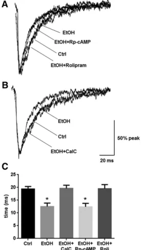
Fig. 4. PKA activation or PKC inhibition mitigate ethanol-induced alterations in mIPSC responses. Whole-cell patch-clamp recordings of cortical neurons were made in the presence of TTX (1mM), CNQX (10mM), and AP-5 (40mM) to isolate GABA mIPSCs after 4-hour exposure to ethanol and/or kinase modulatory drugs. (A) Decay time (decayt1) was significantly decreased by ethanol exposure, an affect that was not affected by coexposure of ethanol with Rp-cAMP; however, coexposure of ethanol with rolipram prevented this decrease. (B) Coexposure of ethanol with CalC also prevented the decrease in decay rate induced by ethanol alone. (C) Quantification of changes in decay t1is shown. (*P , 0.05 compared with control, EtOH plus CalC, or EtOH plus rolipram groups; one-way ANOVA, Bonferroni post-test). Summarized mIPSC metrics are presented in Table 1. AP-5,D-2-amino-5-phosphonopentanoic acid; CNQX, 6-cyno-7-nitroquinoxaline-2,3-dione; Ctrl, control; EtOH, ethanol; Roli, rolipram; TTX, tetrodotoxin.
to 8 per group). No changes in GABAAg2 subunit abundance
were observed in response to ethanol or PKA modulators (Fig. 2, C and D). Nodsubunits were detected in the synaptic fraction (data not shown), although dsubunit abundance is known to decrease after 4-hour ethanol exposure in the extrasynaptic fraction (Carlson et al., 2016).
PKC Activity Mediates Ethanol-Induced Increases in Synaptic GABAA a4 Subunits. Although we previously found increases in P2 fraction levels of GABAAa4 to be caused
by PKC activity (Werner et al., 2011), we next used subcellular fractionation to confirm that these changes occur in the synaptic fraction. Ethanol increased synaptic GABAA a4
levels, an effect that was prevented by inhibiting PKC with CalC (0.3mM; Fig. 3A,P,0.01, one-way ANOVA,F512.53,
P,0.01, Bonferroni post-test,n56 per group). CalC alone had no effect on GABAAa4 subunit abundance. Direct activation of
PKC with the phorbol ester phorbol-12,13-dibutyrate mim-icked the effect of ethanol in increasing synaptic GABAAa4
abundance (Fig. 3B,P,0.01,ttest,n55 to 6 per group).
PKC Inhibition and PKA Activation Prevent Ethanol-Induced Changes in GABA mIPSCs. Finally, we investigated the ramifications of PKA and PKC activity on mIPSC kinetics during 4-hour ethanol exposure. We pre-viously demonstrated that GABA mIPSC decayt1is decreased
after 4-hour ethanol exposure (Werner et al., 2011). Coex-posure of ethanol with Rp-cAMP had no effect on the ethanol-induced decrease in GABA mIPSC decay t1, whereas
coexposure with rolipram prevented the decrease (Fig. 4, A and C,P ,0.001, one-way ANOVA, F5 9.955,P, 0.001, Bonferroni post-test, n 5 11–14 per group). Coexposure of ethanol with CalC prevented the decrease in GABA mIPSC decayt1(Fig. 4, B and C,P,0.001, one-way ANOVA,F5
11.78,P,0.001, Bonferroni post-test,n511–14 per group). There were no other effects on mIPSC kinetics (Table 1).
Discussion
In this study, we demonstrate that PKA and PKC have opposing effects on synaptic GABAA a4 subunit abundance
and function. We elucidate that PKC activation by ethanol
upregulates synaptic GABAA a4 abundance to reduce the
mIPSC decay rate, whereas maintaining PKA activation through phosphodiesterase inhibition prevents these effects. These data further characterize the regulation of a poorly understood GABAA receptor that may be an important
mediator of the chronic effects of ethanol, and these results enhance our understanding of kinase regulation of GABAA
receptors by ethanol (Table 2). Thus, these findings implicate potential therapeutic methods for restoring normal GABAergic functioning after chronic alcohol misuse.
We used a 4-hour in vitro ethanol exposure paradigm that recapitulates changes observed after chronic ethanol con-sumption and withdrawal (Devaud et al., 1997; Kumar et al., 2003; Carlson et al., 2013, 2016). It was not surprising that PKA inhibition during 4-hour ethanol exposure had no effect, because we previously found that PKA abundance (Carlson et al., 2013) and activity (Carlson et al., 2016) are not altered by ethanol exposure for 4 hours, despite the effects of ethanol at 1 hour. Nonetheless, it was possible that earlier PKA activation might produce downstream effects and this possibility was ruled out by testing the effect of PKA in-hibition. Since PKA inhibition had no effect on synaptic GABAA a4 receptor expression, it appears that constitutive
PKA activity does not regulate synaptic GABAAa4 receptors.
The data suggest that PKA activation downregulates synaptic GABAAa4 receptors, because either maintaining PKA
activ-ity through phosphodiesterase inhibition or activating PKA through adenylyl cyclase activation prevented ethanol-induced increases in GABAA a4 subunits. Because GABAA a4bg2 receptors are upregulated in other pathologic condi-tions, including seizure disorders (González and Brooks-Kayal, 2011) and progesterone withdrawal (Gulinello et al., 2002), PKA regulation of these receptors likely has broad implications deserving further exploration.
Our laboratory previously demonstrated that PKC activa-tion by ethanol leads to an upregulaactiva-tion of the abundance of GABAAa4 subunits in vitro after 4 hours, purportedly owing
to an increase in the synaptic population of GABAA a4
receptors (Werner et al., 2011). However, these studies did not isolate synaptic a4 receptors since d subunits were
TABLE 1
GABA mIPSC decay kinetics after exposure to PKA modulators and ethanol
Data are presented as means6S.E.M.
Measure Control Ethanol Ethanol Plus CalC Ethanol Plus Rp-cAMP Ethanol Plus Rolipram
Rise time (ms) 2.461.0 3.161.4 2.860.9 1.961.0 2.660.6 Amplitude (pA) 22.161.2 23.561.0 22.861.9 21.660.9 22.561.1 Frequency (Hz) 1.960.4 1.560.8 2.160.6 1.860.4 2.060.5 Decayt1 19.461.0 12.561.3* 19.661.3 12.461.4* 19.561.6 Decayt2 30.162.3 32.162.6 31.262.5 32.061.9 31.661.4
Number 14 11 11 11 11
*P,0.05 (compared with control, ethanol plus CalC, or ethanol plus rolipram groups, one-way ANOVA, Bonferroni post-test).
TABLE 2
Summary of findings on modulation of GABAAa1 anda4 subunits by ethanol and kinases
Data are presented as means6S.E.M. Downward arrows indicate negative modulation, whereas upward arrows indicate positive modulation. The dash indicates no change.
Subunit Chronic Ethanol PKC PKA
GABAAa1 ↓(Kumar et al., 2003) ↓(Kumar et al., 2010) ↑(Carlson et al., 2013) Extrasynaptic GABAAa4 ↓(Liang et al., 2006)a —(Carlson et al., 2016) ↑(Carlson et al., 2016) Synaptic GABAAa4 ↑(Liang et al., 2006)a ↑(This study) ↓(This study)
detected in the P2 fraction. Our current results, combined with recent findings demonstrating a lack of effect of PKC on extrasynaptic GABAA a4d receptors (Bohnsack et al., 2016;
Carlson et al., 2016), support the hypothesis that PKC effects are specific for the synaptic GABAA a4 receptors in cortical
neurons and that synaptic and extrasynaptic populations of receptors are subject to different methods of regulation. The similarity of ethanol effects in the synaptic fraction and P2 fraction suggests that the P2 fraction largely consists of synaptic components and that the subsynaptic fraction, although func-tionally relevant (Carlson et al., 2016), may not be present in sufficient quantities to confound these results.
Our results suggest that PKA and PKC play oppositional roles in synaptic GABAA a4 receptor regulation, similar to our
previous findings on GABAA a1 receptor regulation (Carlson
et al., 2013). These results are consistent with other studies demonstrating oppositional roles of PKC and PKA activity on GABA receptor functioning (Poisbeau et al., 1999; Brandon et al., 2000; Bohnsack et al., 2016). The observation that PKA activa-tion decreases synaptic a4 abundance could explain why we previously found no difference in whole-cell GABA-evoked current amplitude or GABA dose response after 1 hour of PKA activation, despite an increase in a1 receptor abundance (Carlson et al., 2013). Similarly, the lack of change in GABAA g2 subunit abundance observed in this study is likely attributable to opposing changes ina4bg2 vs.a1bg2 receptors (Kumar et al., 2010). The observation that kinase inhibition in the absence of ethanol had no effect on synaptica4 subunits suggests that these receptors do not undergo constitutive regulation by these path-ways but are only altered after a physiologic insult such as high concentrations of ethanol. These findings are consistent with previous studies in which the PKA RIIb subunit did not constitutively regulatea4 subunits in vivo (Carlson et al., 2014). The more rapid mIPSC decay constants after 4-hour ethanol exposure were consistent with previous findings from our laboratory (Werner et al., 2011). The synaptic GABAA a4
receptors display more rapid decay times than GABAA a1
receptors in recombinant systems (Whittemore et al., 1996; Brown et al., 2002) anda4 knockout mice have longer decay times compared with wild-type mice (Chandra et al., 2006). Thus, the faster decay time after chronic ethanol exposure is consistent with a higher proportion of GABAAa4 receptors in
the synaptic GABA receptor population; however, it will be important to confirm this conclusion in recombinant systems that can isolate synaptica4 receptors physiologically. These results mirror similar studies uncovering reduced decay times after chronic ethanol exposure in the hippocampus (Cagetti et al., 2003; Liang et al., 2006) and during withdrawal from the neuroactive steroid allopregnanolone (Hsu et al., 2003). These changes are correlated with a reduction in the anxiolytic and sleep-inducing effects of ethanol associated with withdrawal and dependence. Thus, the finding that PKC inhibition or PKA activation in the presence of ethanol prevented the faster GABAAmIPSC decay time suggests two possible methods of
preventing pathologic changes associated with ethanol de-pendence. Finally, the correspondence of the functional changes in mIPSCs with the change ina4 subunit expression detected by synaptic fractionation technique further validates the viability of this method of isolating synaptic proteins.
Our results underscore the potential therapeutic relevance for phosphodiesterase inhibition using drugs such as roli-pram. Recent studies in rodent models have demonstrated
decreased drinking behavior in animals given phosphodies-terase inhibitors (Hu et al., 2011; Wen et al., 2012; Blednov et al., 2014; Franklin et al., 2015). Together, these studies suggest that phosphodiesterase inhibition provides a promis-ing target for the treatment of alcohol use disorders. Future in vivo studies examining effects of coadministration of ethanol and rolipram (or administration of rolipram after chronic ethanol) on GABAergic trafficking and GABA-related behav-ior would be a logical extension of these data.
This study expands our understanding of kinase signaling in modulating the GABAergic effects of ethanol. The data sug-gest that PKA activity may prevent alterations of GABAergic inhibition associated with chronic alcohol misuse, whereas PKC activity may facilitate them. These second messenger pathways could provide important targets for treatments to prevent or restore normal GABAAreceptor functioning
asso-ciated with alcohol tolerance, dependence, and withdrawal.
Acknowledgments
The authors thank Todd K. O’Buckley, Raechel McKinley, and Danielle Morrow for technical assistance.
Authorship Contributions
Participated in research design:Carlson, Bohnsack, Morrow.
Conducted experiments:Carlson, Bohnsack.
Performed data analysis:Carlson, Bohnsack.
Wrote or contributed to the writing of the manuscript: Carlson, Bohnsack, Morrow.
References
Blednov YA, Benavidez JM, Black M, and Harris RA (2014) Inhibition of phospho-diesterase 4 reduces ethanol intake and preference in C57BL/6J mice. Front Neurosci8:129.
Bohnsack JP, Carlson SL, and Morrow AL (2016) Differential regulation of synaptic and extrasynaptica4 GABA(A) receptor populations by protein kinase A and protein kinase C in cultured cortical neurons.Neuropharmacology105:124–132. Brandon NJ, Delmas P, Kittler JT, McDonald BJ, Sieghart W, Brown DA, Smart TG,
and Moss SJ (2000) GABAAreceptor phosphorylation and functional modulation in cortical neurons by a protein kinase C-dependent pathway. J Biol Chem275: 38856–38862.
Brown N, Kerby J, Bonnert TP, Whiting PJ, and Wafford KA (2002) Pharmacological characterization of a novel cell line expressing humana(4)b(3)dGABA(A) receptors. Br J Pharmacol136:965–974.
Cagetti E, Liang J, Spigelman I, and Olsen RW (2003) Withdrawal from chronic intermittent ethanol treatment changes subunit composition, reduces synaptic function, and decreases behavioral responses to positive allosteric modulators of GABAAreceptors.Mol Pharmacol63:53–64.
Carlson SL, Bohnsack JP, Patel V, and Morrow AL (2016) Regulation of extra-synaptic GABAAa4 receptors by ethanol-induced protein kinase A, but not protein kinase C activation in cultured rat cerebral cortical neurons.J Pharmacol Exp Ther356:148–156.
Carlson SL, Kumar S, Werner DF, Comerford CE, and Morrow AL (2013) Ethanol activation of protein kinase A regulates GABAAa1 receptor function and traf-ficking in cultured cerebral cortical neurons.J Pharmacol Exp Ther345:317–325. Carlson SL, O’Buckley TK, Thomas R, Thiele TE, and Morrow AL (2014) Altered GABAA receptor expression and seizure threshold following acute ethanol chal-lenge in mice lacking the RIIbsubunit of PKA.Neurochem Res39:1079–1087. Chandra D, Jia F, Liang J, Peng Z, Suryanarayanan A, Werner DF, Spigelman I,
Houser CR, Olsen RW, and Harrison NL, et al. (2006) GABAAreceptora4 subunits mediate extrasynaptic inhibition in thalamus and dentate gyrus and the action of gaboxadol.Proc Natl Acad Sci USA103:15230–15235.
Devaud LL, Fritschy JM, Sieghart W, and Morrow AL (1997) Bidirectional alter-ations of GABA(A) receptor subunit peptide levels in rat cortex during chronic ethanol consumption and withdrawal.J Neurochem69:126–130.
Devaud LL, Purdy RH, Finn DA, and Morrow AL (1996) Sensitization ofg -amino-butyric acidAreceptors to neuroactive steroids in rats during ethanol withdrawal.J Pharmacol Exp Ther278:510–517.
Farrant M and Nusser Z (2005) Variations on an inhibitory theme: phasic and tonic activation of GABA(A) receptors.Nat Rev Neurosci6:215–229.
Franklin KM, Hauser SR, Lasek AW, McClintick J, Ding ZM, McBride WJ, and Bell RL (2015) Reduction of alcohol drinking of alcohol-preferring (P) and high-alcohol drinking (HAD1) rats by targeting phosphodiesterase-4 (PDE4). Psychopharma-cology (Berl)232:2251–2262.
Goebel-Goody SM, Davies KD, Alvestad Linger RM, Freund RK, and Browning MD (2009) Phospho-regulation of synaptic and extrasynaptic N-methyl-d-aspartate receptors in adult hippocampal slices.Neuroscience158:1446–1459.
González MI and Brooks-Kayal A (2011) Altered GABA(A) receptor expression during epileptogenesis.Neurosci Lett497:218–222.
Gulinello M, Gong QH, and Smith SS (2002) Progesterone withdrawal increases the alpha4 subunit of the GABA(A) receptor in male rats in association with anxiety and altered pharmacology - a comparison with female rats.Neuropharmacology43: 701–714.
Hanchar HJ, Chutsrinopkun P, Meera P, Supavilai P, Sieghart W, Wallner M, and Olsen RW (2006) Ethanol potently and competitively inhibits binding of the alcohol antagonist Ro15-4513 to alpha4/6beta3delta GABAAreceptors.Proc Natl Acad Sci USA103:8546–8551.
Hsu FC, Waldeck R, Faber DS, and Smith SS (2003) Neurosteroid effects on GABAergic synaptic plasticity in hippocampus.J Neurophysiol89:1929–1940. Hu W, Lu T, Chen A, Huang Y, Hansen R, Chandler LJ, and Zhang HT (2011)
Inhibition of phosphodiesterase-4 decreases ethanol intake in mice. Psychophar-macology (Berl)218:331–339.
Knoflach F, Benke D, Wang Y, Scheurer L, Lüddens H, Hamilton BJ, Carter DB, Mohler H, and Benson JA (1996) Pharmacological modulation of the diazepam-insensitive recombinant gamma-aminobutyric acidA receptors alpha 4 beta 2 gamma 2 and alpha 6 beta 2 gamma 2.Mol Pharmacol50:1253–1261. Kumar S, Kralic JE, O’Buckley TK, Grobin AC, and Morrow AL (2003) Chronic
ethanol consumption enhances internalization ofa1 subunit-containing GABAA receptors in cerebral cortex.J Neurochem86:700–708.
Kumar S, Porcu P, Werner DF, Matthews DB, Diaz-Granados JL, Helfand RS, and Morrow AL (2009) The role of GABA(A) receptors in the acute and chronic effects of ethanol: a decade of progress.Psychopharmacology (Berl)205:529–564. Kumar S, Suryanarayanan A, Boyd KN, Comerford CE, Lai MA, Ren Q, and Morrow
AL (2010) Ethanol reduces GABAA alpha1 subunit receptor surface expression by a protein kinase Cg-dependent mechanism in cultured cerebral cortical neurons. Mol Pharmacol77:793–803.
Liang J, Suryanarayanan A, Abriam A, Snyder B, Olsen RW, and Spigelman I (2007) Mechanisms of reversible GABAAreceptor plasticity after ethanol intoxication.J Neurosci27:12367–12377.
Liang J, Zhang N, Cagetti E, Houser CR, Olsen RW, and Spigelman I (2006) Chronic intermittent ethanol-induced switch of ethanol actions from extrasynaptic to syn-aptic hippocampal GABAA receptors.J Neurosci26:1749–1758.
Olsen RW and Sieghart W (2009) GABAAreceptors: subtypes provide diversity of function and pharmacology.Neuropharmacology56:141–148.
Papadeas S, Grobin AC, and Morrow AL (2001) Chronic ethanol consumption dif-ferentially alters GABA(A) receptor alpha1 and alpha4 subunit peptide expression
and GABA(A) receptor-mediated 36 Cl(-) uptake in mesocorticolimbic regions of rat brain.Alcohol Clin Exp Res25:1270–1275.
Poisbeau P, Cheney MC, Browning MD, and Mody I (1999) Modulation of synaptic GABAAreceptor function by PKA and PKC in adult hippocampal neurons.J Neurosci19:674–683.
Sundstrom-Poromaa I, Smith DH, Gong QH, Sabado TN, Li X, Light A, Wiedmann M, Williams K, and Smith SS (2002) Hormonally regulateda4b2dGABAAreceptors are a target for alcohol.Nat Neurosci5:721–722.
Tretter V and Moss SJ (2008) GABA(A) receptor dynamics and constructing GABAergic synapses.Front Mol Neurosci1:7.
Wallner M, Hanchar HJ, and Olsen RW (2003) Ethanol enhancesa4b3danda6b3d g-aminobutyric acid type A receptors at low concentrations known to affect hu-mans.Proc Natl Acad Sci USA100:15218–15223.
Wei W, Faria LC, and Mody I (2004) Low ethanol concentrations selectively augment the tonic inhibition mediated by delta subunit-containing GABAAreceptors in hippocampal neurons.J Neurosci24:8379–8382.
Wen RT, Zhang M, Qin WJ, Liu Q, Wang WP, Lawrence AJ, Zhang HT, and Liang JH (2012) The phosphodiesterase-4 (PDE4) inhibitor rolipram decreases ethanol seeking and consumption in alcohol-preferring Fawn-Hooded rats.Alcohol Clin Exp Res36:2157–2167.
Werner DF, Kumar S, Criswell HE, Suryanarayanan A, Fetzer JA, Comerford CE, and Morrow AL (2011) PKCgis required for ethanol-induced increases in GABA(A) receptora4 subunit expression in cultured cerebral cortical neurons.J Neurochem
116:554–563.
Whittemore ER, Yang W, Drewe JA, and Woodward RM (1996) Pharmacology of the human gamma-aminobutyric acidA receptor alpha 4 subunit expressed in Xenopus laevis oocytes.Mol Pharmacol50:1364–1375.
Zhang H and Pandey SC (2003) Effects of PKA modulation on the expression of neuropeptide Y in rat amygdaloid structures during ethanol withdrawal.Peptides
24:1397–1402.