The Immediate Effects of Graston Technique and Self Myofascial Release on Range of Motion and Local Neuromuscular Activity in the Triceps Surae Group.
Emily Gibb
A thesis submitted to the faculty of the University of North Carolina at Chapel Hill in partial fulfillment of the requirements for the degree of Master of Arts in the Department of Exercise & Sport Science in the College of Arts & Sciences.
Chapel Hill 2012
Approved by:
Troy Blackburn, Ph.D, ATC Meredith Petschauer, Ph.D, ATC Barnett Frank, MA, ATC
Table of Contents
CHAPTER I ...1
Variables ...6
Research Objectives ...7
Operational Definitions...9
Delimitations ...9
Limitations ...9
Assumptions...10
Clinical Significance:...11
CHAPTER II...12
Pproperties OF Non-Contractile Soft Tissue: Ligaments, Tendons, and Fascia ....12
Properties of Skeletal Muscle ...12
MUSCLE SPINDLES ...13
MUSCLE STIFFNESS ...14
Soft Tissue Massage and Manual Therapies...16
INSTRUMENT-ASSISTED SOFT TISSUE MASSAGE THERAPIES ...19
Self-Myofascial Release...20
Graston Technique®...22
GT vs. Self-Myofascial Release ...24
CHAPTER III ...27
SUBJECTS...27
Assessments...28
Passive Dorsiflexion Range of Motion Assessment ...28
Spinal Stretch Reflex (SSR)...28
Experimental Protocol ...31
Data Collection, reduction, and Statistical Analysis ...33
CHAPTER IV ...34
CHAPTER V ...40
TABLES...46
FIGURES...51
CHAPTER I INTRODUCTION
Soft tissue based treatments such as stretching and massage have long been a treatment modality employed to address lower extremity functional restrictions in the field of sports medicine and athletic training. Graston Technique® is a type of
instrument-assisted soft tissue massage that aims to identify and treat restrictions in soft tissue, specifically fascia. This technique utilizes several specialized stainless steel tools that have beveled edges and specific treatment surfaces. A clinician must be trained and certified by the Graston company in the use of these instruments in order to perform Graston Technique® on patients. Graston Technique® has carved out a niche in sports medicine and can be found in many athletic training rooms and physical therapy clinics. The use of Graston tools has been growing in popularity over the last several years, and is currently used by over 1,000 outpatient clinics and more than 185 amateur and
to support benefits of this treatment immediately following its administration. In the athletic training setting it is common practice to implement GT prior to a practice or competition. Anecdotally here at The University of North Carolina at Chapel Hill we see that the patients almost always perceive a difference in tissue tightness, pain, and
function immediately following administration of this treatment. However, there is little to no evidence to explain these phenomena. We hoped to provide preliminary research regarding the immediate effects that these treatments have in the population in which they are currently being used.
Self-Myofascial Release (SMR) is a treatment modality that is similarly aimed at correcting dysfunction and restriction in myofascial tissue (Curran, 2008). SMR is a patient self-administered technique that is usually accomplished with the assistance of a foam roller. The use of foam rollers and other types of SMR is commonplace in settings such as fitness centers, athletic facilities, and clinics (Harris, 2002). Similar to GT, patients often report a decrease in perceived tightness, pain and function following
treatment with a foam roller, yet there is no evidence-based explanation for the symptoms these individuals report following either of these treatments.
GT and SMR are often used to treat the same types of soft tissue dysfunction and injury in the collegiate athletic population. However, these treatments differ substantially in terms of both financial and time costs. GT requires a training session and purchase of the tool set. To be certified in the most basic level of GT and purchase a set of tools, the cost is approximately $1,500. GT also requires a clinician’s full attention for one
2
the Graston tools themselves and clinician training. Foam rollers can be purchased for as little as $15, and it is free to educate yourself and potential patients in their proper use. SMR can be administered to more than one patient simultaneously as long as the patients have been educated in its use. These are all important factors in the decision making process regarding the use of these two treatments. Because GT and SMR have similar treatment goals and are often used interchangeably in the clinical setting, it is necessary to compare their effects to enhance clinical decision-making regarding their use. Therefore, this study compared the acute effects of these two treatments on a series of functional outcomes to determine if similar treatment effects can be elicited with the less time intensive and more cost effective SMR technique.
It is generally accepted that soft tissue treatments such as massage and stretching do not enact acute plastic tissue modifications. Instead, it has been proposed that there may be a change in neurophysiological properties that occurs as a result of these types of treatment. In general, previous literature has observed an acute decrease in reflex activity and muscle stiffness that allows for a greater range of motion following stretching
demonstrated that repetitive stretching did have an effect on increasing joint range of motion over a longer period of time compared to a jogging exercises protocol, but that these effects did not have any carry-over effect immediately following treatment (McNair, 1990). GT and SMR are similar in duration and goals of treatment, thus we would expect that they also would fail to cause an acute change in tissue length. Based on previous research, it is unlikely that the acute effects of GT and SMR reported by patients result from a permanent mechanical length change of the myofascial tissue. If we assume that these acute effects are not a result of a mechanical change in tissue, we must
hypothesize that there is some other factor (or set of factors) being affected that is responsible for these changes. Perhaps, rather than a change in tissue length or structure, there is an impact of these treatments on the neurological elements of the
musculotendinous unit which results in the perceived acute effects. This hypothesis can be supported by the findings of previous massage literature, in which massage has been shown to decrease motor neuron pool excitability and muscle stiffness (Goldberg, 1992; Hunter, 2006; Morelli, 1990). There have been many studies that documented the effect of different types of massage (cross friction massage, petrissage, effluerage, etc) on reflex activity and EMG measures. For example, Morelli et al, 1990, concluded that there was a significant decrease in the H-reflex amplitude of the triceps surae during massage in healthy subjects. This study demonstrated that reflex amplitude was decreased during the treatment, but did not have a carry-over effect in the long term. The decrease in activity was only present during the treatment. No decrease was seen in the ten-minute
4
following the treatment. Based on the results of this study, Morelli et al concluded that massage may be compared to other therapeutic modalities that are commonly used in treatment of a hyperexcitable motor neuron pool (Morelli, 1990). Several other studies have come to similar conclusions regarding the decrease of reflex activity and excitability during and immediately following types of massage treatment (Hunter, 2006; Lee, 2009; Weerapong, 2005). Because both GT and SMR are types of massage, we expected to see similar changes in motor neuron pool excitability and stiffness following their use.
Muscle stiffness is defined as the ratio of the change in force to the change in length of a muscle (McNair, 1990). It is a result of both the neural and mechanical (length/tension) contributions to a muscle’s resting state. The mechanical contributions, as stated above, are unlikely to be altered permanently by either of these massage-based treatments. The neural aspect of muscle stiffness is thought to be directly related to the muscle spindle mechanoreceptors within a musculotendinous unit during a stretching episode (Chalmers, 2004). Weerapong et al (2006) suggests that any change in muscle stiffness seen following massage-based interventions may be a result of a change in the level of neural activation (as opposed to a change in tissue length). Therefore, we expected to see a decrease in neural activation and stiffness as a result of our massage-type interventions.
motion could be a result of a decrease in muscle stiffness and motor neuron pool excitability. Several studies have documented that range of motion is increased immediately following a massage or stretching session (Feland, 2001; Marek, 2005; Schilling, 2000). Because of these previous findings, we were interested in determining if there is an immediate effect of our two interventions on dorsiflexion range of motion.
The effect of soft tissue mobilization techniques such as GT and SMR on these neurological aspects of the muscular system should be examined to assist in providing a better understanding of the acute effects these techniques have on muscle function. Both GT and SMR are intended to minimize soft tissue restriction caused by adhesions, overuse, or injury. They are both becoming more prevalent in clinical settings today, yet there is limited research to support their use or to distinguish their relative efficacy. Because there is a significant disparity in the financial and time costs of the two modalities, it is clinically and financially prudent to conduct a study to determine the overall acute effects of GT and SMR application on soft tissue, and whether these effects differ from one another in manipulating muscle stiffness and reflex sensitivity.
Specifically, it is necessary to evaluate if there is a change in neural factors associated with muscle function immediately following treatment. If this is so, it will further guide our clinical and financial decision making with regard to the continued use of GT and SMR.
6
properties of the target tissues, we did not expect to see a permanent significant change in dorsiflexion range of motion. However, an immediate change in range of motion may occur as a result of a reduction in motor neuron pool excitability and stiffness.
Additionally, we administered a Likert scale survey both pre and post treatment to assess the difference in “tightness” that is perceived by the subject.
The purpose of this study was to compare the effects of GT and SMR on passive dorsiflexion ROM, muscle stiffness, spinal stretch reflex amplitude, and patient
perception of “tightness” immediately post-treatment. The effects of these modalities on these outcome variables is clinically relevant because currently there is no strong
evidence base to establish a rationale for either of their use to enact immediate effects. Additionally, because there is a large gap in the financial and time investment
accompanying these two modalities, it is indicated to determine if there is a difference in their effects. We hypothesized that there would be a difference in perceived tightness, ROM, muscle stiffness, and SSR activity following treatment with both GT and SMR as compared to the control condition, but that there would be no differences between the two interventions.
VARIABLES
Independent Variables
i. Intervention (GT, SMR, Control) ii. Time (Pre, Post)
Dependent Variables
1. Bent knee
ii. Perceived treatment efficacy 1. “Tightness” VAS score iii. Spinal stretch reflex sensitivity
1. Amplitude
a. Electromyographic activity of the triceps surae group.
b. Plantarflexion peak force resulting from the spinal stretch reflex
iv. Muscle Stiffness
RESEARCH OBJECTIVES
RQ1: What are the effects of Graston Technique® and Self-Myofascial Release on patient perception of tightness in the triceps surae group immediately following treatment?
RH1: There will be a decrease in patient perception of tightness immediately following treatment of the triceps surae group with Graston Technique® and Self-Myofascial Release.
8
RH2: There will be a decrease in muscle stiffness immediately following treatment of the triceps surae group with Graston Technique® and Self-Myofascial Release.
RQ3: What are the effects of Graston Technique® and Self-Myofascial Release on spinal stretch reflex amplitude, latency, and peak plantarflexion force in the triceps surae group immediately following treatment?
RH3: There will be decrease in spinal stretch reflex amplitude, an increase in latency, and a decrease in force production immediately following treatment of the triceps surae group with Graston Technique® and Self-Myofascial Release.
RQ4: What are the effects of Graston Technique® and Self-Myofascial Release in passive dorsiflexion range of motion immediately following treatment?
RH4: There will be an increase in passive ankle dorsiflexion range of motion immediately following treatment of the triceps surae group with Graston Technique® and Self-Myofascial Release.
RQ5: Do Graston Technique® and Self-Myofascial Release have differential effects on patient perception of tightness, muscle stiffness, spinal stretch reflex sensitivity, and passive dorsiflexion range of motion immediately following treatment to the triceps surae group?
spinal stretch reflex, and passive dorsiflexion range of motion immediately following treatment to the triceps surae group.
OPERATIONAL DEFINITIONS
GT: Treatment with Graston® tools performed by a clinician according to a
specific protocol that was predetermined by the clinician and described in Table 3.
SMR: Patient operated treatment with a foam roller that will follow a protocol established by the clinician and described in Table 4.
Passive Dorsiflexion ROM: Measurement of ankle dorsiflexion with patient seated on a table; as measured with a standard goniometer. The patient will be prone and measurements will be taken with the patient’s knee flexed to 90º of flexion. The patient will be taken to the end range of dorsiflexion by the clinician. Perceived Difference: Patient feedback about the amount of “tightness” they feel following treatment with GT and/or SMR in the form of a visual analog scale.
DELIMITATIONS
a. Using subjects that have varied ankle ROM b. Using subjects that have different levels of initial
restrictions in their triceps surae complex LIMITATIONS
10
c. Inability to standardize application of force during GT treatments both between patients and between treatment sessions
d. Inability to control patient application of force during SMR treatments.
e. Inability to determine whether there is a mechanical change in tissue length (physical lengthening of tissue) following treatment because we will not be monitoring the subjects for an extended period after our final measurements.
ASSUMPTIONS
a. Participants can accurately describe their perceived change following treatment
b. Order of measurement of outcome variables received will not significantly impact the results of this study
c. Crossover effect between treatments will be minimal due to the washout period between interventions
d. Participants will apply a relatively similar amount of pressure during the SMR.
CLINICAL SIGNIFICANCE:
CHAPTER II
REVIEW OF THE LITERATURE
PROPERTIES OF NON-CONTRACTILE SOFT TISSUE: LIGAMENTS, TENDONS, AND FASCIA
Generally, the term soft tissue refers to several types of connective tissue that make up the human body, including ligaments, tendons, fascia, and skeletal muscle. Dysfunction in these tissues can be caused by acute mechanisms of injury or by overuse mechanisms of injury. An acute mechanism of injury is more likely to result in a
structural defect of that tissue (i.e. complete sprain of ligament, rupture of tendon). These types of dysfunction in soft tissue are not the primary focus of this study, although acute injuries can be a mitigating factor in development of eventual overuse tissue dysfunction. These soft tissues are greatly affected by movement of the body because they span joints and are responsible for creating and stabilizing movement. Of primary interest in this investigation is dysfunction of an overactive myofascial tissue and its associated tendon. (Marieb, 2007; Seeley, 2006; Tortora, 1999)
PROPERTIES OF SKELETAL MUSCLE
is called cross bridging. The amount of cross bridging that is achieved during a muscle contraction influences the force and quality of that contraction (Tortora, 1999).
Each muscle has a layer of connective tissue that surrounds it called fascia. This fascia is a continuation of the epimysium, which is the outer layer of muscle tissue. There are two types of fascia, deep fascia and superficial fascia. Deep fascia is found between muscles; superficial fascia is found between muscles and skin (Marieb, 2007). Because it is part of the muscle unit, fascia is subject to all forces that act upon that muscle. Thus, if there is a spasm or trigger point in the muscle, the fascia in proximity to the affected muscle is similarly restricted in that area. For this reason, we refer to some types of massage as a treatment for myofascial dysfunction.
Muscle tissue can become disrupted as a result of acute and chronic injury. Acute injury often consists of a tearing or partial tearing of the muscle fibers. (Hubbard, 1996) As with non-contractile tissue, we are less interested in this type of injury for the purpose of this study.
MUSCLE SPINDLES
14
perturbations or forces acting on the muscle that may cause injury (Kuitunen, 2002). If the muscle spindles are overactive, perhaps as a result of an altered or pathological length tension relationship of the target muscle, it is possible that the spindle’s over-activity could be causing hyper-reflexive contraction of the muscle (Newham, 1997) (Chalmers, 2004). This reflex contraction, if maintained over a period of time due to over-active spindles, could be the “tightness” that is anecdotally reported by active individuals in the clinical setting who seek treatment via GT and administer SMR (Hubbard, 1996; Rivner, 2001). Part of the rationale of using GT and SMR treatments in the clinical setting is to affect the length tension relationships of injured or “tight” muscles. Perhaps we are actually affecting the neural drive to a muscle, and that is what creates a sensation of relief of tightness following these treatments. Past research on the effects of massage has shown consistent decreases in motor neuron pool excitability (Goldberg, 1992; Morelli, 1990; Weerapong, 2005). Because GT and SMR are types of massage therapy, we theorize that they will have similar treatment effects on neural factors.
MUSCLE STIFFNESS
mechanical factors of that muscle. The neural drive to a muscle will determine the amount of contraction and cross bridging of fibers that occurs. (Sheean, 2002; Taylor, 1990) If there is too much neural stimulation, perhaps as a result of overactive afferent mechanoreceptors in that muscle (i.e. muscle spindles), it is possible that there may be an associated increase in cross bridging and a tightness of the muscle that does not allow it to be at its optimum functional level. Similarly, the mechanical length of a muscle can affect the ability of that muscle to contract and maintain an optimum amount of cross bridges.(Taylor, 1990) If the muscle is stretched past its ideal functional length, there will not be enough overlap of fibers to create a strong contraction. If there is too much overlap of these fibers, the muscle will not have the available space it needs to further contract and produce force (Taylor, 1990). It is more likely that the second condition, also referred to as active insufficiency, will contribute to a sensation of “tightness” of the muscle. It is generally accepted that the level of stiffness a muscle has at a given time will determine that muscle’s ability to absorb or dampen forces, which can in turn be related to a
16
SOFT TISSUE MASSAGE AND MANUAL THERAPIES
Basic soft tissue massage and manual therapy techniques are sometimes the first treatment for pain and limited range of motion due to soft tissue dysfunction. Massage therapists have long been present in the field of athletics and orthopedics to address both pathological and non-pathological concerns related to soft tissue. Massage therapy is a respected and accepted method of care for soft tissue injuries among clinicians.
(Weerapong, 2005) There are varying and conflicting theories on why massage is effective at treating these types of injuries.
In massage literature, there is a great base of support for the patient subjective response to massage. This may sometimes be referred to as the placebo effect. A great deal of experimental and inquiry studies have found that patients tend to feel an increase in function and a decrease in pain level following different types of massage treatment. (Gam, 1998; Hernandex-Reif, 2001) Some researchers have determined that this may simply be a human response to receiving tactile treatment, while others believe suggest there may be deeper physiological reasons for this treatment effect. In particular, massage has been shown to cause a decrease in anxiety and depression immediately following its use (Field, 1998). Regardless, this phenomenon is commonly reported following soft tissue massage treatments, and could be a possible reason for a positive patient response following any type of massage application.
result of an increase in temperature (Wiktorsson-Moller, 1983). Although there is some research to support the increase in tissue compliance following temperature increase, it has also been documented that most of the increase in temperature is very superficial in nature and does not have a significant effect on deeper muscles or other structures (Drust, 2002). It should be noted, however, that the triceps surae can be particularly influenced by increases in tissue temperature due to the superficial nature of the musculature and tendon (Wiktorsson-Moller, 1983).
18
tolerance as opposed to a change in physical length of a muscle-tendon unit (Schilling, 2000; Wiktorsson-Moller, 1983).
Of particular interest to this investigation is the effect of massage on neural activity and excitability. Many studies have documented a decrease in and reflex activity following the implementation of massage (Field, 1998; Goldberg, 1992; Hunter, 2006; Morelli, 1990; Sefton, 2011; Weerapong, 2005). This relates to our study because we are interested in seeing if either GT or SMR treatments affect the sensitivity of the spinal stretch reflex. We theorize that GT and SMR may have this treatment effect because past massage literature has shown consistent decreases in motor neuron pool excitability (Goldberg, 1992; Morelli, 1990; Weerapong, 2005). Because GT and SMR are types of massage, we would expect them to have similar treatment effects on neural factors. The majority of these studies documented a decrease in motor neuron pool excitability primarily for the duration of the massage, but did not see a great deal of carry-over or long term effect. Morelli et al, 1990, described an experiment that involved measuring the H-reflex (measure of MNP activity) during a massage. They described a consistent
similarities to the use of transverse friction massage and the use of a treatment such as Graston Technique® and hypothesize that the treatment effects of these two modalities would likely be similar. Although there is a great deal of research that documents the decrease in motor neuron pool excitability during massage, there is also literature that supports the opposite position. Many studies have failed to conclude that there is a significant change in neural factors during or following massage (Hunter, 2006;
Newham, 1997). Hunter et al, 2006, concluded that lower limb massage did not change neuromuscular recruitment and activity, but rather caused a change in muscle
architecture. Regardless, the majority of research that has looked at the effects of massage on neural factors draws the conclusion that further research is necessary.
INSTRUMENT-ASSISTED SOFT TISSUE MASSAGE THERAPIES
20
concentric knee extension were used as the outcome measures. The Stick was not found to make a significant difference in any of these measures, but the authors did express that there was a trend towards improvement in the 20-yard dash measure after self-massage with The Stick (Mikesky, 2002).
Of note, however, are studies that have implemented the use of tools for soft tissue massage in a manner similar to GT. Melham et al. conducted a case study on a patient with ankle pain and scar tissue adhesions that were restricting his ankle range of motion. An intervention of augmented soft tissue mobilization (ASTM) was implemented using specifically designed tools that were very similar to the GT tools, in that they had different shapes and edge beveling designed for certain treatment areas or designed to exert an increased amount of pressure on a focal area of tissue. The patient was treated 2 times per week for 7 weeks. He reported feeling relief, and there were clinically obvious gains in range of motion as well as a decrease in palpable tissue density, but MRI images of the target soft tissue did not reveal a significant change in the amount of scar tissue present as compared to baseline after the completion of the treatment. With regard to a long-term study of this nature, it is possible that the patient would have experienced a relief in symptoms and an increase in range of motion simply due to time, but perhaps the ASTM had a positive effect on the targeted soft tissue. However, the immediate effects of the treatment were not addressed in this study. (Melham, 1998)
SELF-MYOFASCIAL RELEASE
and their surrounding fascia. Fascia, the tissue that surrounds the other tissues of the body to provide support, can become adhered to those tissues which it covers, causing
restrictions that can limit range of motion. Much as GT is aimed at releasing these adhesions with metal tools, a foam roller is intended for this same use by way of self-myofascial release (SMR) (Curran, 2008).
Over time fascial restrictions can cause limitations in range of motion. These restrictions can be caused either by a physical limit to motion, or theoretically by impedance of the neuromuscular efficiency due to a fascial restriction (Barnes, 1997). The treatment involves holding a sustained pressure over the target area for 90-120 seconds, after which a release of the tissue should occur (due to a break up of a true tissue adhesion) that allows the pressure to be pushed deeper into the soft tissue. This release is due to the break up of a true tissue adhesion. After this process is repeated several times, the initial restriction should be less palpable, if it still exists at all (Barnes, 1997). The goal of myofascial release is to create permanent tissue changes and elongation (Barnes, 1997).
22
myofascial release to attempt to decrease the restriction. This involves remaining stationary on a restricted spot for 90-120 seconds and allowing for a release to occur within the soft tissue. Once release has occurred, the patient moves on to find other restrictions in the soft tissue, again by rolling and changing body position atop the foam roller (Curran, 2008).
GRASTON TECHNIQUE®
The GT company has established protocols for specific regions of the body. Overall, these protocols are intended to be of use for more than one intervention session in order to establish long-term changes in the target soft tissue. GT markets their treatment as a way to eliminate fascial and scar tissue restrictions that may be causing pain or
preventing full range of motion of a joint to occur. The company describes its treatment as being aimed at the potential fascial restrictions that can arise surrounding muscle, tendon, and ligamentous structures in the body. According to the manual used in the basic clinician training session, GT is meant to expand on the principles of transverse or cross friction massage. They maintain that this technique can be appropriate for either a chronic or an acute injury. The manual used in the M1 training session, which is the basic session that all clinicians trained in GT begin with, does not include specific recommendations for the number of interventions necessary to produce the desired tissue changes. According to the description of the treatment that is available on the GT commercial website, adhesions and scar tissue will be eliminated “over time”
are seen “over time” as opposed to immediately. Again, there is no specific number of treatments outlined for the desired effect to occur. Regardless of the specific region that the treatment is targeted at, GT recommends that the clinician follow the same basic outline to govern each treatment. In the M1 manual, these steps are described as the basic components of GT. Initially it is recommended that the patient participate in a short soft tissue warm up session. This may include the use of heat modalities such as ultrasound or moist heat packs. This warm up may also be in the form of a cardiovascular session on a bike or treadmill to establish more systemic tissue heating. Following this warm up session, the clinician will administer the GT. It is recommended by the GT company that this treatment session is followed with both stretching and strengthening exercises, as well as some form of cryotherapy to prevent soreness in the target area. The GT company does not endorse or recommend a specific stretching or strengthening program to follow each treatment, but merely indicates that there is physiological rationale for their
inclusion following each treatment with GT tools (Graston, 2010).
24
alteration to the mechanoreceptor activity in the local area of target tissue. There is no research to support the immediate benefits or effects of GT on any joint or region of the body.
The research that does exist regarding GT is largely made up of case studies and small-scale experiments (Burke, 2006; Hammer, 2008; B. Looney, Srokose, DC., Fernandez-de-las-Penas, C., Cleland, JA. , 2011). Burke et al evaluated the efficacy of GT on carpal tunnel syndrome and determined that it was in fact effective, but the
immediate post treatment effects of the treatment were not addressed. A test protocol that called for 2 treatments per week for the first 4 weeks, and 1 treatment per week for the last 2 weeks was used in this study. (Burke, 2006) Hammer et al investigated the use of GT on patients with plantar fasciitis and related heel pain. Again, the treatment
intervention was established as a series of 8 treatments over the course of 3-8 weeks. This treatment was aimed at the triceps surae and other lower leg musculature. Static
stretching repetitions and cryotherapy were also included in this intervention. 70% of patients in this study experienced relief of pain by the end of the intervention period, but no mention is made of their perceived symptom difference immediately following
treatment with GT. (B. Looney, Srokose, DC., Fernandez-de-las-Penas, C., Cleland, JA. , 2011)
GT VS. SELF-MYOFASCIAL RELEASE
must perform GT every time it is administered. This treatment session may last up to 15-20 minutes including preparation and post-treatment cleaning of tools and patient table. Conversely, SMR only requires a clinician to instruct the patient during the first treatment session. Following the first session, the patient can perform this treatment on his or her own. Obviously, this presents a vast difference in need for clinician time and availability. In a clinic or athletic training room setting where clinician time is limited, or the patient population vastly overwhelms the number of clinicians, this is an important factor to take into consideration. Additionally, it is not just clinician time and availability that must be taken into account, but also the fatigue and potential wear put on the clinician from administering GT. Although part of the rationale behind the design of the GT tools is to decrease the stress on the clinician from performing the treatment (GT M1 Manual), there is obviously more stress on the clinician from a GT treatment as opposed to a patient administered SMR treatment. Again, in a clinical setting where multiple patients may require soft tissue intervention, this may become a factor.
26
between the two modalities, we cannot be sure if we are sacrificing a potential valuable intervention option by selecting SMR based solely on price.
Despite these two main differences in our intervention modalities of interest, they are used similarly in a clinical setting. Often, a patient will receive GT or SMR before going to practice or to a game. Both treatment modalities are marketed to the active public as being used for the treatment of muscle and connective tissue dysfunction, and utilize similar principles (the direct pressure of a device to the target treatment area). SMR and GT are sometimes used interchangeably for patients with myofascial dysfunction.
CHAPTER III METHODOLOGY
SUBJECTS
Twenty healthy individuals (10 males, 10 females) were recruited from the
undergraduate population at UNC-CH to participate in this study. These twenty participants ranged in age from 18 – 25 years to reflect a young, active population. Inclusion criteria for participation in this study required that each subject be recreationally active, participating in at least 3 periods of exercise or physical activity per week each lasting 30 minutes or more. Exclusion criteria included a history of neurological disorder, or lower extremity surgery or injury within the three months prior to participation. Additionally, we excluded subjects who were currently experiencing lower extremity injury because of the possibility of range of motion, muscle stiffness, and neural activity changes that could be the result of edema, active inflammation, or pain. The purpose of these exclusion criteria was to eliminate potential sources of confounding variables from a pre-existing or recent injury that may influence range of motion, muscle stiffness, or reflex activity in our subjects.
The subjects (n = 20) were separated into two groups (n1 = 10, n2 = 10) using a
28
the order in which the two interventions were administered. Group 1 received GT treatment during session 1 and performed the SMR protocol during treatment session 2. Group 2 performed the SMR protocol in treatment session 1 and received GT in treatment session 2.
ASSESSMENTS
Passive Dorsiflexion Range of Motion Assessment
Prone on a table. Measurements were taken with the participant’s knee in 90 degrees of flexion. A standard digital inclinometer was used for this assessment. With the ankle and knee placed at 90 degrees of flexion, the inclinometer was placed atop the participant’s foot and zeroed. The clinician then passively brought the ankle into the maximum available amount of dorsiflexion, stopping at the first point of restriction. The degree value of
dorsiflexion was read with the inclinometer still positioned atop the ankle. Three trials of this measure were performed and averaged for statistical analyses.
Spinal Stretch Reflex (SSR)
30 Muscle Stiffness
The methodology for the muscle stiffness procedure was based on that of Blackburn et al. (J. Blackburn, Padua, DA., Guskiewicz, KM., 2008) Muscle stiffness data were collected using a force plate. EMG data were also collected during these measures, with the same electrode and muscle arrangement as was used in the SSR testing. The subject was positioned in a loading device with the hip, knee, and ankle joints in 90° of flexion. The metatarsal heads were positioned on a wooden block, which was fixed to the force plate. First, we obtained maximal voluntary isometric contraction (MVIC) values for each subject. To do this, we positioned the subject’s ankle at 90° by placing a block under the calcaneus. The subject’s knee was secured with a webbing strap that was attached to a block that was secured to the force plate underneath the subject’s seat. This strap will secure the subject’s knee and shank so the contraction is truly isometric. The subject was then asked to
plantarflex as forcefully as possible against the wooden block. This test was repeated five times per subject. From these data we determined the average MVIC force for each subject. We used 30% of each subject’s MVIC as the applied load for the stiffness testing protocol.
limb. A perturbation was manually applied in an inferior direction on top of the loading device, directly over the subject’s knee and with the force directed downward through the vertical/long-axis of the shank. The perturbation was applied by a single clinician, in an effort to maintain perturbation force consistent throughout testing. The perturbation
forcefully lengthened the triceps surae and caused oscillatory flexion/extension motion about the ankle joint. Additionally, these measures were practiced during pilot testing. We
performed 5 repetitions of this testing. The reaction force of the metatarsal heads on the force plate was analyzed and muscle stiffness of the triceps surae was calculated by finding the time difference between the first two oscillatory peaks (t2-t1) and using it to calculate damped oscillatory frequency (f) using the equation f = 1/(t2-t1). Once we obtained the frequency, we used it to find the stiffness (k) in the equation k = 4n2mf2 (J. Blackburn, Norcross, MF., Padua, DA., 2011). To calculate muscle stiffness, we evaluated the damped oscillatory motion about the ankle joint using the GRFv. Because the motion about the ankle is measured in this manner, we can calculate stiffness using the frequency of the oscillatory motion.
The intra-session reliability of muscle stiffness measurements has been shown in previous studies to have an intra-class correlation coefficient of up to 0.89. In our study we were as consistent as possible with all set-up to further ensure reliability between testing sessions. (J. Blackburn, Padua, DA., Guskiewicz, KM., 2008)
EXPERIMENTAL PROTOCOL
32
prior to participation. After informed consent was obtained, participants warmed-up on a stationary cycle ergometer for 5 min. Passive dorsiflexion range of motion, triceps surae stiffness, and spinal stretch reflex characteristics were then assessed as described in detail above. Participants then remained in the reflex testing position quietly for 6 minutes after which these measures were repeated. These measures provided “control data”, with the difference between these measures representing the changes in the dependent variables attributable to the passage of time and repeated measurement (i.e. pre-test sensitization). The second set of measurements also served as the pre-test values for the first treatment.
Following collection of control data the participant completed a ten point likert scale indicating his or her perceived amount of “tightness” pre-treatment. After completing this survey, the participant received GT or performed a SMR protocol on the triceps surae complex. Participants were shown an instructional video prior to the treatment on the day they performed the SMR protocol in an attempt to standardize the performance of this treatment. The specific treatment protocols are outlined in Tables 1, 3 and 4. The intervention the participant received during treatment session 1 was dependent on group assignment. Following the intervention, passive dorsiflexion range of motion, muscle stiffness, and stretch reflex amplitudes and forces were measured again, as well as the ten point Likert scale.
performed the SMR protocol based on group assignment. Following the intervention, all measures were re-assessed concluding treatment session 2.
DATA COLLECTION, REDUCTION, AND STATISTICAL ANALYSIS
All data were sampled using The Motion Monitor software (Innovative Sports Training, Chicago, IL, USA) and processed with custom-developed software (LabVIEW, National, San Antonio, TX, USA). EMG and load cell data were sampled at 1000 Hz. Load cell data were lowpass filtered at 10 Hz using a 4th order, zero-phase lag Butterworth filter.
EMG data were bandpass filtered at 20-350 Hz and smoothed using a 20 ms root-mean-square sliding window function. Change scores between pre to post test on all dependent variables (VAS survey, dorsiflexion range of motion, muscle stiffness, spinal stretch reflex amplitude) were calculated. We then ran dependent-samples T-tests to demonstrate that the control sessions did not differ from one another. The data from the two control sessions were averaged for each subject and the mean value was used for comparison of the control
CHAPTER IV RESULTS
Twenty healthy participants (age = 21.8±2.8; mass = 69.2±14.5), ten men (age = 22.6±2.9; mass = 76.7±11.0) and ten women (age = 21±2.7; mass = 61.8±14.1), who fit our inclusion criteria, completed this study. All participants completed both sessions of testing in their entirety.
LIKERT SCALE DATA (PARTICIPANT PERCEIVED CHANGE IN TIGHTNESS) The Likert scale survey was administered to each participant immediately prior to and following the performance of both the Graston Technique® and SMR treatments. The
differences between the scores for the two treatments were calculated using a dependent samples T-test. Results indicated a significantly different decrease in perceived level of tightness following the Graston Technique® treatment (M= -2.5, SD = 1.732) over the SMR treatment (M = -1.65, SD = .988), t (19) = -2.203, p = .040.
participant reaction to the Graston Technique® treatment protocol, as it has not been previously documented in research literature and is part of the primary basis of our hypotheses for this larger study.
DORSIFLEXION RANGE OF MOTION
The first statistics run on the DF ROM data were to determine whether or not there was a difference in participant dorsiflexion from session one to session two. This test was performed to assure that any ultimate differences in dorsiflexion motion were not a result of a difference in condition when reporting for individual sessions. To do this, we ran a dependent samples T-Test between the control scores for session one and session two for dorsiflexion range of motion. The results indicated that there was no difference between participant scores on the Graston Technique® test session (M= 21.78, SD = 4.806) and the SMR test session (M = 21.85, SD = 5.317), t (19) = .125, p = .902.
36
us to combine these two control change scores to create one average control score for use in the analysis of variance that was performed on the range of motion data (Figure 6).
Finally, a repeated measures analysis of variance was performed to compare the changes in dorsiflexion between control, Graston Technique® and SMR sessions. The results of this analysis and post hoc testing using Tukey’s HSD showed that there existed a
significant main effect of treatment on dorsiflexion range of motion (F (2,38) = 11.949, p <.001)) between control change scores (M = 0.830, SD = .65877) and both Graston
Technique® change scores (M = 1.816, SD= 1.387) and SMR change scores ( M = 2.0, SD = 1.562) (Figure 3). There was no significant difference between the change scores for GT and SMR. 95% confidence intervals were also analyzed (control [-.225, .391], GT [1.167, 2.465], SMR [1.269, 2.732].
REFLEX FORCE
A repeated measures analysis of variance was used to analyze the reflex force data for the control change scores, GT change scores, and SMR change scores. The results of this analysis did not demonstrate a significantly different effect of treatment on reflex force (F (2,38) = .106, p =. 9)) between control change scores (M = 4.52, SD = 13.8), Graston Technique® change scores (M = 4.12, SD= 19.7) and SMR change scores (M = 6.5, SD = 15.45).
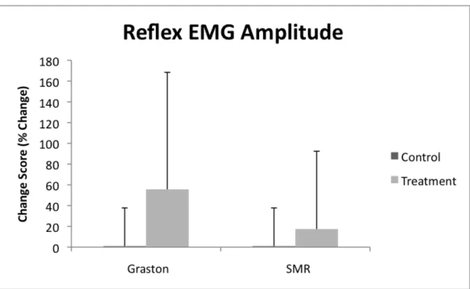
REFLEX EMG AMPLITUDE
As with the other outcome variables, it was necessary to first run a dependent samples T-Test to determine that there was no difference between control scores between sessions one and two. The results of this analysis demonstrated that there existed no significant
difference between participant scores on the Graston Technique® test session (M= 28.68, SD = 112.9 and the SMR test session (M = 21.86, SD = 112.2), t (19) = .191, p = .851. After this procedure, the control scores for sessions one and two were averaged to create one overall control score for reflex EMG amplitude.
38
experienced a decrease in reflex EMG amplitude and 8 participants experienced an increase in reflex EMG amplitude. Using SPSS statistical software we identified 5 cases that were determined to be outliers. There did not appear to be a commonality among these cases (sex, treatment order, etc) that could have caused them to be outliers. These five cases were removed from the data set and the analysis of variance was performed again with these cases excluded. The results of the second ANOVA (Figure 5) also failed to show a significant difference of effect of treatment on reflex EMG amplitude (F (2,28) = .1.713, p = .199)) between control change scores (M = 1.01, SD = 36.8), Graston Technique® change scores (M = 55.7, SD= 112.67) and SMR change scores (M = 17.4, SD = 74.9).
REFLEX BACKGROUND EMG & REFLEX HAMMER FORCE
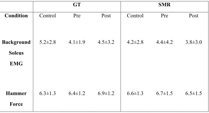
These data were analyzed to confirm that the lack of change in reflex EMG amplitude and force data were not caused by a difference in background EMG and reflex hammer force across conditions. To compare these data we ran a repeated measures analysis of variance across conditions. There was no difference in either background EMG or reflex hammer force across conditions (Table 5), therefore we can conclude that the lack of a significant difference in our primary reflex outcome measures (reflex force and EMG amplitude) were not a result of a difference in background EMG and reflex hammer force.
MUSCLE STIFFNESS
Prior to completing muscle stiffness testing for each condition in each testing session a set of MVIC trials were performed. When compared following completion of data
CHAPTER V DISCUSSION
To our knowledge, this is the first study to compare the effects of Graston Technique® and self-myofascial release on range of motion and neural factors in the triceps surae group. Our primary findings support several of our research hypotheses. We were correct in predicting that there would be a change in patient perception of tightness following administration of the Graston Technique® treatment, however we were incorrect to also predict a significant difference in perception of tightness following the SMR treatment. The change seen in the general collegiate population through the use of our Likert scale tool was mirrored in the results of this study. This supports the basic rationale behind this project in that there seems to exist a general belief that Graston Technique® is more effective than other treatments in reducing tightness and increasing mobility. The basis for the theory behind the other outcome measures included in this research was based on the assumption that this was the case.
not supported by the results of our reflex force and EMG data. We must, therefore, look to alternate explanations to explain the significant differences found in patient perception of change and dorsiflexion range of motion.
There are several possible explanations for the fact that participants seemed to feel a greater change in “tightness” following the GT session versus the SMR session. First, it is important to recognize that one treatment (GT) is clinician controlled with regards to applied pressure and application of treatment, whereas one treatment (SMR) is patient controlled and is therefore less regulated. Although the two treatment protocols were created by the primary investigator with the intention of being as similar as possible, the inherent differences in the two massage modalities make exact symmetry of treatment impossible. During the GT treatment session the clinician identified restricted areas through the protocol described in Table 3 and treated those areas accordingly. During the SMR protocol, the participant was asked to identify restricted areas based on their subjective feelings during the treatment and then to treat those areas accordingly based on the protocol outlined in Table 4. It is
42
patient perception is the possible placebo effect created by the implementation of a treatment modality by a clinician (as opposed to a self-imposed treatment session). The placebo effect is most accurately described as a psychobiological phenomenon that can be attributed to different mechanisms. (Benedetti, 2005) In general it describes an improvement brought about not by a specific treatment or therapy but by the patient’s expectation of improvement simply because they receive treatment. (Benedetti, 2005; Brown, 1998) Based on the theory behind the placebo effect, it is logical to conclude that a patient would perceive his or herself to have a greater level of improvement following a clinician-administered treatment than a self-administered one. We must also consider that there may have been differences in participant level of activity prior to and between each testing session. Because this was a small scale pilot-type study, there were no restrictions on level of activity (extreme exercise, etc) between the testing sessions. This may have had an effect on the outcome of the second treatment session – particularly if a participant had performed a great deal of physical activity in the days prior to one testing session but not the first. There may have also existed
differences in the patient populations that we were unable to screen for due to the small scope of this study. These things could include tensile tissue strength, ability to tolerate discomfort, and inherent flexibility. It is logical to conclude that any of these things could have affected the outcome of the study if they differed between participants.
previously, the muscle stiffness data could not be analyzed so we are unable to draw conclusions regarding that outcome variable.
If we exclude neural changes as a possible reason for the increase in range of motion, we are still left with several possible causes. First of all, during the course of the stiffness, MVIC, and reflex procedures there was a great deal of manipulation of the ankle joint. It is possible that during these procedures we affected the stretch tolerance of the muscle unit without actively lengthening the muscle tissue. Previous literature has found that after a period of passive stretching there is an increase in stretch tolerance without an increase in tissue length or a change in neural properties. (Folpp, 2006; Halbertsma, 1994; S. S.
44 LIMITATIONS
We acknowledge that there were several limitations associated with our study. First and foremost, the inability to draw conclusions from the muscle stiffness data was a large limitation because it was intended to be a third of our outcome data. Secondly, the reliability of the reflex hammer was not as high as we hoped it would be and created a great deal of variability in our data set. Additionally, the inability to control the amount of pressure exerted on the foam roller by the participants during the SMR treatment could have greatly influenced the results of the likert survey. It is also important to note that the GT manual and instructional material call for use of GT on a long-term basis and do not make mention of any intended or reported effects of immediate use. In this study, we used GT as it is commonly used in the clinic and as we have seen it to be effective immediately on patients. The way that GT was used in this research protocol was correct with regards to technique and patient instruction, but was not in line with the longer term protocol that the GT company markets.
CONCLUSIONS
46 TABLES
Table 1: PROCEDURES
Treatment Session One Treatment Session Two Group One
(N=10; 5 males, 5 females)
1. 5 Min Bike
2. ROM, MVIC, MS, SSR measured 3. 5 minute waiting
period
4. ROM, MVIC, MS, SSR measured 5. Likert Survey 6. SMR
7. Likert Survey, ROM, MVIC, MS, SSR measured
1. 5 Min Bike
2. ROM, MVIC, MS, SSR measured 3. 5 minute waiting
period
4. ROM, MVIC, MS, SSR measured 5. Likert Survey 6. GT
7. Likert Survey, ROM, MVIC, MS, SSR measured Group Two
(N=10; 5 males, 5 females)
1. 5 Min Bike
2. ROM, MVIC, MS, SSR measured 3. 5 minute waiting
period
4. ROM, MVIC, MS, SSR measured 5. Likert Survey 6. GT
7. Likert Survey, ROM, MVIC, MS, SSR measured
1. 5 Min Bike
2. ROM, MVIC, MS, SSR measured 3. 5 minute waiting
period
4. ROM, MVIC, MS, SSR measured 5. Likert Survey 6. SMR
Table 2: SUMMARY
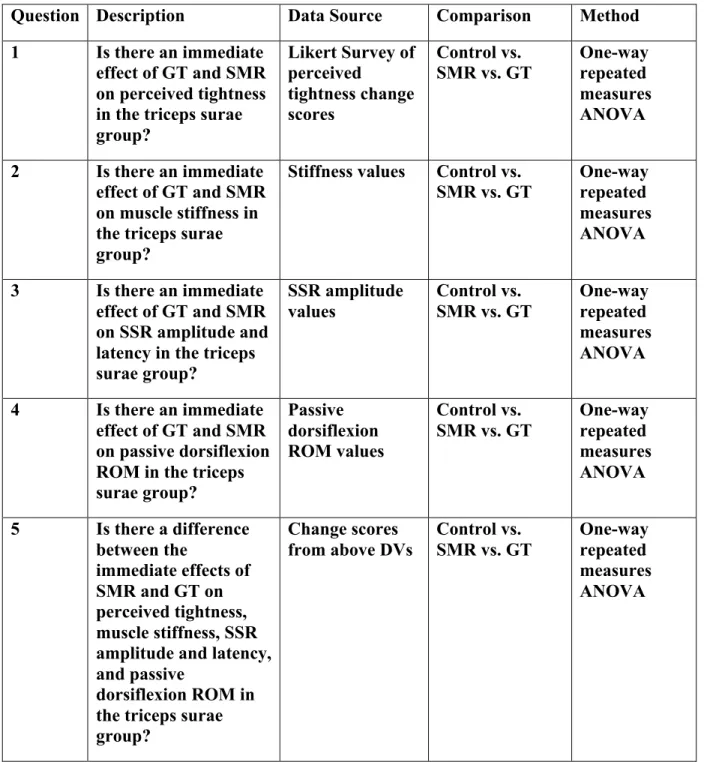
Question Description Data Source Comparison Method 1 Is there an immediate
effect of GT and SMR on perceived tightness in the triceps surae group?
Likert Survey of perceived
tightness change scores
Control vs. SMR vs. GT
One-way repeated measures ANOVA
2 Is there an immediate effect of GT and SMR on muscle stiffness in the triceps surae group?
Stiffness values Control vs.
SMR vs. GT One-way repeated measures ANOVA
3 Is there an immediate effect of GT and SMR on SSR amplitude and latency in the triceps surae group?
SSR amplitude values
Control vs. SMR vs. GT
One-way repeated measures ANOVA
4 Is there an immediate effect of GT and SMR on passive dorsiflexion ROM in the triceps surae group?
Passive dorsiflexion ROM values
Control vs. SMR vs. GT
One-way repeated measures ANOVA
5 Is there a difference between the
immediate effects of SMR and GT on perceived tightness, muscle stiffness, SSR amplitude and latency, and passive
dorsiflexion ROM in the triceps surae group?
Change scores from above DVs
Control vs. SMR vs. GT
48 Table 3: GRASTON TREATMENT PROTOCOL Graston Treatment 6 Minutes Total
1 Min General scan with GT-1 to
entire body of gastroc/soleus
1 Min Targeted scan with GT-4 to
lateral head gastroc and medial head gastroc
2 Min Treatment of identified
restrictions with GT-3
1 Min Treatment to MT junction
and achilles tendon with GT-6
1 Min General wrap-up scan of
Table 4: SMR TREATMENT PROTOCOL SMR Treatment 6 Minutes Total
1 Min General scan with GT-1 to
entire body of
gastroc/soleus using foam roller
1 Min Targeted scan with GT-4 to
lateral head gastroc and medial head gastroc
2 Min Treatment of identified
restrictions with 30 second hold over area (3-4 areas total)
1 Min Treatment to MT junction
and achilles tendon
1 Min General wrap-up scan of
50
Table 5: Background Soleus EMG & Hammer Force Data
GT SMR
Condition Control Pre Post Control Pre Post
Background Soleus
EMG
5.2±2.8 4.1±1.9 4.5±3.2 4.2±2.8 4.4±4.2 3.8±3.0
Hammer Force
FIGURES
56 Figure 6: Change Score Set-Up
CNTRL SESSION 1 CNTRL SESSION 2 GT SMR
⇓ ⇓ ⇓ ⇓ ⇓ ⇓ ⇓ ⇓ ⇓ ⇓ ⇓ ⇓
PAIRED SAMPLES T TEST ⇓ ⇓
P < .05 ⇓ ⇓
AVERAGE CNTRL ⇓ ⇓
⇓ ⇓ ⇓
⇓ ⇓ ⇓
REFERENCES
Barlow, A., Clarke, R., Johnson, N., Seabourne, B., Thomas, D., Gal, J. (2004). Effect of massage of the hamstring muscle on performance of the sit and reach test. Br J Sports Med, 38.
Barnes, M. (1997). The basic science of myofascial release: morphologic change in connective tissue. Journal of Bodywork and Movement Therapies, 1(4), 231-238. Benedetti, F., Mayberg, H., Wager, T., Stohler, C., Zubieta, JK. . (2005).
Neurobiological Mechanisms of the Placebo Effect. The Journal of Neuroscience, 25(45).
Blackburn, J., Norcross, MF., Padua, DA. (2011). Influences of hamstring stiffness and strength on anterior knee joint stability. Clinical Biomechanics, 26.
Blackburn, J., Padua, DA., Guskiewicz, KM. (2008). Muscle Stiffness and Spinal Stretch Reflex Sensitivity in the Triceps Surae. Journal of Athletic Training, 43(1), 29-36.
Brown, W. A. (1998). The placebo effect. Scientific American.
Burke, J., et al. . (2006). A Pilot Study Comparing Two Manual Therapy Interventions for Carpal Tunnel Syndrome. Journal of Manipulative and Physiological
Therapeutics, 30(1), 50-61.
Chalmers, G. (2004). Re-examination of the possible role of Golgi tendon organ and muscle spindle reflexes in proprioceptive neuromuscular facilitation muscle stretching. Sports Biomech, 3(1), 159-183.
Chan, A., Myrer, JW., Measom, GJ., Draper, DO. . (1998). Temperature Changes in Human Patellar Tendon in Response fo Therapeutic Ultrasound. Journal of Athletic Training, 33(2), 130-135.
Curran, P., Fiore, R., Crisco, J. (2008). A Comparison of the Pressure Exerted on Soft Tissue by 2 Myofascial Rollers. Journal of Sport Rehabilitation, 17, 432-442. Drust, B., Atkinson, G., Gregson, W., French, D., Binningsley, D. (2002). The Effects of
58
Feland, J., Myrer, JW., Merrill, RM. . (2001). Acute Changes in Hamstring Flexibility: PNF versus static stretch in senior athletes. Physical Therapy in Sport, 2, 186-193. Field, T. (1998). Massage Therapy Effects. American Psychologist, 53(12), 1270-1281. Folpp, H., Deall, S., Harvey, LA., Gwinn, T. (2006). Can apparent increases in muscle
extensibility with regular stretch be explained by changes in tolerance to stretch? Australian Journal of Physiotherapy, 52.
Gam, A., et al. . (1998). Treatment of myofascial trigger-points with ultrasound combined with massage and exercise - a randomised controlled trial. Pain, 77, 73-79.
Goldberg, J., Sullivan, J., Seaborne, D. (1992). The Effect of Two Intensities of Massage on H-Reflex Amplitude. Physical Therapy, 72(6), 50-59.
Graston. (2010). M1 - Basic Training.
Halbertsma, J., Goeken, LNH. (1994). Stretching Exercises: Effect on Passive
Extensibility and Stiffness in Short Hamstrings of Healthy Subjects. Arch Phys Med Rehabil, 75.
Hammer, W. (2008). The effect of mechanical load on degenerated soft tissue. Journal of Bodywork and Movement Therapies, 12, 246-256
.
Harris, R. E., Clauw, D.J. . (2002). The Use of Complementary Medical Therapies in the Management of Myofascial Pain Disorders. Current Pain and Headache Reports, 6, 370-374.
Hermens, H., B. Freriks, et al. (2000).
Hernandex-Reif, M., Field, T., Krasnegor, J., Theakston, H. (2001). Lower back pain is reduced and range of motion increased after massage therapy. Intern J.
Neuroscience, 106, 131-145.
Howitt, S., Wong, J., Zabukovec, S. . (2006). The conservative treatment of Trigger Thumb using Graston Techniques and Active Release Techniques. J Can Chiropr Assoc, 50(4), 249-254.
Hubbard, D. R. (1996). Chronic and Recurrent Muscle Pain: Pathophysiology and Treatment, and Review of Pharmacologic Studies. Clinical Overview and Pathogenesis, 123-143.
Hunter, A., Watt, J., Watt, V., Galloway, S. (2006). Effect of lower limb massage on electromyography and force production of the knee extensors. Br J Sports Med, 40, 114-118.
Irnich, D. e. a. (2001). Randomised trial of acupuncture compared with conventional massage and "sham" laser acupuncture for treatment of chronic neck pain. BMJ, 322.
Kuitunen, S., Komi, P., Kyrolainen, H. . (2002). Knee and Ankle Joint Stiffness in Sprint Running. Medicine & Science in Sports & Exercise, 166-173.
Lee, H., Wu, SK., You, JY. . (2009). Quantitative application of transverse friciton massage and its neurological effects on flexor carpi radialis. . Manual Therapy, 14, 501-507.
Lewek, M., Breslin, R., Hlad, L., Lanton, A., St. John, J. (2010). Non-paretic quadriceps activity influences paretic quadriceps activity post-stroke. Clinical
Neurophysiology, 121, 1962-1967.
Looney, B., Srokose, DC., Fernandez-de-las-Penas, C., Cleland, JA. (2011). Graston Instrument Soft Tissue Mobilization and Home Stretching for the Management of Plantar Hell Pain: A Case Series. Journal of Manipulative and Physiological Therapeutics, 34(2), 138-142.
Looney, B., Srokose, DC., Fernandez-de-las-Penas, C., Cleland, JA. . (2011). Graston Instrument Soft Tissue Mobilization and Home Stretching for the Management of Plantar Heel Pain: A Case Series. Journal of Manipulative and Physiological Therapeutics, 34(2), 138-142.
Magnusson, S. (1998). Passive properties of human skeletal muscle during stretch maneuvers. Scand J Med Sci Sports, 8.
Magnusson, S. S., E., Aagaard, P, Sorensen, H., Kjaer, M. . (1996). A mechanism for altered flexibility in human skeletal muscle. Journal of Physiology, 497(1). Marek, S., et al. . (2005). Acute Effects of Static and Proprioceptive Neuromuscular
Facilitation Stretching on Muscle Strength and Power Output. Journal of Athletic Training, 40(2), 94-103.
60
McNair, P., Stanley, S. (1990). Effect of passive stretching and jogging on the series elastic muscle stiffness and range of motion of the ankle joint. Br J Sports Med, 30, 313-318.
Melham, T., Sevier, TL., Malnofski, MJ., Wilson, JK., Helfst, RH. . (1998). Chronic ankle pain and fibrosis successfully treated with a new noninvasive augmented soft tissue mobilization technique (ASTM): a case report. Medicine & Science in Sports & Exercise, 30(6), 801-804.
Mikesky, A., Bahamonde, R., Stanton, K., Alvey, T., Fitton, T. (2002). Acute Effects of The Stick on Strength, Power, and Flexibility. Journal of Strength and
Conditioning Research, 16(3), 446-450.
Morelli, M., Seaborne, D., Sullivan, J. (1990). Changes in H-Reflex Amplitude During Massage of Triceps Surae in Healthy Subjects. JOSPT, 12(2), 55-59.
Newham, D., Lederman, E. . (1997). Effect of manual therapy techniques on the stretch reflex in normal human quadriceps. Disability and Rehabilitation, 19(8), 326-331. Rivner, M. H. (2001). The Neurophysiology of Myofascial Pain Syndrome. Current Pain
and Headache Reports, 5, 432-440.
Safran, M., Garrett, W., Seaber, A., Glisson, R., Ribbeck, R. . (1988). The role of warmup in muscular injury prevention. American Journal of Sports Medicine, 16(2).
Schilling, B., Stone, MH. . (2000). Stretching: Acute Effects on Strength and Power Performance. Strength and Conditioning Journal, 22(1), 44-47.
Seeley, R., Stephens, TD., Tate, P. (2006). Anatomy & Physiology (Vol. 7th). Boston: McGraw Hill.
Sefton, J., Yarar, C., Carpenter, DM., Berry, JW. (2011). Physiological and clinical changes after therapeutic massage of the neck and shoulders. Manual Therapy, 16, 487-494.
Sheean, G. (2002). The pathophysiology of spasicity. European Journal of Neurology, 9, 3-9.
Sinkjaer, T., Toft, E., Andreassen, S., Hornemann, B. (1988). Muscle Stiffness in Human Ankle Dorsiflexors: Intrinsic and Reflex Components. Journal of Neurophysiology, 60(3), 1110-1121.
Threlkeld, A. (1992). Effects of Manual Therapy on Connective Tissue. Physical Therapy, 72(12), 893-902.
Tortora, G. (1999). Principles of Human Anatomy (Vol. 8th). New York: John Wiley & Sons, Inc.
Weerapong, P., Hume, P., Kolt, G. (2005). The Mechanisms of Massage an Effects on Performance, Muscle Recovery, and Injury Prevention. Sports Med, 36(3), 235-256.
Wiktorsson-Moller, M., Oberg, B., Ekstrand, J., Gillquist, J. . (1983). Effects of
warming up, massage, and stretching on range of motion and muscle strength in the lower extremity. . The American Journal of Sports Medicine, 11(4), 249-252. Ylinen, J. e. a. (2009). Effect of stretching on hamstring muscle compliance. J Rehabil