Received 6 May 2016
|
Accepted 27 Oct 2016
|
Published 3 Jan 2017
Therapeutic microparticles functionalized with
biomimetic cardiac stem cell membranes and
secretome
Junnan Tang
1,2,3,4,
*, Deliang Shen
1,4,
*, Thomas George Caranasos
5,
*, Zegen Wang
6
, Adam C. Vandergriff
2,3
,
Tyler A. Allen
2,3
, Michael Taylor Hensley
2,3
, Phuong-Uyen Dinh
2,3
, Jhon Cores
2,3
, Tao-Sheng Li
7
,
Jinying Zhang
1,4
, Quancheng Kan
8
& Ke Cheng
2,3,6,9
Stem cell therapy represents a promising strategy in regenerative medicine. However, cells
need to be carefully preserved and processed before usage. In addition, cell transplantation
carries immunogenicity and/or tumourigenicity risks. Mounting lines of evidence indicate
that stem cells exert their beneficial effects mainly through secretion (of regenerative factors)
and membrane-based cell–cell interaction with the injured cells. Here, we fabricate a
synthetic cell-mimicking microparticle (CMMP) that recapitulates stem cell functions in
tissue repair. CMMPs carry similar secreted proteins and membranes as genuine cardiac
stem cells do. In a mouse model of myocardial infarction, injection of CMMPs leads to the
preservation of viable myocardium and augmentation of cardiac functions similar to cardiac
stem cell therapy. CMMPs (derived from human cells) do not stimulate T-cell infiltration in
immuno-competent mice. In conclusion, CMMPs act as ‘synthetic stem cells’ which mimic
the paracrine and biointerfacing activities of natural stem cells in therapeutic cardiac
regeneration.
DOI: 10.1038/ncomms13724
OPEN
1Department of Cardiology, The First Affiliated Hospital of Zhengzhou University, Zhengzhou, Henan 450052, China.2Department of Molecular Biomedical
M
ultiple types of adult stem cells, such as mesenchymal
stem cells, cardiac stem cells (CSCs), and endothelial
progenitor cells have entered clinical investigations
worldwide
1–6. Differentiation of injected cells into the host tissues
has been reported. However, these sporadic events could not
explain the therapeutic benefits seen in animal models and
human trials
7,8. Later on, the field realized that one important
mode of therapeutic action is the secretion of paracrine factors by
injected stem cells that act like ‘mini-drug pumps’ to promote
endogenous repair
9,10. Moreover, stem cell membranes are not
null in the repair process: contact with the injected stem cells
triggers intracellular protective/regenerative pathways in the host
cells
11,12. On the basis of these two aspects, we proposed a
‘core-shell’ design of a therapeutic microparticle (MP) which mimicked
CSC-conditioned media with growth factors
Microparticles (MP)
CSC cell membrane (CM) vesicles
Cell-mimicking MP (CMMP)
Bind, release, repair Injection into mice
with heart attack
Therapeutic outcomes
Pump function Scar size
Angiogenesis
Viable myocardium
Apoptosis
Injured cardiomyocytes
VEGF
0 20 40 60 80
Time (h)
Cum
la
tiv
e
r
e
le
a
se
%
IGF-1
0 20 40 60 80
C
u
m
lat
iv
e r
e
le
as
e
%
HGF
0 20 40 60 80
Ctrl. MP1
CMMP Ctrl. MP1
CMMP Ctrl. MP1
CMMP
C
u
m
lat
iv
e r
e
leas
e
%
CSC MP
CMMP
CSC membrane cloaking
CD105 CD90
--- iso -- Ctrl. MP1 -- Ctrl. MP2 --CMMP -- CSC
Diameter
Ctrl. MP
1
Ctrl. MP
2 CM
MPCSC
0 5 10 15 20 25
μ
m
b
c
f
g
h
i
j
k
l
Control MP1
CMMP
d
e
a
20 μm
20 μm 10 μm
10 μm
CMMP:
factor+ /membrane+
Control MP1: factor+ /membrane–
Control MP2: factor–/membrane+
CD1 05
CD90 CD45 CD34 cki t
0 2 4 6 8 10 20 40 60 80
100 CMMP
CSC Ctrl. MP2
%
120 96 72 48 24 0
144 168
Time (h) 120 96 72 48 24 0
144 168
Time (h)120 96 72 48 24 0
144 168
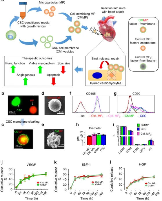
Figure 1 | Physiochemical and biological properties of CMMPs.(a) Overall biochemical design and study model of CMMPs. MPs (that is, Control MP1)
were fabricated from PLGA and conditioned media of human CSCs, then MPs were cloaked with membrane fragments of CSCs to form CMMPs. Control
MP2was fabricated by cloaking empty PLGA particles with CSC membranes. The therapeutic potential of CMMPs was tested in a mouse model of
myocardial infarction. (b,c) Texas red succinimidyl ester-labelled MPs (b, red) were cloaked with the membrane fragments of green fluorescent
DiO-labelled CSCs (b, green) to form CMMP (c, red particle with green coat). Scale bar, 20mm. (d,e) SEM revealed the CSC membrane fragments on
CMMPs (e) but not on Control MP1(non-cloaked MP) (d). Scale bar, 10mm. (f,g) Major human CSC markers CD105 (f) and CD90 (g) were positive on
CMMPs and Control MP2but not on non-cloaked Control MP1, indicating the successful membrane cloaking on CMMPs. (h) CMMPs, Control MP1and
Control MP2have similar sizes to those of CSCs.n¼3 for each group. (i) CMMPs and Control MP2carried similar surface antigens as CSCs did.n¼3 for
each group. (j–l) Similar release profile of CSC factors (namely vascular endothelial growth factor (VEGF), insulin-like growth factor (IGF)-1 and hepatocyte
growth factor (HGF)) was observed in CMMPs and Control MP1, indicating membrane cloaking did not affect the release of CSC factors from CMMPs and
Control MP1.n¼3 for each time point. All data are mean±s.d. Comparisons between any two groups were performed using two-tailed unpaired Student’s
stem cell biointerfacing during regeneration. This particle, named
cell-mimicking MP (CMMP), contained control-released stem
cell factors in its polymeric core and was cloaked with stem cell
membrane fragments on the surface. Our hypothesis is that
CMMP can exert similar regenerative outcomes as real CSCs but
are superior to the later since they are more stable during storage
and do not stimulate T-cell immune reaction since they are not
real cells.
In the present study, we report for the first time a poylmer MP
which emulates CSC functions during tissue repair. In a mouse
model of myocardial infarction, injection of CMMPs led to
preservation of viable myocardium and augmentation of cardiac
functions similar to CSC therapy. CMMPs (derived from human
cells) did not stimulate T-cell infiltration in immuno-competent
mice, suggesting their excellent safety profile. Although our first
application targeted the heart, the CMMP strategy represents a
platform technology that can be applied to multiple stem cell
types and the repair of various organ systems.
Results
Physiochemical and biological properties of CMMPs
. The
biochemical design and work model of CMMPs were outlined in
Fig. 1a. Briefly, Control MP
1were fabricated from
poly(lactic-co-glycolic acid) (PLGA) and conditioned media of human CSCs
which were isolated from human hearts using the cardiosphere
method as previously described
13,14(Supplementary Fig. 1). The
conditioned media contains various growth factors secreted by
CSCs
10. CSCs have been tested and proven safe and effective in
Phase I/II clinical trials
1–3. After that, MPs (Texas red
succinimidyl ester-labelled; Fig. 1b, red) were cloaked with the
membrane fragments of CSCs (green fluorescent DiO-labelled;
Fig. 1b, green) to become the final product CMMP (Fig. 1c, red
particle with green coat). Fluorescent imaging revealed there is no
specific DiO outer layer fluorescence on Texas red succinimidyl
ester-labelled MPs (Control MP
1) after 30 min co-culture
(Supplementary Fig. 2). Scanning electron microscopy (SEM)
revealed the effective CSC membrane cloaking on CMMPs
(Fig. 1e) but not on non-cloaked MPs (Control MP
1; Fig. 1d). As
another control particle, Control MP
2was fabricated by cloaking
empty PLGA particles with CSC membranes. We fabricated
CMMPs, Control MP
1and Control MP
2with sizes similar to
those of real CSCs (Fig. 1h). As an indicator of successful
membrane cloaking, flow cytometry analysis confirmed the
expression of major human CSC markers (for example, CD105,
CD90) on CMMPs and Control MP
2but not on Control MP
1(Figs 1f,g and 2). Overall, both CMMPs and Control MP
2carried
similar surface antigens as CSCs did (Fig. 1i). Membrane cloaking
did not affect the release of CSC factors (namely vascular
endothelial growth factor, insulin-like growth factor-1 and
hepatocyte growth factor) from CMMPs and Control MP
1(Fig. 1j–l; Supplementary Fig. 3). Snap freezing in
80
°
C and
thawing in water did not affect the membrane coating
(Supplementary Fig. 4a), size (Supplementary Fig. 4b,c) or
surface antigen expression of CMMPs (Supplementary Fig. 4d–
f). These results confirmed CMMPs recapitulated the secretome
and surface antigen profile of genuine CSCs. In contrast, Control
MP
1contained CSC secretome but not the membranes of
CSCs, while Control MP
2carried the membranes of CSCs
successfully.
0
CSC CMMP
CSC CMMP 500
1,000 1,500
2,000 CD105 PE
F
lu
o
. in
te
n
s
it
y
(
a
.u
.)
0 200 400 600 800 1,000
CD90 FITC
Fl
uo.
i
n
te
ns
it
y
(
a
.u
.)
CSC CMMP
CSC CMMP
CD105 PE
CD90 FITC
#
# 50 μm
50 μm
50 μm 50 μm
a
b
Figure 2 | Fluorescence densities of CD105 and CD90 expressions on CMMPs and CSCs.(a) Fluorescent images of CSCs (left panel) and CMMPs (right panel) labelled with CD105-PE conjugated antibody. Quantitative analysis of fluorescent intensities of CSCs (blue bar) and CMMPs (green bar).
(b) Fluorescent images of CSCs (left panel) and CMMPs (right panel) labelled with CD90-FITC conjugated antibody. Quantitative analysis of fluorescent
intensities of CSCs (blue bar) and CMMPs (green bar).n¼6 for each group. All data are mean±s.d. # indicatesPo0.05 when compared with CMMP
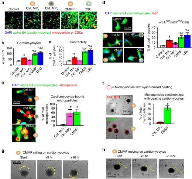
CMMPs promote cardiomyocyte functions
in vitro
. An
important potency indicator of CSCs is their ability to promote
the functions of
in vitro
-cultured cardiomyocytes. CMMPs,
Control MP
1, Control MP
2, or CSCs (red, Fig. 3a) were
co-cul-tured with neonatal rat cardiomyocytes (NRCMs; stained for
alpha-sarcomeric actin (green), Fig. 3a) in plain Iscove’s modified
Dulbecco’s medium. Solitary NRCM culture was included as the
negative control. While Control MP
1(red bar, Fig. 3b) increased
the numbers of NRCMs as compared with those from Control
MP
2(pink bar, Fig. 3b) or solitary NRCM culture (white bar,
Fig. 3b), the greatest NRCM numbers were seen in those
co-cultured with CMMPs (green bar, Fig. 3b) and genuine CSCs
(blue bar, Fig. 3b). Furthermore, CMMPs and Control MP
1robustly promoted NRCM contractility (Fig. 3c) and proliferation
(as indicated by Ki67-positive nuclei, Fig. 3d). Both CMMPs and
Control MP
2could firmly bind to cardiomyocytes, as cells did,
while most non-cloaked Control MP
1floated in the media
(Fig. 3e). Such binding was confirmed by CMMPs’ synchronized
movement with adjacent beating cardiomyocytes (Fig. 3f;
Supplementary Movies 1 and 2). Moreover, time-lapse imaging
revealed the rolling (Fig. 2g; Supplementary Movie 3) and
tra-velling (Fig. 3h; Supplementary Movie 4) of CMMPs on attached
cardiomyocytes, suggesting the biointerfacing between CMMPs
and cardiomyocytes. Such dynamic activities were not seen in
non-cloaked Control MP
1. These
in vitro
cell-based assays suggest
the regenerative potential of CMMPs in the heart.
Cardiomyocytes-bound microparticles
Ctrl. MP
1
Ctrl. MP
2
CMMP 0
20 40 60 80 100
%
o
f
to
ta
l
m
icropart
ic
les
CMMP
Contractility
0 20 40 60 80 100
% o
f
to
ta
l m
y
o
cyt
es
Cardiomyocytes
0 50 100 150
n per HP
F
Microparticles synchronized with beating cardiomyocytes
Ctrl. MP 1
CMMP 0
20 40 60 80
%
o
f
to
ta
l
m
icropart
ic
les
Ctrl. MP1
Control
DAPI alpha-SA (cardiomyocytes) microparticle orCSCs
a
*&
DAPI alpha-SA (cardiomyocytes) microparticle
e
CMMP
b
= Microparticles with synchronized beating
f
αSAPOS/ki67POSCells
ControlCtrl. MP 1
Ctrl. MP 2
CMMP CSC
Control Ctrl. MP
1
Ctrl. MP 2
CMMPCSC Control Ctrl. MP
1
Ctrl. MP 2
CMMP CSC
0 10 20 30 40
%
o
f to
ta
l m
y
o
c
y
te
s
Ctrl. MP1 Control
DAPI alpha-SA (cardiomyocytes) ki67
g
CMMP rolling on cardiomyocytesh
CMMP moving on cardiomyocytesc
#
d
# Ctrl. MP1
CMMP
200 μm
50 μm
50 μm
50 μm
Ctrl. MP1
Start +5 hr +10 hr Start +5 hr +10 hr
20 μm
20 μm
CSC CMMP
Ctrl. MP2 Ctrl. MP2
Ctrl. MP2
CSC
#
*&
*&#
*&
*&# *&#
*&#
*&# *&#
Figure 3 | Effects of CMMPs on NRCMs functionsin vitro.(a) Representative images of cardiomyocytes stained with alpha sarcomeric actin (green)
co-cultured with Control MP1, Control MP2, CMMPs or CSCs (red). Scale bar, 200mm. (b) Quantitative analysis reflected that Control MP1(red bar)
increased the numbers of NRCMs as compared with those from Control MP2(pink bar) or solitary NRCM culture (white bar), but the greatest NRCM
numbers were seen in those co-cultured with CMMPs (green bar) and genuine CSCs (blue bar).n¼5 for each group. (c) Higher NRCM contractility was
seen in those cultured with CMMPs (green bar) and CSCs (blue bar) compared with those cultured with Control MP1(red bar).n¼5 for each group.
(d) Representative images and quantitative analysis of NRCMs stained with alpha sarcomeric actin (green) and proliferation marker Ki67 (red), treated
with Control MP1, Control MP2, CMMPs or CSC.n¼5 for each group. Scale bar, 50mm. (e) Representative images and quantitative analysis of CMMP
(red) or Control MP1(red), Control MP2(red) binding to NRCMs (green).n¼3 for each group. Scale bar, 50mm. (f) Representative movie screenshots and
quantitation of Control MP1’s and CMMP’s synchronized movement with adjacent beating cardiomyocytes.n¼3 for each group. Scale bar, 50mm.
(g,h) Time-lapse imaging revealed the rolling (g) and travelling (h) of CMMPs on attached cardiomyocytes. Yellow arrows indicated the rolling or moving
directions. Scale bar, 20mm. All data are mean±s.d. * indicatesPo0.05 when compared with Control group;#indicatesPo0.05 when compared with
Control MP1group; & indicatedPo0.05 when compared with Control MP2. Comparisons between any two groups were performed using two-tailed
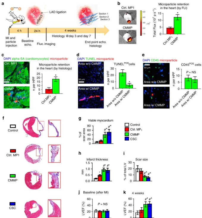
CMMP therapy in immunodeficient mice with heart attack
. To
test the therapeutic potential of CMMPs, we employed a mouse
model of myocardial infarction (heart attack) by left anterior
descending artery (LAD) ligation (Fig. 4a). CMMPs or Control
MP
1were intramyocardially injected immediately after LAD
ligation. Negative or positive control animals received injection of
vehicle (PBS) or CSCs, respectively.
Ex vivo
fluorescent imaging
at Day 3 revealed that more CMMPs were retained in the heart
after injection than Control MP
1(Fig. 4b) were. This was further
confirmed by histology (Fig. 4c). This was consistent with
CMMP’s superior binding to cardiomyocytes
in vitro
(as seen in
Fig. 3). In addition,
ex vivo
fluorescent imaging indicated that the
majority of CMMPs remained in the heart after injection, while
‘washed away’ CMMP signal could be found in the lung and the
liver (Supplementary Fig. 5), consistent with the notion that the
needle injection can cause vessel damage and the venous drainage
brings the particles to the lungs
15. The off-target expression in the
liver may represent the leakage of CMMPs into the LV cavity
during injection. Nevertheless, the majority of CMMPs remain in
the heart after injection.
In vivo
degradation of CMMPs was evident as only a negligible
amount of particles remained in the heart at Day 28
(Supplementary Fig. 6). A cohort of animals was killed at Day
7 for assessment of myocardial tissue apoptosis and infiltration of
macrophages in CMMP-treated animals. TdT-mediated dUTP
nick end labelling (TUNEL) staining revealed the anti-apoptosis
effects of CMMP: less apoptotic nuclei were detected in areas with
the presence of CMMPs (green nuclei, Fig. 4d). CMMP treatment
did not cause the exacerbation of inflammation: the tissue
densities of CD45-positive cells were indistinguishable in areas
with or without CMMPs (Fig. 4e). Masson’s trichrome staining 4
weeks after treatment (Fig. 4f; red
¼
healthy myocardium and
blue
¼
scar tissue) revealed Control MP
1treatment (red bars,
Fig. 4g–i) exhibited a certain degree of heart morphology
protection compared with Control PBS injections (white bars,
Fig. 4g–i). However, the greatest protective effects were seen in
the animals treated with CMMPs (green bars, Fig. 4g–i). Such
protective effects were similar to those injected with CSCs (blue
bars, Fig. 4g–i). The
bona fide
efficacy indicator for stem cell
therapy is the ability to ameliorate ventricular dysfunction or
even boost cardiac function over time, gauged by
echocardio-graphy. Left ventricular ejection fractions (LVEFs) were measured
at baseline (4 h post infarct) and 4 weeks afterwards. LVEFs were
indistinguishable at baseline for all groups (Fig. 4j), indicating a
similar degree of initial heart injury. Over the 4 week period,
the LVEFs in control (PBS or saline)-treated animals continued
deteriorating (white bar, Fig. 4k) while the Control MP
1-treated
animals exhibited a trend of LVEF augmentation (red bar,
Fig. 4k) but did not reach statistical significance. CMMP
treat-ment robustly boosted cardiac function with the highest LVEFs at
4 weeks (green bar, Fig. 4k). Such treatment effects were
indisti-nguishable from those of CSC treatment with real CSCs (blue
bars, Fig. 4k). Histological analysis indicated that such functional
benefits by CMMP treatment were accompanied by
remuscular-ization (Fig. 5a), proliferation of endogenous cardiomyocytes
(Fig. 5b), augmentation of blood flow (Fig. 5c), and increase of
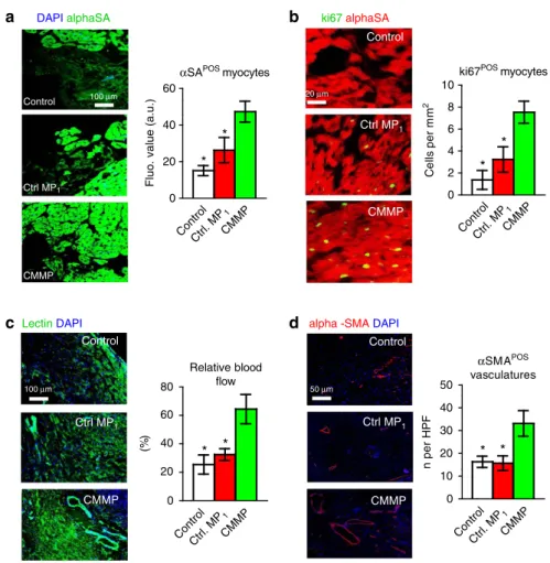
vessel density (Fig. 5d) in the post-MI heart.
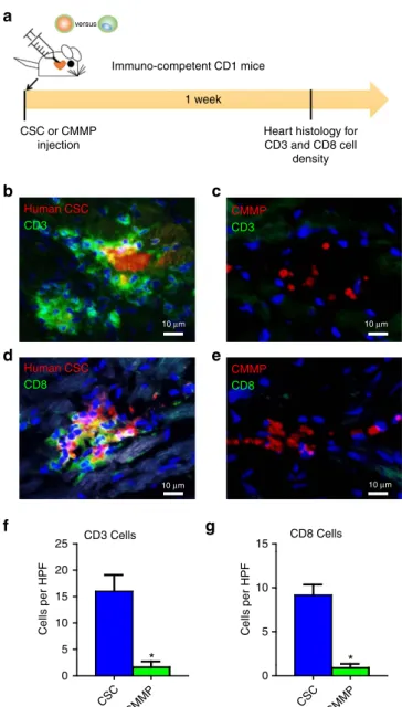
CMMP injection does not promote T-cell infiltration in
nor-mal mice
. To evaluate the local T-cell immune response to
CMMPs, immune-competent CD1 mice were intramyocardially
injected with human CSCs or CMMPs. Animals were killed 7
days after injection for assessment of immune rejection in the
heart, as gauged by CD3
þand CD8
þT cell infiltration (Fig. 6a).
CMMP (red) injection elicits negligible T-cell rejection as very
few CD3
þ(green) or CD8
þ(green) T cells were detected in the
heart (Fig. 6c,e). In contrast, severe rejection was detected in
mouse hearts treated with human CSCs: injected CSCs (red) were
surrounded by clusters of CD3
þ(green) or CD8
þ(green) T cells
(Fig. 6b,d). Quantitative analysis also confirmed that CMMP
stimulated negligible local T-cell infiltration as compared with the
severe T-cell stimulation by human CSCs (Fig. 6f,g).
Discussion
The last one and a half decades witnessed the booming of stem
cell therapies for multiple diseases
16–18. Deviating from the initial
perspective that stem cells exert their therapeutic effects through
direct cell differentiation and tissue replacement, the paradigm
has shifted as emerging evidence suggests that most adult stem
cell types exert their beneficial effects through paracrine
mechanisms (soluble factors)
19–21. In addition, studies further
suggest that cell–cell contact between the injected cells and the
host cells plays an important role in tissue regeneration
11. PLGA,
as a biocompatible and biodegradable polymer, has provided a
safe and non-toxic building block for various control-release
systems
22. Previous studies have demonstrated the success of
coating PLGA nanoparticles with cell membranes from red blood
cells
23, platelets
24and cancer cells
25,26. Inspired by these findings,
we designed CMMPs and demonstrated the therapeutic effects of
CMMPs in an experimental myocardial infarction model. The
comparison between CMMP and CSCs is outlined in
Suppleme-ntary Table 1. CMMP represents a synthetic MP functionalized
with both stem cell membranes and secretome, harnessing the
power of these two major components of stem cell-induced
rege-neration. Moreover, CMMP overcomes several major limitations
of live stem cells as therapy products. First, stem cells need to be
carefully cryo-preserved and thawed before they can be sent to
the clinic. As living organisms, how the cells are prepared and
processed can greatly affect the therapeutic outcomes. Second,
stem cell transplantation carries certain risks (for example,
tumourogenecity and immunogenicity if allogeneic or xenogeneic
cells were used). CMMPs will most likely be delivered
intramyocardially via direct muscle injection. Such injection
normally requires open-chest surgery. However, percutaneous
options are becoming available with the implementation of the
NOGA mapping systems
27. Moreover, our future studies will
explore the potential of vascular delivery of CMMPs (for example,
intracoronary, intravenous) with the focus on targeting CMMPs
to the injury and promoting extravasation through the
mechanism of angiopellosis
28,29. One caveat of our study is that
with the existing assay it is difficult to conclude whether
cardiomyocytes (or their progenitors) really are proliferating
and leading to remuscularization after CMMP injection.
Although this proof-of-concept study targets the heart, CMMP
represents a platform technology that is generalizable to other
stem cell types and the repair of various other organ systems.
Methods
Derivation and culture of human CSCs
.
Institutional review board approval was obtained for all procedures, and informed consent was achieved from all patients. Human CSCs were derived from donor human hearts as previously described5,13. Briefly, myocardial tissues were minced into small pieces (about 2 mm3), thenwashed with PBS and digested with collagenase solution (Sigma, St. Louis, MO, USA). The tissue fragments were cultured as ‘cardiac explants’ on plates coated with 0.5 mg ml1fibronectin (Corning, Corning, NY, USA) in Iscove’s modified Dulbecco’s medium (Invitrogen, Carlsbad, CA, USA) supplemented with 20% fetal bovine serum (Corning), 0.5% Gentanicin (Gibco, Life Technologies, California, USA), 0.1 mM 2-mercaptoethanol (Invitrogen, Carlsbad, CA, USA) and 1% L-glutamine (Invitrogen, Carlsbad, CA, USA). Within 1–2 weeks, a layer of stromal-like flat cells, and phase-bright round cells, emerged from the cardiac explant with phase bright cells over them. These cardiac explant-derived cells were collected using TryPLE Select (Gibco), and then seeded at a density of 2104
cells ml1in UltraLow Attachment flasks (Corning) for cardiosphere formation.
CMMP Ctrl. MP1
Microparticle retention in the heart (by FLI)
0
Ctrl.MP 1
CMMP
Ctrl.MP 1
CMMP
5 10 15
T
o
ta
l F
lu
x
(
1
0
7 p s
–1
)
a
b
*
Ctrl. MP1
CMMP
0 5 10 15 25
c
d
DAPI TUNEL microparticle
Area w/ CMMP Area w/o CMMP
Are a w
/o CM MP
Area w / CMMP
0 10 20 30
TUNELPOScells
Area w/o
CMMP
Area w/ CMMP
0 5 10 15
CD45POS cells
n per H
P
F
e
DAPI CD45 microparticle
Area w/o CMMP
Area w/ CMMP
*
*
P = NS
P = NS Infarct thickness
0.0 0.5 1.0 1.5
mm
Viable myocardium
0 20 40 60
80 Control
Ctrl. MP1
CMMP CSC
% o
f
scar
ar
ea
4 weeks
0 20 40 60
LV
E
F
(
%
)
Baseline (after MI)
0 20 40 60
LVEF (%
)
Scar size
0 10 20 30
% o
f t
o
ta
l L
V
Control
Ctrl. MP1
CMMP
CSC
f
g
h
i
j
k
#
*
#*
* #* #*
#
*
#*
#
*
#*
50 μm 50 μm
50 μm
5.0
Section 1 Section 2 Section 3
4.0
×102
3.0
2.0
1.0
LAD ligation
4 h
MI and particle injection
Baseline
echo. Fluo. imaging
20
n per HPF
n per HPF
Microparticle retention in the heart (by histology)
DAPIalpha-SA(cardiomyocytes)microparticle
End point echo. histology
24 h 4 weeks
Histology @day 3 and day 7 Versus
Figure 4 | CMMPs ameliorate ventricular dysfunction and promote cardiac repair in a mouse model of heart attack.(a) Schematic showing the overall
design of animal experiments to test the therapeutic benefits of CMMPs in a mouse model of myocardial infarction. (b) Representativeex vivofluorescent
imaging of mouse hearts and quantitative analysis of fluorescent intensities at Day 3 after Control MP1or CMMP injections.n¼3 animals per group.
* indicatedPo0.05 when compared with Control MP1group. (c) Representative microscopic images and quantitative analysis of mouse hearts
(myocytes stained with alpha sarcomeric actin (green)) 3 days after injection of Control MP1(red) or CMMPs (red).n¼3 animals per group. Scale bar,
50mm. * indicatedPo0.05 when compared with Control MP1group. (d) Representative fluorescent micrographs and quantitative analysis showing the
presence of TUNELþ apoptotic cells (green) in CMMP-treated hearts at Day 7.n¼3 animals per group. Scale bar, 50mm. * indicatedPo0.05.
(e) Representative fluorescent micrographs showing the presence of CD45þ cells (green) in the hearts treated with or without CMMPs (red) at Day 7.
n¼3 animals per group. Scale bar, 50mm. NS indicatedP40.05. (f) Representative Masson’s trichrome-stained myocardial sections 4 weeks after
treatment with Control PBS, Control MP1,CMMPs or CSCs. In this staining blue¼scar tissue and red¼viable myocardium. Snapshots¼high magnification
images of the red box area. (g–i) Quantitative analyses of viable myocardium (g), infarct thickness (h) and scar size (i) from the Masson’s trichrome
images.n¼5 animals per group. (j,k) LVEF was measured by echocardiography at baseline (4 h post-MI) and 4 weeks afterward in Control PBS, Control
MP1, CMMP and CSC groups.n¼7 animals per group. * indicatedPo0.05 when compared with Control group;#indicatedPo0.05 when compared with
Control MP1group; NS indicatedP40.05. All data are mean±s.d. Comparisons between any two groups were performed using two-tailed unpaired
cardiospheres. Cardisophere-derived CSCs were generated by seeding collected cardiospheres on fibronectin-coated plates. All cultures were incubated in 5% CO2 at 37°C.
Fabrication of Control MPs and CMMPs
.
CSC factor-loaded PLGA micro-particles (Control MP1) were fabricated by a water/oil/water (w/o/w) emulsion technique. Briefly, human CSC conditioned media as the internal aqueous phase with polyvinyal alcohol (0.1% w/v) was mixed in methylene chloride (DCM) containing PLGA as the oil phase. The mixture was then sonicated on ice for 30 s using a sonicator with a Microtip probe (Misonix, XL2020, Farmingdale, NY, USA). After that, the primary emulsion was immediately introduced into water with polyvinyal alcohol (0.7% w/v) to produce a w/o/w emulsion. The secondary emulsion was emulsified for 5 min on a high-speed homogenizer. The w/o/w emulsion was continuously stirred overnight at room temperature to promote solvent evaporation. The solidified MPs, namely Control MP1, were then centrifuged, washed three times with water, lyophilized and stored at 80°C. To prepare CMMPs, DiO (Invitrogen)-labelled CSCs went through three freeze/thaw cycles. After which, the disrupted CSCs were sonicated forB5 min at room temperature along with the Control MP1. After that, the particles were washed three times in PBS by centrifugation. Control MP2was fabricated by cloaking empty PLGA particles with CSC membranes. Successful membrane coating was confirmed using fluorescent microscopy.Protein release studies
.
Total protein contents in MPs were determined using the following method. Approximately 10 mg freeze-dried microparticles were dissolved in 1 ml DCM for 60 min. Then, 1 ml PBS was added into solution followed by agitation for 10 min to extract protein from DCM into PBS. After centrifugation,the concentration of protein in the aqueous phase was determined by a BCA Protein Assay Kit (Thermo Fisher Scientific, Waltham, MA, USA). For release studies, MPs were incubated in PBS at 37°C. Supernatant was collected at various time points and the concentrations of proteins were determined by commercially available ELISA kits (R & D Systems, Minneapolis, MN, USA)
Scanning electron microscopy
.
The morphology of microparticles was studied by SEM (Philips XL30 scanning microscope, Philips, The Netherlands). Freeze-dried microparticles were mounted on aluminium stubs with double-sided tape and coated with a thin layer of gold. The coated specimen was then scanned and photographed under the microscope at an acceleration voltage of 15 kV.Flow cytometry
.
To characterize the phenotypes of Control MP1, Control MP2, CMMP, and CSC, flow cytometry was performed using a CytoFLEX Flow Cytometer (Beckman Coulter, Brea, CA) and analysed using FCS Express software (De Novo Software, Los Angeles, CA). Briefly, cells were incubated with FITC, PE, or APC-conjugated antibodies against CD105 (10ml per 40ml of sample, FAB 10971P, R&D Systems), CD90 (10ml per 40ml of sample, BD 555595), CD45 (10ml per 40ml of sample, BD 555482), CD34 (10ml per 40ml of sample, BD 555821) and CD117 (5ml per 45ml of sample, c-kit; BD 550412) from BD company (Franklin Lakes, NJ) for 60 min. Isotype-identical antibodies from BD Company served as negative controls.Immunocytochemistry
.
Control MP1, Control MP2, CMMP, and CSC were pre-labelled with red-fluorescent Texas red succinimidyl ester (1 mg ml1(Invitrogen, Carlsbad, California)). NRCM or NRCM co-cultured with pre-labelled
Control Lectin DAPI
0 20 40 60 80
Relative blood flow
(%
)
alpha -SMA DAPI
CMMP
Control
0 10 20 30 40 50
αSMAPOS
vasculatures
n per H
P
F
0 2 4 6 8 10
ki67POS myocytes
C
e
lls
per
m
m
2
Ctrl MP1 Ctrl MP1
CMMP DAPI alphaSA
Control
CMMP Ctrl MP1
Control Ctrl. MP
1
CMMP Control Ctrl. MP
1
CMMP
Control Ctrl. MP
1
CMMP Control Ctrl. MP
1
CMMP
0 20 40 60
αSAPOS myocytes
Fluo. value (a.u
.)
a
c
d
*
*
ki67 alphaSAControl
CMMP
b
100 μm 20 μm
100 μm 50 μm
*
*
*
*
*
*
Ctrl MP1
Figure 5 | Injection of CMMPs promotes angiomyogenesis.(a) Representative images showing alpha sarcomeric actin (aSA)-positive cardiomyocyte
nuclei (green) in control PBS-, Control MP1- or CMMP-treated hearts at 4 weeks. The numbers ofaSA-positive nuclei were quantified.n¼3 animals per
group. Scale Bar, 100mm. (b) Representative images showing Ki67-positive cardiomyocyte nuclei (green) in control PBS-, Control MP1- or CMMP-treated
hearts at 4 weeks. The numbers of Ki67-positive nuclei were quantified.n¼3 animals per group. Scale Bar, 20mm. (c) Representative images showing
lectin-labelled blood vessels (green) in control PBS- and Control MP1- or CMMP-treated hearts at 4 weeks. The lectin fluorescent intensities were
quantified.n¼3 animals per group. Scale Bar, 100mm. (d) Representative images showing arterioles stained with alpha smooth muscle actin (aSMA, red)
in control PBS-, Control MP1- or CMMP-treated hearts at 4 weeks. The numbers ofaSMA positive vasculatures were quantified.n¼3 animals per group.
Scale Bar, 50mm. * indicatesPo0.05 when compared with CMMP group. All data are mean±s.d. Comparisons among more than two groups were
Control MP1, Control MP2, CMMP, and CSC were plated onto fibronectin-coated chamber slides (BD Biosciences) and subsequently fixed with 4% paraformalde-hyde before immunocytochemistry (ICC) staining. Slides were stained with the antibodies againsta-SA (1:100, a7811, Sigma) or ki67 (1:100, ab15580, Abcam) and detected by FITC- or Texas Red-conjugated secondary antibodies (1:100). Nuclei were stained with DAPI. Images were taken with an epi-fluorescent microscope (Olympus IX81).
Mouse model of myocardial infarction
.
All animal work was compliant with the Institutional Animal Care and Use Committee at North Carolina State University.The method to induce myocardial infarction in mice was based on previous studies30. Briefly, male SCID Beige mice were anaesthetized with 3% isofluorane combined with 2% oxygen inhalation. Under sterile conditions, the heart was exposed by a minimally invasive left thoracotomy and acute myocardial infarction (AMI) was produced by permanent ligation of the LAD coronary artery. Immediately after AMI induction, the heart was randomized to receive one of the following four treatment arms: (1) ‘Control (PBS)’ group: intramyocardial injection of 50ml PBS into the heart immediately after AMI; (2) ‘Control MP1’ group: intramyocardial injection of 1105Control MP1in 50ml PBS into the heart immediately after AMI; (3) ‘CMMP’ group: intramyocardial injection of 1105
CMMPs in 50ml PBS into the heart immediately after AMI; (4) ‘CSC’ group: intramyocardial injection of 1105CSCs in 50ml PBS into the heart immediately after AMI. To enable visualization of Control MP1or CMMP in a cohort of animals, we pre-labelled the Control MP1or CMMP with Texas Red-X succinimidyl ester (1 mg ml1(Invitrogen, Carlsbad, California)).
Ex vivofluorescent imaging for biodistribution of CMMPs
.
Seven days afterinjection, a cohort of mice receiving CMMPs were killed; their heart, lung, spleen, liver, and kidneys were removed for biodistribution studies.Ex vivofluorescent imaging was performed with an IVIS XenogenIn VivoImager (Caliper Life-sciences, Waltham, MA).
Heart morphometry
.
After the echocardiography study at 4 weeks, all animals were killed and hearts were collected and frozen in optimum cutting temperature (OCT) compound. Specimens were sectioned at 10mm thickness from the apex to the ligation level with 100mm intervals. Masson’s trichrome staining was per-formed as described by the manufacturer’s instructions (HT15 Trichrome Staining (Masson) Kit; Sigma-Aldrich). Images were acquired with a PathScan Enabler IV slide scanner (Advanced Imaging Concepts, Princeton, NJ). From the Masson’s trichrome stained images, morphometric parameters including viable myocardium, infarct thickness and scar size were measured in each section with NIH ImageJ software. The percentage of viable myocardium as a fraction of the scar area (infarcted size) was quantified as described31–33. Three selected sections werequantified for each animal.
Cardiac function assessment
.
All animals underwent transthoracic echocardio-graphy under 1.5% isofluorane-oxygen mixture anaesthesia in supine position at 4 h and 4 weeks. The procedure was performed by an animal cardiologist blind to the experimental design using a Philips CX30 ultrasound system coupled with an L15 high-frequency probe. Hearts were imaged in 2D in long-axis views at the level of the greatest LV diameter. EF was determined by using the formula (LVEDV–LVESV/LVEDV)100%.Histology
.
For immunohistochemistry staining, heart cryosections were fixed with 4% paraformaldehyde, permeabilized and blocked with Protein Block Solution (DAKO, Carpinteria, CA) containing 0.1% saponin (Sigma, St Louis, MO), and then incubated with the following antibodies overnight at 4°C: mouse anti-alpha sarcomeric actin (1:100, a7811, Sigma), rabbit anti-CD45 (1:100, ab10559, Abcam, Cambridge, United Kingdom), mouse anti-Actin,a-Smooth Muscle antibody (1:100, A5228, Sigma), rabbit anti-Ki67 (1:100, ab15580, Abcam), rabbit anti-CD3 (1:100, ab16669, Abcam) and mouse anti-CD8 alpha (1:100, mca48r, abd Serotec, Raleigh, NC ). FITC- or Texas-Red secondary antibodies (1:100) were obtained from Abcam Company and used for the conjunction with these primary anti-bodies. For assessment of cell apoptosis, heart cryosections were incubated with TUNEL solution (Roche Diagnostics GmbH, Mannheim, Germany) and counter-stained with DAPI (Life Technology, NY, USA). For assessment of angiogram, heart cryosections were incubated with Lectin (FL-1171, Vector laboratories, Burlingame, CA, USA). Images were taken by an Olympus epi-fluorescence microscopy system.Immunogenicity studies for human CSCs and CMMPs
.
Immuno-competent male CD1 mice were anaesthetized with 3% isofluorane combined with 2% oxygen inhalation. Under sterile conditions, the heart was exposed by a minimally invasive left thoracotomy, and the heart was randomized to receive one of the two treat-ments: (1) ‘CMMP’ group: intramyocardial injection of 1105CMMPs in 50mlPBS into the heart; (2) ‘CSC’ group: intramyocardial injection of 1105human CSCs in 50ml PBS into the heart. To enable visualization of CMMPs or CSCs, they were pre-labelled with red fluorophore.
Statistical analysis
.
All results are expressed as mean±s.d. Comparison between two groups was performed with two-tailed Student’st-test. Comparisons among more than two groups were performed using one-way ANOVA followed bypost hocBonferroni test. Differences were considered statistically significant when thePvalueo0.05.
Human CSC
CD8
Human CSC
CD3
CMMP
CD8
CMMP
CD3
10 μm
10 μm 10 μm
10 μm
0
CSC
CMMP CSC CMMP
5 10 15 20 25
Ce
ll
s
p
e
r
HP
F
0 5 10 15
C
e
lls
per
H
P
F
a
b
d
c
e
f
g
CD8 CellsCD3 Cells
*
*
versus
Immuno-competent CD1 mice
1 week
CSC or CMMP injection
Heart histology for CD3 and CD8 cell
density
Figure 6 | CMMP injection does not stimulate local T-cell immune response in immunocompetent mice.(a) Schematic showing the overall animal study design to assess the local T-cell immune reaction induced by
human CSCs or CMMPs derived from human CSCs. (b,c) Representative
fluorescent images showing the presence of infiltrated CD3þ T cells
(green) in CSCs (red, b)- or CMMPs (red, c)-injected hearts at Day 7. Scale
bar, 10mm. (d,e) Representative fluorescent images showing the presence
of infiltrated CD8þ T cells (green) in CSCs (red,b) - or CMMPs (red,c
)-injected hearts at Day 7. Scale bar, 10mm. (f,g) Quantitative analysis of
CD3þ and CD8þ T cells in CSCs (blue or CMMPs (green
bar)-injected hearts at Day 7.n¼3 animals per group. All data are mean±s.d.
Comparisons between any two groups were performed using two-tailed
unpaired Student’st-test. * IndicatedPo0.05 when compared with CSC
Data availability
.
The authors declare that all data supporting the findings of this study are available within the article and its Supplementary Information files or from the corresponding author on reasonable request.References
1. Makkar, R. R.et al.Intracoronary cardiosphere-derived cells for heart regeneration after myocardial infarction (CADUCEUS): a prospective, randomised phase 1 trial.Lancet379,895–904 (2012).
2. Bolli, R.et al.Cardiac stem cells in patients with ischaemic cardiomyopathy (SCIPIO): initial results of a randomised phase 1 trial.Lancet378,1847–1857 (2011).
3. Malliaras, K.et al.Intracoronary cardiosphere-derived cells after myocardial infarction: evidence of therapeutic regeneration in the final 1-year results of the CADUCEUS trial (CArdiosphere-Derived aUtologous stem CElls to reverse ventricUlar dySfunction).J. Am. Coll. Cardiol.63,110–122 (2014). 4. Chen, S. L.et al.Effect on left ventricular function of intracoronary
transplantation of autologous bone marrow mesenchymal stem cell in patients with acute myocardial infarction.Am. J. Cardiol.94,92–95 (2004). 5. Cheng, K.et al.Relative roles of CD90 and c-kit to the regenerative efficacy of
cardiosphere-derived cells in humans and in a mouse model of myocardial infarction.J. Am. Heart Assoc.3,e001260 (2014).
6. Katritsis, D. G.et al.Transcoronary transplantation of autologous mesenchymal stem cells and endothelial progenitors into infarcted human myocardium.Catheter. Cardiovasc. Interv.65,321–329 (2005).
7. Bai, X.et al.Both cultured and freshly isolated adipose tissue-derived stem cells enhance cardiac function after acute myocardial infarction.Eur. Heart J.31,
489–501 (2010).
8. Forrester, J. S., Price, M. J. & Makkar, R. R. Stem cell repair of infarcted myocardium: an overview for clinician.Circulation108,1139–1145 (2003). 9. Avolio, E.et al. Ex vivomolecular rejuvenation improves the therapeutic
activity of senescent human cardiac stem cells in a mouse model of myocardial infarction.Stem Cells32,2373–2385 (2014).
10. Li, T. S.et al.Direct comparison of different stem cell types and subpopulations reveals superior paracrine potency and myocardial repair efficacy with cardiosphere-derived cells.J. Am. Coll. Cardiol.59,942–953 (2012). 11. Xie, Y.et al.Importance of cell-cell contact in the therapeutic benefits of
cardiosphere-derived cells.Stem Cells32,2397–2406 (2014). 12. Ho, Y. S.et al.Cardioprotective actions of TGFbRI inhibition through
stimulating autocrine/paracrine of survivin and inhibiting Wnt in cardiac progenitors.Stem Cells34,445–455 (2016).
13. Cheng, K.et al.Functional performance of human cardiosphere-derived cells delivered in an in situ polymerizable hyaluronan-gelatin hydrogel.Biomaterials
33,5317–5324 (2012).
14. Cheng, K.et al.Human cardiosphere-derived cells from advanced heart failure patients exhibit augmented functional potency in myocardial repair.JACC Heart Fail.2,49–61 (2014).
15. Al Kindi, A., Ge, Y., Shum-Tim, D. & Chiu, R. C. Cellular cardiomyoplasty: routes of cell delivery and retention.Front Biosci.13,2421–2434 (2008). 16. Segers, V. F. & Lee, R. T. Stem-cell therapy for cardiac disease.Nature451,
937–942 (2008).
17. Lindvall, O. & Kokaia, Z. Stem cells for the treatment of neurological disorders.
Nature441,1094–1096 (2006).
18. Fox, I. J.et al.Stem cell therapy. Use of differentiated pluripotent stem cells as replacement therapy for treating disease.Science345,1247391 (2014). 19. Hodgkinson, C. P., Bareja, A., Gomez, J. A. & Dzau, V. J. Emerging concepts in
paracrine mechanisms in regenerative cardiovascular medicine and biology.
Circ. Res.118,95–107 (2016).
20. Walter, J., Ware, L. B. & Matthay, M. A. Mesenchymal stem cells: mechanisms of potential therapeutic benefit in ARDS and sepsis.Lancet Respir. Med.2,
1016–1026 (2014).
21. Lanzoni, G.et al.Concise review: clinical programs of stem cell therapies for liver and pancreas.Stem Cells31,2047–2060 (2013).
22. Hu, C. M., Fang, R. H., Luk, B. T. & Zhang, L. Polymeric nanotherapeutics: clinical development and advances in stealth functionalization strategies.
Nanoscale6,65–75 (2014).
23. Luk, B. T.et al.Interfacial interactions between natural RBC membranes and synthetic polymeric nanoparticles.Nanoscale6,2730–2737 (2014). 24. Hu, C. M.et al.Nanoparticle biointerfacing by platelet membrane cloaking.
Nature526,118–121 (2015).
25. Fang, R. H., Kroll, A. V. & Zhang, L. Nanoparticle-based manipulation of antigen-presenting cells for cancer immunotherapy.Small11,5483–5496 (2015).
26. Fang, R. H.et al.Cancer cell membrane-coated nanoparticles for anticancer vaccination and drug delivery.Nano Lett.14,2181–2188 (2014).
27. Gyo¨ngyo¨si, M. & Dib, N. Diagnostic and prognostic value of 3D NOGA mapping in ischemic heart disease.Nat. Rev. Cardiol.8,393–404 (2011). 28. Cheng, K.et al.Brief report: mechanism of extravasation of infused stem cells.
Stem Cells30,2835–2842 (2012).
29. Allen, T. A.et al.Angiopellosis as an alternative mechanism of cell extravasation.Stem Cells. doi: 10.1002/stem.2451 (2016).
30. Andrade, J. N.et al.Rapid and efficient production of coronary artery ligation and myocardial infarction in mice using surgical clips.PLoS ONE10,e0143221 (2015).
31. Cheng, K.et al.Magnetic targeting enhances engraftment and functional benefit of iron-labeled cardiospherederived cells in myocardial infarction.
Circ. Res.106,1570–1581 (2010).
32. Shen, D.et al.Effects of matrix metalloproteinases on the performance of platelet fibrin gel spiked with cardiac stem cells in heart repair.Stem Cells Transl. Med.5,793–803 (2016).
33. Vandergriff, A. C.et al.Magnetic targeting of cardiosphere-derived stem cells with ferumoxytol nanoparticles for treating rats with myocardial infarction.
Biomaterials35,8528–8539 (2014).
Acknowledgements
This work was supported by funding from National Institute of Health HL123920, NC State University Chancellor’s Faculty Excellence Program, NC State Chancellor’s Innovation Fund, University of North Carolina General Assembly Research Opportu-nities Initiative grant and National Natural Science Foundation of China 81370216, 81570274, 31670895 and U1404802, Science and Technology Innovation Team Support Project of Henan Province 14IRTSTHN018, Innovation Team of Science and Technol-ogy Project of Henan Province 164200510012. J.T. is supported by China Scholarship Council. The study is also partially supported by the Cooperative Research Grant(s) of Atomic Bomb Disease Institute at Nagasaki University, Japan.
Author contributions
J.T., D.S., T.G.C., J.Z., Q.K. and K.C. designed research, performed biochemical, cellular and animal experiments, analysed the data and drafted the paper. Z.W., T.A.A., A.C.V., M.T.H., P.-U.D. and J.C. performed cellular andin vitroexperiments. T.-S.L., J.Z., Q.K., and K.C. directed the research and provided financial support.
Additional information
Supplementary Informationaccompanies this paper at http://www.nature.com/ naturecommunications
Competing financial interests:The authors declare no competing financial interests.
Reprints and permissioninformation is available online at http://npg.nature.com/ reprintsandpermissions/
How to cite this article:Tang, J.et al.Therapeutic microparticles functionalized with biomimetic cardiac stem cell membranes and secretome.Nat. Commun.8,
13724 doi: 10.1038/ncomms13724 (2017).
Publisher’s note:Springer Nature remains neutral with regard to jurisdictional claims in published maps and institutional affiliations.
This work is licensed under a Creative Commons Attribution 4.0 International License. The images or other third party material in this article are included in the article’s Creative Commons license, unless indicated otherwise in the credit line; if the material is not included under the Creative Commons license, users will need to obtain permission from the license holder to reproduce the material. To view a copy of this license, visit http://creativecommons.org/licenses/by/4.0/