Cover Page
The following handle holds various files of this Leiden University dissertation:
http://hdl.handle.net/1887/59503
Author: Booij, T.H.
Phenotypic screening
with 3D cell-based assays
Phenotypic screening with 3D cell-based assays Tijmen Booij
September 2017
ISBN: 978-94-028-0871-1
© 2017, Tijmen Booij. All rights reserved. No part of this thesis may be reproduced or transmitted in any form, by any means, electronic or mechanical, without prior written permission from the author.
Phenotypic screening
with 3D cell-based assays
Proefschrift
Ter verkrijging van de graad van Doctor aan de Universiteit Leiden,
op gezag van Rector Magnificus prof.mr. C.J.J.M. Stolker,
volgens besluit van het College voor Promoties
te verdedigen op woensdag 20 December 2017
klokke 10:00 uur
door
Tijmen Harmen Booij
Promotor
Prof. Dr. Bob van de Water (Universiteit Leiden/LACDR)
Prof. Dr. Dorien J.M. Peters (Leids Universitair Medisch Centrum)
Co-promotor
Dr. Leo S. Price (Universiteit Leiden/OcellO B.V.)
Promotiecommissie
Prof. Dr. Hubertus Irth (Universiteit Leiden/LACDR) (voorzitter)
Prof. Dr. Joke A. Bouwstra (Universiteit Leiden/LACDR) (secretaris)
Overige leden
Prof. Dr. Ad P. IJzerman (Universiteit Leiden/LACDR)
Prof. Dr. Huib Ovaa (Leids Universitair Medisch Centrum)
Prof. Dr. Roos Masereeuw (Universiteit Utrecht)
Prof. Dr. Paul Jennings (VU Amsterdam)
The investigations described in this thesis were performed at Division of Toxicology of the Leiden Academic Centre for Drug Research, Leiden University, Leiden,
the Netherlands and at the Human Genetics department, Leiden University Medical Center, Leiden, the Netherlands
CHAPTER 1
General introduction
CHAPTER 2
Getting the most out of 3D cell based assays with high content
image analysis and phenotypic profiling
CHAPTER 3
Development of a 3D tissue culture-based high-content
screening platform that uses phenotypic profiling to
discriminate selective inhibitors of receptor tyrosine kinases
CHAPTER 4
High-throughput phenotypic screening of kinase inhibitors to
identify drug targets for polycystic kidney disease
CHAPTER 5
Phenotypic profiling of 3D-cultured micro-tissues to identify
selective inhibitors of cyst growth
CHAPTER 6
In vitro
3D phenotypic drug screen identifies celastrol as an
effective
in vivo
inhibitor of polycystic kidney disease
CHAPTER 7
General discussion and future perspectives
CHAPTER 8
Appendices
11
35
55
83
113
145
173
1
General introduction
chapter 1
12
Challenges in drug research & development
Over the past decades, it has become clear that the increasing investments in pharma-ceutical research and development (R&D) have not translated into an anticipated in-crease in approved new drugs. With new drugs being discovered at a steady rate by the pharmaceutical industry and the exponentially rising R&D costs, it is becoming more difficult to obtain a return of R&D investments and to fund new research. It is possible that the current R&D strategies are exhausted and that the only solution to this pro-blem is a radical change towards more innovative strategies to improve success rates of
new drugs.1 Additionally, when older patents of important blockbuster drugs expire, the
pharmaceutical industry will be required to address R&D productivity to remain viable.
As a brief summary of the steps in current drug discovery, novel drug candidates are typically discovered after the identification of a new drug target. Drug targets are often proteins that are aberrantly active or inactive in a pathophysiological process, and modulation of these targets, or their signalling cascade, could therefore be used to alleviate or reverse disease symptoms. Not surprisingly, the identification of new drug targets typically requires many years of intensive study of disease-associated cellular signalling pathways. After a disease target has been identified, potential drug candidates that bind to the target protein, often many thousands, can be synthesised and assayed on cell culture models for the disease. This pre-selection on cell culture
models is required in order to preselect a small number of molecules for in vivo efficacy
measurement and clinical development (figure 1).
This particular drug discovery workflow is target-based: it relies on known
informa-tion of disease targets.2 A drawback associated with this strategy is the assumption
that modulation of a single protein target is sufficient for the alleviation of disease symptoms: many diseases are much more complex than this and require a broader tar-geting approach, which can for example be overcome by treatment with multiple drugs. Another consequence of this target-driven drug discovery is that only the most potent inhibitors or activators for certain potentially druggable targets will progress through drug development. While these molecules may be curative for a certain disease, many diseases do not require complete abolishing of one molecular target, but rather require fine-tuning of multiple target proteins, which is often the most challenging to achieve. Importantly, entirely abolishing a single disease target can also lead to drug side effects. Antineoplastic drugs (chemotherapeutics) are an example of this: while these drugs mostly interfere with cell division or promote programmed cell death and there-by inhibit tumour growth, they can also have similar effects on healthy cells, with side effects as a result.
In addition to the increasing R&D costs, high drug attrition rates pose a large challenge. Most drugs initially introduced in the drug development pipeline will never reach the clinical evaluation stage. However, even for drugs that enter clinical trials,
the average success rate is only around 11%.3 Because this phase in drug development
ensure that new drugs do not fail during this phase, or after the clinical trials. How-ever, many drugs that are introduced in clinical trials suffer from lack of efficacy or
toxicity that was not predicted.4 Additionally, the success rate of clinical trials highly
differs between therapeutic areas. For example, drugs for cardiovascular indications have an approximate 20% chance of success, while this is only 8% for drugs targeting
diseases of the central nervous system.3 These differences are likely, at least in part,
attributable to our knowledge of the etiology of the disease, the complexity of the biology and the predictive value of disease models. Also after drug approval by the Food and Drug Administration (FDA, USA) or European Medicines Agency (EMA, Europe),
unexpect-ed toxicity or lack of clinical benefit remains an important factor for drug withdrawal.3
Therefore, in order to make better and safer drugs and to achieve higher success rates in the clinic, it is necessary that better drugs are pre-selected before these reach clini-cal development.
Physiological relevance of 2D in vitro disease models to predict drug efficacy
In order to pre-select better drugs for clinical development, it is necessary to have a more detailed look at the earlier stages of the R&D pipeline. After the identification of a druggable target for a disease, potential drug candidates that modulate the activity of the target can be developed, and can eventually be tested in a biological model.
When potential drug candidates are first investigated in cell-based biological models (in vitro), generally their efficacy is assessed on two-dimensional (2D) cell culture models, which are often named monolayers (figure 2). In such a cell culture system, cells that are relevant to the investigated disease or process are cultured on culture plastic in growth medium, supplemented with animal serum and often antibiotics. These cell models are generally easy to maintain and cheap to use, the latter of which is extremely important when testing many thousands of candidate molecules at once, often referred to as high-throughput screening (HTS). However, in recent years it has become increasingly clear that for many diseases, these monolayer cultures often fail to predict drug efficacy in animal models (in vivo) or in clinical trials.
A large problem associated with this poor translation is that many drug
candi-dates that appear to be successful in such an in vitro model, fail in the later, more
14
expensive, drug development stages. Conversely, it is possible that potentially good drugs fail to show desirable effects in monolayer cell cultures, causing these to be fil-tered out and never progress into further development. A key concept in this problem is that monolayer cultures cannot adequately recapitulate the complex conditions in the body, since tissues are comprised of many cell types that interact within a three-dimen-sional environment. Hence, the tissue architecture that is observed in the body cannot be adequately recapitulated using monolayer cell culture models, since these fail to reflect the tissue architecture and its relevance in various disease processes. This, coupled to the implementation of the three R’s (reduction, refinement, replacement) to
reduce the use of animals in drug testing5 means that more relevant in vitro evaluation
models are required to select better drugs.
In order to provide a background for the different disease areas described through-out this dissertation, the following subchapters give a brief overview on two neoplastic disorders, cancer and polycystic kidney disease (PKD), where tissue architecture is es-sential for the pathophysiology and thereby providing a rationale for the development
of new in vitro disease models on which to test candidate drugs.
CANCER
Disease background
Cancer is a neoplastic disorder that is characterized by abnormal cell proliferation and
is among the most common causes of death worldwide.6 The process by which healthy
cells can transform into cancer cells and form a tumour is a multistep process by which cells need to acquire properties that confer a proliferative advantage, such as self- stimulatory growth signals, insensitivity to anti-growth signals, the ability to evade apoptosis and avoid immune destruction and the ability to induce angiogenesis. These FIGURE 2 Cells cultured as a monolayer are poor simulators of human biology.
A) Schematic representation of cells cultured as a monolayer on culture plastic.
Red lines represent the cortical cytoskeleton and nuclei are shown as blue spheres.
B) Immunofluorescence image of 2D-cultured mIMCD3 cells (BD Pathway 855, 10x
factors, amongst many more, contribute to the origin and growth of a primary tumour7-8 and are generally the result of acquired mutations that confer a growth advantage.
Importantly, the main cause of cancer-related deaths generally is not the presence of the primary tumour, but rather the metastasis of tumour cells to distant sites, where the growth of tumours can interfere with normal organ function. In order for metastasis to occur, tumour cells need to acquire migratory properties. These properties can then allow the cells to escape from the primary tumour into the blood stream (or lymphatic system), and eventually extravasate at a distant site. This metastatic cascade is illu- strated in figure 3, and describes the process of invasion of tumour cells into the extra-cellular matrix, the intravasation into the blood stream and eventual extravasation at
a distant site.9 In general, the cause of this migratory phenotype is the loss of cell-cell
contacts and changes in cell-matrix contacts and the secretion of matrix-remodelling enzymes. Collectively, this switch in cellular behaviour is often termed epithelial- to mesenchymal transition (EMT). It is currently becoming clearer that the tumour
extra-cellular matrix10-11 and many immune cells12-15 play a role in carcinogenesis.
Important-ly, the causes of cancer are highly diverse, and range from genetic predisposition to
DNA damage to diet.16 Specific signalling pathways that are involved in this process are
therefore highly variable and also depend on the tumour type and its underlying mu-tations. It is therefore not feasible and also not the scope of this chapter to discuss all these properties in detail, and the reader is referred to other relevant literature.7-9, 16-18 FIGURE 3 Schematic representation of the metastatic cascade showing the processes of invasion, intravasation and extravasation at a distant site to establish a metastasis.
16
Current therapeutic strategies and limitations
Surgery is often the first line of treatment against a primary tumour that has not yet metastasized, sometimes supplemented with radiation- or chemotherapy, if required. Whether this strategy is successful depends highly on the tumour type, its underly-ing mutations and the tumour stage. For tumours that have already metastasized, surgery on its own is often not sufficient to cure the patient, and it is therefore often supplemented with radiation-, chemo- or immunotherapy. These additional therapies generally function to inhibit tumour cell proliferation, taking advantage of the tumours’ defective DNA repair mechanisms, or to eradicate tumour cells by the immune system. However, such therapies, with the possible exception of immunotherapy, usually have side effects related to their effects on healthy cells. For example, the chemothera-peutic drug cisplatin (cis- diamminedichloroplatinum(II), CDDP) is a molecule that
intercalates directly into the tumour cells’ DNA,19 thereby preventing cell division and
tumour growth. However, the use of this molecule is limited by its nephrotoxic effects,20
which are, at least in part, attributable to active cisplatin uptake in the kidneys by high
affinity copper uptake protein 1 (Ctr1)21 and organic cation transporter OCT2 (SLC22A2),22
causing the local increase in intracellular cisplatin concentrations that is responsible for its nephrotoxic side effects. While molecules such as cisplatin can be effective at preventing the growth of tumours and their metastases, many such molecules do not effectively eradicate 100% of the tumour cells. Such drugs have mostly been developed using 2D-cultured, immortalized (and rapidly proliferating), tumour cell lines, most of which have retained little resemblance to the tumour they were originally derived from. Critically, because tumours are comprised of more than one cell type, some cell types are often unaffected by these proliferation-inhibiting drugs, which can in turn be responsible for tumour re-growth and therapy resistance.
Another strategy to improve patient survival is to prevent cancer metastasis, by block-ing processes such as angiogenesis, cancer cell invasion into the surroundblock-ing matrix, intravasation or extravasation. Especially in this context, conventional 2D cell culture
models represent a poor representation of the in vivo situation, since they lack the
presence of extracellular matrix to model these processes. As one of the main topics of this thesis, we describe the development of a more physiologically relevant cell culture assay that can be used to study cancer cell invasion (the first step of the metastatic cascade) and to test treatments to prevent this process.
POLYCYSTIC KIDNEY DISEASE
Genetic background
Polycystic kidney disease (PKD) is a genetic disorder in which fluid-filled cysts devel-op in the kidneys (figure 4). In principal, these cysts develdevel-op in all segments of the
nephron,23 the kidney’s smallest functional unit, but have been described to originate
more often from the collecting duct.24 As more and more cysts develop and grow over
an autosomal dominant (ADPKD) and autosomal recessive (ARPKD) form. ADPKD is the
most common form of the disease, which affects approximately 1 in 2500 people,25 and
thereby also places a large burden on society. The autosomal recessive form is much less
prevalent (only approximately 1 in 20000 people26), but has a more severe nature. This
specific form of PKD is often referred to as childhood PKD, because kidney function de-clines much faster than in the autosomal dominant form. Approximately half of the
new-borns that survive the neonatal period will develop ESRD within the first decade of life.27
In the case of ADPKD, a heterozygous genetic defect in either the PKD1 gene on
chro-mosome 16, or the PKD2 gene on chromosome 4, underlies cyst formation, although
the precise mechanism by which these mutations can cause the development of cysts remains largely unknown. One hypothesis that supports the slow progression of ADPKD
is that heterozygous mutation of PKD1 or PKD2 is not sufficient to cause the formation
of cysts, and inactivation of the second allele during life is required. This is supported
by the finding that homozygous Pkd1 or Pkd2 inactivation in mice is embryonic lethal.28
Additionally, renal injury may be an important contributor29 to initiate cyst formation, as
it is known that the presence of cysts can obstruct neighbouring tubules, likely leading
to a cystic snowball effect that aggravates the cyst formation.30
PKD1 encodes the protein polycystin-1, which is a 467kDa transmembrane
18
ceptor-like molecule thought to be involved in mechanosensing31 and cell-cell and
cell-matrix interactions.32 Moreover, this protein was recently identified as a receptor
for various WNT ligands.33 Polycystin-2 is a 110kDa polypeptide encoded by the PKD2
gene, and this protein is known as a non-selective cation channel that is permeable
to calcium ions.34 Polycystin-2 is often named transient receptor potential polycystic
2 (TRPP2). ADPKD as a result from mutations in PKD2 is generally milder, since cysts
develop later.35 The polycystin proteins can bind to each other36 and form a functional
complex37-38 which is thought to be involved in the translation of mechanical stimuli to
an influx of Ca2+ into the cell. This process is thought to be mediated by the localization
of this complex to the tubular cells primary cilium, an organelle protruding from the cell membrane. However, the polycystin proteins localize also to different parts of the
cell, such as the endoplasmic reticulum and cell-cell and cell-matrix contacts,39-41 where
they likely perform different functions ranging from mechanotransduction to regulating planar cell polarity (PCP).
For the autosomal recessive form of PKD, mutations in the PKHD1 gene are
re-sponsible for the early onset and rapid progression of cystic kidney disease. It is
estimated that such mutations are carried by approximately 1:70 people.27, 42 The PKHD1
gene encodes for the protein fibrocystin, also known as polyductin. Fibrocystin is a recep-tor-like protein for which ligands are currently unknown, and it can form a complex with
polycystin-2.43 Due to the lower prevalence of ARPKD, the following sections of this
sub-chapter will focus instead on ADPKD. For more insight into the mechanistic background of
ARPKD and its similarities with ADPKD, the reader is referred to other literature.26
Signalling alterations in ADPKD
Inactivating mutations in the genes responsible for ADPKD result in dysregulated
cellu-lar signalling pathways, with reduced intracellucellu-lar Ca2+ levels as a central mediator. This
reduction of Ca2+ can in turn activate calcium-inhibitable adenylyl cyclase (AC), to
stim-ulate 3’-5’-cyclic adenosine monophosphate (cAMP) production. Conversely, reduced
Ca2+ levels can also inhibit calcium-dependent phosphodiesterases (PDEs) to prevent
cAMP breakdown. Alternatively, abnormal activation of the vasopressin V2 receptor (V2R) by antidiuretic hormone arginine vasopressin (AVP) can also drive the accumu-lation of cAMP through the activation of AC. This mechanism, together with the
alter-ations in calcium homeostasis, has a central role in the pathophysiology of ADPKD.44
cAMP is an important second messenger, and the increased levels lead to many changes in cellular signalling, including increased activity of B-Raf, ERK, mTOR and PI3K pathways and of various proteins involved in the cell cycle and fluid transport (figure 5).44-47 Together, these changes lead to increased cell proliferation, dedifferentiation and increased fluid secretion. These signalling alterations likely lead to the initiation of cyst formation and the consequent expansion that eventually causes renal failure.
Current therapeutic strategies
fre-quent disease-associated symptoms such as hypertension,48-50 cyst infection,51 and
pain.45, 52-53 The patient will ultimately require renal transplantation, when the kidney
function has deteriorated to the point of ESRD. However, as kidneys for transplanta-tion are not always available, there have been many efforts to find a medicinal treat-ment to prevent disease progression. Due to the genetic background of the disease, it is amenable that genetic screening to allow early detection of the disease combined with pharmacological treatment that delays progression of the disease is sufficient to allow for a lifetime without disease symptoms. However, due to the extensive pathway deregulations in PKD (figure 5), the identification of effective drug treatments has been problematic.
Currently, the only therapy approved in the EU to slow disease progression is
tol-vaptan (marketed under the name Jinarc). This V2R inhibitor slowed down increases
FIGURE 5 Schematic representation of known signalling alterations in ADPKD. Figure based on Torres et al, 2007 and Torres, 2010.45, 110 Adenosine monophosphate (AMP),
20
in kidney volume and the decline in renal function in a recent Phase III clinical trial.54
Blockade of the V2R prevents the binding of AVP to the receptor and prevents the con-sequent activation of AC, thereby delaying the growth of cysts. However, treatment with tolvaptan is also correlated with extensive side-effects that could limit patient compli-ance. These side-effects are largely related to the pharmacological action of tolvaptan and require patients to consume excessive amounts of water due to increased urine production. In addition, even though liver injury as a result of tolvaptan treatment is infrequent, patients receiving long-term tolvaptan treatment may be at risk of serious irreversible liver injury.55-57 This illustrates that novel therapies are still needed.
In the past, there have been several clinical trials for mTOR inhibitors such as sirolim-us (rapamycin) or everolimsirolim-us. While mTOR inhibitors have often been proven effective in in vivo PKD models,58-61 there have also been conflicting results.62 In line with this, the clinical trials that have been performed for such inhibitors, have failed to show a
clinical benefit.63-68 However, it is possible that renal targeting of mTOR inhibitors like
rapamycin can improve therapeutic response due to local increases in concentration.69
Other therapies currently undergoing clinical evaluation include somatostatin ana-logues (ALADIN trial), niacinamide, epidermal growth factor (EGF) inhibitors, and trip-tolide, (although a recent study with triptrip-tolide, NCT00801268, has been terminated due to high patient drop-out). A more detailed overview of the clinical trials undertaken
for ADPKD has recently been published elsewhere.70
Limitations of 2D in vitro models
Even though 2D in vitro models for PKD have been useful to investigate signalling
path-ways, there are limits to their usefulness in evaluating the effects of potential therapeu-tics. Principally, the main pathophysiological characteristic, the growth of cysts, cannot be simulated in 2D, since a cyst is a three-dimensional structure. Very importantly, when test molecules are provided to 2D-cultured monolayers, the cells will be exposed
to the test molecules on their apical side (figure 6A), whereas a closed cyst is more
likely to take up a test molecule through its basolateral side (figure 6B). These
differ-ences in administration route could in turn lead to differdiffer-ences in intracellular
concen-trations, depending on transporter localization differences between apical and basal
membranes.
Therefore, while the signalling pathways in PKD have often been investigated on 2D cell culture models, pharmacological treatment evaluation for PKD has traditionally been pursued in animal models, as treatment efficacy cannot adequately be measured
using 2D cell culture. With the desire to reduce animal experimentation in mind,5 new,
more relevant, in vitro cell culture models for PKD need to be developed to facilitate
preclinical testing of potential drugs. The development of relevant in vitro assays for
3D cell culture models in drug discovery
In order to overcome problems traditionally associated with 2D cell cultures and to
improve physiological relevance of in vitro cell models, the past couple of decades
have witnessed the development of three-dimensional (3D) cell culture models. These models were developed in order to better model disease biology and bridge the gap
between 2D in vitro models and the in vivo situation.71 A great example that cells, when
confined to a monolayer, display unnatural behaviour was proven when Bissell and colleagues showed that non-cancerous breast epithelial cells developed similarly to breast carcinoma cell lines when grown in 2D monolayers. However, when these cells were grown in reconstituted basement membrane (BM), non-cancerous cells respond-ed to the presence of the BM by growth arrest, lumen formation and correct cell
polar-ity, whereas the cancer cell lines were not growth-inhibited by the presence of BM.72
Since then, several groups have proven large differences between 2D- and 3D-cultured cells, such as increased metabolic enzyme expression in liver cells which, may have
profound consequences for in vitro toxicity assessment.73-74 Additionally, the growth
of tumour cells in 3D is known to enhance in vivo-like gene expression and structural
properties.75-78
3D cell culture models in drug screening
Over the years, many different technologies have been optimized to allow compound screening on 3D cultured micro-tissues. Broadly, these techniques can be divided into scaffold-free and scaffold-based technologies, while the first category is largely comprised of multicellular tumour spheroids suspended in media, the second cate- gory comprises all matrix-embedded models such as the models used throughout this thesis.
22
to a multicellular spheroid because cell-cell interactions dominate the cell-substrate
interactions.79-80 These cultures are known to be easy to prepare and provide
physiolog-ically relevant responses.81 The cultures can be easily adapted in most labs, since there
are many commercially available solutions, such as the hanging-drop microtiter plate (HDM) technology developed by InSphero AG. This technology can be readily scaled
up to 9682 and even 384 well plate formats.83 Additionally, spheroids can be grown
in the ultra-low attachment 96-well or 384-well plates commercially available from
Corning as used here.84 These 3D culture systems therefore have large potential in 3D
high-throughput screening. Drawbacks of using such systems is that cells are required to produce their own extracellular matrix as they receive no support from the suspen-sion media, and the limitation to the number of spheroids that are present in each well. Additionally, such a spheroid model may not recapitulate some aspects of tissue biolo-gy, since processes such as tubulogenesis, fibrosis and many more are not dependent on spheroid formation in the body. As an upside, the presence of suspension media rather than a gel makes it easier to collect 3D cultured cells for other techniques in mo-lecular biology such as western blotting for the detection of proteins.
In contrast to the scaffold-free techniques, scaffold-embedded cell cultures are often used to prepare 3D cultures. These scaffolds are discussed further in more detail below, but mostly function to provide an extracellular matrix (ECM) to the cultured cells. Advantages of these scaffold-embedded cell cultures are that they are generally com-patible with regular 96- and 384 well plates, and that the morphology of the cultured multicellular structures is not limited to the formation of spheroids. Additionally, the scaffold can often be modified to accommodate different cell types and behaviour, and multicellular structures can grow in all planes of the scaffold. While the increased number of structures in each scaffold can be considered as an advantage, it also
poses new challenges for the analysis of results, as will be discussed in more detail in
chapter 2. However, due to the presence of a scaffold, which is often rich in proteins, these 3D culture techniques are often not easily compatible with standard techniques such as western blotting, nucleic acid (RNA/DNA) extraction or immunofluorescent labelling.
ECM-mimicking scaffolds for 3D cell culturing
It is important to realise that the ECM that cells grow in are not ‘passive’, but highly contribute to cellular signalling. Consequently, a large effort was directed to the de-
velopment of different ECM-mimics that simulate biological ECM properties.85-88 Cell
be-haviour in 3D cell systems can be influenced by different matrix types. Importantly, the choice of matrix will depend on the cell type and aim of the study. In order to provide 3D cultured cells with more physiologically relevant microenvironments, hydrogels com-prise a highly convenient and highly popular material for 3D culturing. However, many different types of hydrogel exist, and these gels can either be purely natural or
syn-thetic.85, 88 Natural hydrogels are derived from natural sources and thereby inherently
nat-ural background, they can also suffer from batch-to-batch variability, which can induce
biological variation in cultured cells.85 The most popular types of natural gels include
agarose, collagen (often collagen type I derived from rat tail tendons), fibronectin,
lami-nin, silk fibroin, fibrin and matrigel,89 the latter containing natural ECM components
and proteins.90 An advantage of these natural materials is that by altering the
concen-tration it is possible to alter the gel rigidity or pore size, making it easy to modify the
gel properties, which in turn alters cell behaviour.91 Synthetic hydrogels, in contrast, are
comprised of non-natural molecules, which have the advantage that the chemical
com-position of the resulting gels is highly reproducible and very well-defined.85 An example
of a synthetic hydrogel is poly-ethylene glycol (PEG), which can support cell viability.85,
92-95 These gels, however, do not always have desirable biological properties and ge-
nerally lack important factors to support cell viability and in vivo-like cell growth. In
order to improve their biocompatibility, synthetic hydrogels may be readily altered to incorporate, for example, relevant integrin-binding domains by incorporating ECM pro-teins or relevant peptide sequences. In addition, it is possible to generate 3D matrices using a combination of natural and synthetic polymers, and these can be designed to resemble the natural ECM. For a more detailed review on the different synthetic and
natural hydrogels and their properties, the reader is referred elsewhere .85-86, 90
Towards more relevant screening assays
Even though the currently available 3D culturing techniques have greatly improved
physiological relevance of in vitro models, these models still have to be incorporated
for routine drug screening.96 Currently, the most common use of 3D cell culture assays
is to validate observations made in 2D cultured cells. This can range from the valida-tion of certain signalling cascades to validating selected hits obtained from compound screens. However, when validating hits from compound screens, it is possible that po-tentially good molecules have been filtered out due to screening in 2D cell cultures. It is therefore important to use more physiologically relevant assays in (primary) compound screening.
24
characteristics.97-98 Even though, in 3D cultures, these cell lines may display more in
vi-vo-like behaviour, the relevance to a functional organ may be improved by the
incorpo-ration of multiple (non-immortalized) cell types. On the other hand, for screening large molecule libraries, this addition of multiple cell types may induce undesirable variation into the model, making efficacy readouts less consistent.
In recent years, there also has been an increased interest in the use of tissue- derived stem cells and induced pluripotent stem cells (iPSC) for drug screening. Indeed,
such cell types can be used to more accurately simulate organ function,99-101 but often
also require extensive differentiation procedures, which means that they can be more expensive to work with. Additionally, these cells often require fresh patient-tissue sup-plies, meaning that they are not always readily available for screening and may also vary more between different sources. However, such stem cells do open the door for more personalized medicine, and it is possible that, eventually, these cell types will become more popular than cultured cell lines.
Alongside the selection of the correct cell type, it is important to also select the correct matrix to culture cells in, so that they can organize in a functional way to form tissue-like structures. An interesting recent development has been the use of de-cellu-larized organs, where the cells have been removed and the extracellular matrix can be
solubilized to form hydrogels. This approach has been applied for multiple organs,102
such as colon,103 heart,104 kidney,105 liver,106 lung107 and skin.108 In its current status how-ever, it is not likely that this can be applied in routine screening purposes. Another use for these de-cellularized organs can be found in organ replacement therapy, since de-cellularized organs can potentially be re-seeded with a patient’s own cells to again
form functional organs.109
Aim and outline of this thesis
In chapter 2, we discuss, in addition to the importance of screening in biologically
relevant in vitro models, the importance of having a biologically relevant assay
read-out. As many groups that make use of 3D cultured cells still use relatively simple mea-surements to assess compound efficacy, such as biochemical toxicity meamea-surements, or single phenotypic parameter readouts of 3D cultures, the complexity that is provided inherently by 3D cultures can be exploited by the application of phenotypic profiling of drug effects.
inhibitors. These findings were subsequently confirmed after in vitro enzyme activity measurements.
In chapter 4, we describe the development of a 3D cell culture-based high-through-put screening platform for PKD. This screening platform was applied to screen a tyro-sine kinase inhibitor library of 273 small molecules with pre-described targets, to iden-tify new druggable targets for PKD. Using multiparametric phenotypic classification of compound effects, we could discriminate desirable compound effects from poten-tially toxic molecules. Using this strategy, we identified many molecules that targeted kinases that are known to be involved in PKD, such as mTOR, CDK and Chk, but we failed to identify PI3K inhibitors as effective molecules, even though this pathway is known to be dysregulated in PKD. Additionally, we found targets that have not been
previously described in PKD, such as Syk. In this chapter, we show that using in vitro
models with high pathophysiological relevance coupled to phenotypic profiling can be used to predict and validate molecular targets.
In chapter 5, we further applied the methodology developed in chapter 4 to screen a kinase inhibitor library to investigate pathways involved in cystogenesis. We
discovered that most active molecules overlapped in target specificity with chapter 4.
However, in order to discriminate potentially non-specific molecules, we screened the entire molecule library using a tumour cell invasion model. This strategy allowed us to prioritize molecules that affected cystogenesis but not tumour cell phenotype.
In chapter 6, we applied the 3D cell culture model developed in chapter 4 to screen a SPECTRUM compound library containing over 2320 molecules to find potentially new therapeutics against PKD. We found that 81 of the 2320 molecules potently inhibited cyst growth, and using multiparametric phenotypic measurements we excluded poten-tially cytotoxic molecules. We selected two molecules, pyrvinium pamoate, an
antihel-mintic drug, and celastrol, a triterpenoid derived from Tripterygium Wilfordii, for in vivo
evaluation in an iKspCre-Pkd1lox,lox mouse model of PKD. In contradiction with the effects
observed in vitro, we did not observe beneficial effects of pyrvinium pamoate on kidney
volume and function. However, we discovered that cyst growth was markedly reduced after treatment with celastrol. In addition, celastrol prevented the associated decline in renal function and also ameliorated tissue fibrosis that normally accompanies cyst growth.
Chapter 7 provides a general discussion to discuss the conclusions in this work and the implications of the work presented in this thesis.
References
1. Munos B: Lessons from 60 years of pharmaceutical innovation. Nat Rev Drug
Discov 8(12): 959-968, 2009
2. Swinney DC, Anthony J: How were new medicines discovered? Nat Rev Drug Discov
26
3. Kola I, Landis J: Can the pharmaceutical industry reduce attrition rates? Nat Rev
Drug Discov 3(8): 711-715, 2004
4. Waring MJ, Arrowsmith J, Leach AR, Leeson PD, Mandrell S, Owen RM, Pairaudeau
G, Pennie WD, Pickett SD, Wang J, Wallace O, Weir A: An analysis of the attrition of
drug candidates from four major pharmaceutical companies. Nat Rev Drug Discov
14(7): 475-486, 2015
5. Lindsjo J, Fahlman A, Tornqvist E: ANIMAL WELFARE FROM MOUSE TO
MOOSE--IMPLEMENTING THE PRINCIPLES OF THE 3RS IN WILDLIFE RESEARCH. J Wildl Dis
52(2 Suppl): S65-77, 2016
6. Jemal A, Bray F, Center MM, Ferlay J, Ward E, Forman D: Global cancer statistics. CA
Cancer J Clin 61(2): 69-90, 2011
7. Hanahan D, Weinberg RA: The hallmarks of cancer. Cell 100(1): 57-70, 2000
8. Hanahan D, Weinberg RA: Hallmarks of cancer: the next generation. Cell 144(5):
646-674, 2011
9. Nieto MA, Huang RY, Jackson RA, Thiery JP: EMT: 2016. Cell 166(1): 21-45, 2016
10. Pietras K, Ostman A: Hallmarks of cancer: interactions with the tumor stroma. Exp
Cell Res 316(8): 1324-1331, 2010
11. Tarin D: Role of the host stroma in cancer and its therapeutic significance. Cancer
Metastasis Rev 32(3-4): 553-566, 2013
12. Corthay A: Does the immune system naturally protect against cancer? Front
Immunol 5 197, 2014
13. Lakshmi Narendra B, Eshvendar Reddy K, Shantikumar S, Ramakrishna S: Immune
system: a double-edged sword in cancer. Inflamm Res 62(9): 823-834, 2013
14. Silver DJ, Sinyuk M, Vogelbaum MA, Ahluwalia MS, Lathia JD: The intersection of cancer, cancer stem cells, and the immune system: therapeutic opportunities.
Neuro Oncol 18(2): 153-159, 2016
15. Candeias SM, Gaipl US: The Immune System in Cancer Prevention, Development
and Therapy. Anticancer Agents Med Chem 16(1): 101-107, 2016
16. Blackadar CB: Historical review of the causes of cancer. World J Clin Oncol 7(1):
54-86, 2016
17. Ravegnini G, Sammarini G, Hrelia P, Angelini S: Key Genetic and Epigenetic
Mechanisms in Chemical Carcinogenesis. Toxicol Sci 148(1): 2-13, 2015
18. Jia LT, Zhang R, Shen L, Yang AG: Regulators of carcinogenesis: emerging roles
beyond their primary functions. Cancer Lett 357(1): 75-82, 2015
19. Chválová K, Brabec V, Kašpárková J: Mechanism of the formation of DNA–protein
cross-links by antitumor cisplatin. Nucleic Acids Res 35(6): 1812-1821, 2007
20. Miller RP, Tadagavadi RK, Ramesh G, Reeves WB: Mechanisms of Cisplatin
Nephrotoxicity. Toxins (Basel) 2(11): 2490-2518, 2010
21. Pabla N, Murphy RF, Liu K, Dong Z: The copper transporter Ctr1 contributes to
cisplatin uptake by renal tubular cells during cisplatin nephrotoxicity. Am J Physiol
Renal Physiol 296(3): F505-511, 2009
22. Ciarimboli G, Ludwig T, Lang D, Pavenstadt H, Koepsell H, Piechota HJ, Haier J, Jaehde U, Zisowsky J, Schlatter E: Cisplatin nephrotoxicity is critically mediated via
23. Devuyst O, Burrow CR, Smith BL, Agre P, Knepper MA, Wilson PD: Expression of aquaporins-1 and -2 during nephrogenesis and in autosomal dominant polycystic
kidney disease. Am J Physiol 271(1 Pt 2): F169-183, 1996
24. Verani RR, Silva FG: Histogenesis of the renal cysts in adult (autosomal dominant)
polycystic kidney disease: a histochemical study. Mod Pathol 1(6): 457-463, 1988
25. Willey CJ, Blais JD, Hall AK, Krasa HB, Makin AJ, Czerwiec FS: Prevalence of
autosomal dominant polycystic kidney disease in the European Union. Nephrol
Dial Transplant, 2016
26. Sweeney WE, Jr., Avner ED: Molecular and cellular pathophysiology of autosomal
recessive polycystic kidney disease (ARPKD). Cell Tissue Res 326(3): 671-685,
2006
27. Sweeney WE, Jr., Avner ED: Pathophysiology of childhood polycystic kidney
diseases: new insights into disease-specific therapy. Pediatr Res 75(1-2): 148-157,
2014
28. Lu W, Peissel B, Babakhanlou H, Pavlova A, Geng L, Fan X, Larson C, Brent G, Zhou J: Perinatal lethality with kidney and pancreas defects in mice with a targetted
Pkd1 mutation. Nat Genet 17(2): 179-181, 1997
29. Kurbegovic A, Trudel M: Acute kidney injury induces hallmarks of polycystic kidney
disease. Am J Physiol Renal Physiol 311(4): F740-F751, 2016
30. Leonhard WN, Zandbergen M, Veraar K, van den Berg S, van der Weerd L, Breuning M, de Heer E, Peters DJ: Scattered Deletion of PKD1 in Kidneys Causes a Cystic
Snowball Effect and Recapitulates Polycystic Kidney Disease. J Am Soc Nephrol
26(6): 1322-1333, 2015
31. Forman JR, Qamar S, Paci E, Sandford RN, Clarke J: The remarkable mechanical
strength of polycystin-1 supports a direct role in mechanotransduction. J Mol Biol
349(4): 861-871, 2005
32. Polycystic kidney disease: the complete structure of the PKD1 gene and its protein.
The International Polycystic Kidney Disease Consortium. Cell 81(2): 289-298, 1995
33. Kim S, Nie H, Nesin V, Tran U, Outeda P, Bai CX, Keeling J, Maskey D, Watnick T,
Wessely O, Tsiokas L: The polycystin complex mediates Wnt/Ca(2+) signalling. Nat
Cell Biol 18(7): 752-764, 2016
34. Giamarchi A, Padilla F, Crest M, Honore E, Delmas P: TRPP2: Ca2+-permeable
cation channel and more. Cell Mol Biol (Noisy-le-grand) 52(8): 105-114, 2006
35. Harris PC, Bae KT, Rossetti S, Torres VE, Grantham JJ, Chapman AB, Guay-Woodford LM, King BF, Wetzel LH, Baumgarten DA, Kenney PJ, Consugar M, Klahr S, Bennett WM, Meyers CM, Zhang QJ, Thompson PA, Zhu F, Miller JP: Cyst number but not the rate of cystic growth is associated with the mutated gene in autosomal dominant
polycystic kidney disease. J Am Soc Nephrol 17(11): 3013-3019, 2006
36. Giamarchi A, Feng S, Rodat-Despoix L, Xu Y, Bubenshchikova E, Newby LJ, Hao J, Gaudioso C, Crest M, Lupas AN, Honore E, Williamson MP, Obara T, Ong AC, Delmas P: A polycystin-2 (TRPP2) dimerization domain essential for the function
28
37. Xu GM, Gonzalez-Perrett S, Essafi M, Timpanaro GA, Montalbetti N, Arnaout MA,
Cantiello HF: Polycystin-1 activates and stabilizes the polycystin-2 channel. J Biol
Chem 278(3): 1457-1462, 2003
38. Newby LJ, Streets AJ, Zhao Y, Harris PC, Ward CJ, Ong AC: Identification, characterization, and localization of a novel kidney polycystin-1-polycystin-2
complex. J Biol Chem 277(23): 20763-20773, 2002
39. Scheffers MS, van der Bent P, Prins F, Spruit L, Breuning MH, Litvinov SV, de Heer E, Peters DJ: Polycystin-1, the product of the polycystic kidney disease 1 gene,
co-localizes with desmosomes in MDCK cells. Hum Mol Genet 9(18): 2743-2750, 2000
40. Huan Y, van Adelsberg J: Polycystin-1, the PKD1 gene product, is in a complex
containing E-cadherin and the catenins. J Clin Invest 104(10): 1459-1468, 1999
41. Wilson PD, Geng L, Li X, Burrow CR: The PKD1 gene product, “polycystin-1,” is a tyrosine-phosphorylated protein that colocalizes with alpha2beta1-integrin in
focal clusters in adherent renal epithelia. Lab Invest 79(10): 1311-1323, 1999
42. Sweeney WE, Avner ED: Polycystic Kidney Disease, Autosomal Recessive. In: edited by Pagon RA, MP Adam, HH Ardinger, SE Wallace, A Amemiya, LJH Bean, TD Bird, N Ledbetter, HC Mefford, RJH Smith and K Stephens, Seattle WA, University of Washington, Seattle. GeneReviews is a registered trademark of the University of Washington, Seattle, 1993,
43. Wang S, Zhang J, Nauli SM, Li X, Starremans PG, Luo Y, Roberts KA, Zhou J: Fibrocystin/polyductin, found in the same protein complex with polycystin-2,
regulates calcium responses in kidney epithelia. Mol Cell Biol 27(8): 3241-3252,
2007
44. Devuyst O, Torres VE: Osmoregulation, vasopressin, and cAMP signaling in
autosomal dominant polycystic kidney disease. Curr Opin Nephrol Hypertens
22(4): 459-470, 2013
45. Torres VE, Harris PC, Pirson Y: Autosomal dominant polycystic kidney disease.
Lancet 369(9569): 1287-1301, 2007
46. Boca M, Distefano G, Qian F, Bhunia AK, Germino GG, Boletta A: Polycystin-1 induces resistance to apoptosis through the phosphatidylinositol 3-kinase/Akt
signaling pathway. J Am Soc Nephrol 17(3): 637-647, 2006
47. Yamaguchi T, Nagao S, Wallace DP, Belibi FA, Cowley BD, Pelling JC, Grantham JJ: Cyclic AMP activates B-Raf and ERK in cyst epithelial cells from
autosomal-dominant polycystic kidneys. Kidney Int 63(6): 1983-1994, 2003
48. Schrier RW, Johnson AM, McFann K, Chapman AB: The role of parental hypertension in the frequency and age of diagnosis of hypertension in offspring with
autosomal-dominant polycystic kidney disease. Kidney Int 64(5): 1792-1799, 2003
49. Chapman AB, Schrier RW: Pathogenesis of hypertension in autosomal dominant
polycystic kidney disease. Semin Nephrol 11(6): 653-660, 1991
50. Ecder T, Schrier RW: Hypertension in autosomal-dominant polycystic kidney
disease: early occurrence and unique aspects. J Am Soc Nephrol 12(1): 194-200,
51. Sallee M, Rafat C, Zahar JR, Paulmier B, Grunfeld JP, Knebelmann B, Fakhouri F: Cyst infections in patients with autosomal dominant polycystic kidney disease.
Clin J Am Soc Nephrol 4(7): 1183-1189, 2009
52. Bajwa ZH, Sial KA, Malik AB, Steinman TI: Pain patterns in patients with polycystic
kidney disease. Kidney Int 66(4): 1561-1569, 2004
53. Bajwa ZH, Gupta S, Warfield CA, Steinman TI: Pain management in polycystic
kidney disease. Kidney Int 60(5): 1631-1644, 2001
54. Torres VE, Chapman AB, Devuyst O, Gansevoort RT, Grantham JJ, Higashihara E, Perrone RD, Krasa HB, Ouyang J, Czerwiec FS: Tolvaptan in patients with autosomal
dominant polycystic kidney disease. N Engl J Med 367(25): 2407-2418, 2012
55. Watkins PB, Lewis JH, Kaplowitz N, Alpers DH, Blais JD, Smotzer DM, Krasa H, Ouyang J, Torres VE, Czerwiec FS, Zimmer CA: Clinical Pattern of Tolvaptan-Associated Liver Injury in Subjects with Autosomal Dominant Polycystic Kidney
Disease: Analysis of Clinical Trials Database. Drug Saf 38(11): 1103-1113, 2015
56. Wu Y, Beland FA, Chen S, Liu F, Guo L, Fang JL: Mechanisms of tolvaptan-induced
toxicity in HepG2 cells. Biochem Pharmacol 95(4): 324-336, 2015
57. Baur BP, Meaney CJ: Review of tolvaptan for autosomal dominant polycystic kidney
disease. Pharmacotherapy 34(6): 605-616, 2014
58. Ravichandran K, Zafar I, Ozkok A, Edelstein CL: An mTOR kinase inhibitor slows
disease progression in a rat model of polycystic kidney disease. Nephrol Dial
Transplant 30(1): 45-53, 2015
59. Shillingford JM, Murcia NS, Larson CH, Low SH, Hedgepeth R, Brown N, Flask CA, Novick AC, Goldfarb DA, Kramer-Zucker A, Walz G, Piontek KB, Germino GG, Weimbs T: The mTOR pathway is regulated by polycystin-1, and its inhibition
reverses renal cystogenesis in polycystic kidney disease. Proc Natl Acad Sci U S A
103(14): 5466-5471, 2006
60. Tao Y, Kim J, Schrier RW, Edelstein CL: Rapamycin markedly slows disease
progression in a rat model of polycystic kidney disease. J Am Soc Nephrol 16(1):
46-51, 2005
61. Zafar I, Belibi FA, He Z, Edelstein CL: Long-term rapamycin therapy in the Han:SPRD
rat model of polycystic kidney disease (PKD). Nephrol Dial Transplant 24(8):
2349-2353, 2009
62. Belibi F, Ravichandran K, Zafar I, He Z, Edelstein CL: mTORC1/2 and rapamycin in
female Han:SPRD rats with polycystic kidney disease. Am J Physiol Renal Physiol
300(1): F236-244, 2011
63. Stallone G, Infante B, Grandaliano G, Bristogiannis C, Macarini L, Mezzopane D, Bruno F, Montemurno E, Schirinzi A, Sabbatini M, Pisani A, Tataranni T, Schena FP, Gesualdo L: Rapamycin for treatment of type I autosomal dominant polycystic
kidney disease (RAPYD-study): a randomized, controlled study. Nephrol Dial
30
64. Ruggenenti P, Gentile G, Perico N, Perna A, Barcella L, Trillini M, Cortinovis M, Ferrer Siles CP, Reyes Loaeza JA, Aparicio MC, Fasolini G, Gaspari F, Martinetti D, Carrara F, Rubis N, Prandini S, Caroli A, Sharma K, Antiga L, Remuzzi A, Remuzzi G: Effect of Sirolimus on Disease Progression in Patients with Autosomal Dominant
Polycystic Kidney Disease and CKD Stages 3b-4. Clin J Am Soc Nephrol 11(5):
785-794, 2016
65. Liu YM, Shao YQ, He Q: Sirolimus for treatment of autosomal-dominant polycystic
kidney disease: a meta-analysis of randomized controlled trials. Transplant Proc
46(1): 66-74, 2014
66. He Q, Lin C, Ji S, Chen J: Efficacy and safety of mTOR inhibitor therapy in patients with early-stage autosomal dominant polycystic kidney disease: a meta-analysis
of randomized controlled trials. Am J Med Sci 344(6): 491-497, 2012
67. Serra AL, Poster D, Kistler AD, Krauer F, Raina S, Young J, Rentsch KM, Spanaus KS, Senn O, Kristanto P, Scheffel H, Weishaupt D, Wuthrich RP: Sirolimus and kidney
growth in autosomal dominant polycystic kidney disease. N Engl J Med 363(9):
820-829, 2010
68. Walz G, Budde K, Mannaa M, Nurnberger J, Wanner C, Sommerer C, Kunzendorf U, Banas B, Horl WH, Obermuller N, Arns W, Pavenstadt H, Gaedeke J, Buchert M, May C, Gschaidmeier H, Kramer S, Eckardt KU: Everolimus in patients with autosomal
dominant polycystic kidney disease. N Engl J Med 363(9): 830-840, 2010
69. Shillingford JM, Leamon CP, Vlahov IR, Weimbs T: Folate-conjugated rapamycin
slows progression of polycystic kidney disease. J Am Soc Nephrol 23(10):
1674-1681, 2012
70. Yu ASL, El-Ters M, Winklhofer FT: Clinical Trials in Autosomal Dominant Polycystic Kidney Disease. In: edited by Li X, Brisbane AU, : The Authors., 2015,
71. Pampaloni F, Reynaud EG, Stelzer EH: The third dimension bridges the gap
between cell culture and live tissue. Nat Rev Mol Cell Biol 8(10): 839-845, 2007
72. Petersen OW, Ronnov-Jessen L, Howlett AR, Bissell MJ: Interaction with basement membrane serves to rapidly distinguish growth and differentiation pattern of
normal and malignant human breast epithelial cells. Proc Natl Acad Sci U S A
89(19): 9064-9068, 1992
73. Takahashi Y, Hori Y, Yamamoto T, Urashima T, Ohara Y, Tanaka H: 3D spheroid
cultures improve the metabolic gene expression profiles of HepaRG cells. Biosci
Rep 35(3), 2015
74. Ramaiahgari SC, den Braver MW, Herpers B, Terpstra V, Commandeur JN, van de Water B, Price LS: A 3D in vitro model of differentiated HepG2 cell spheroids with improved liver-like properties for repeated dose high-throughput toxicity studies.
Arch Toxicol 88(5): 1083-1095, 2014
75. Ghosh S, Spagnoli GC, Martin I, Ploegert S, Demougin P, Heberer M, Reschner A: Three-dimensional culture of melanoma cells profoundly affects gene expression
profile: a high density oligonucleotide array study. J Cell Physiol 204(2): 522-531,
76. Kenny PA, Lee GY, Myers CA, Neve RM, Semeiks JR, Spellman PT, Lorenz K, Lee EH, Barcellos-Hoff MH, Petersen OW, Gray JW, Bissell MJ: The morphologies of breast cancer cell lines in three-dimensional assays correlate with their profiles of gene
expression. Mol Oncol 1(1): 84-96, 2007
77. Birgersdotter A, Baumforth KR, Porwit A, Sundblad A, Falk KI, Wei W, Sjoberg J, Murray PG, Bjorkholm M, Ernberg I: Three-dimensional culturing of the Hodgkin
lymphoma cell-line L1236 induces a HL tissue-like gene expression pattern. Leuk
Lymphoma 48(10): 2042-2053, 2007
78. Birgersdotter A, Sandberg R, Ernberg I: Gene expression perturbation in vitro--a
growing case for three-dimensional (3D) culture systems. Semin Cancer Biol 15(5):
405-412, 2005
79. Hirschhaeuser F, Menne H, Dittfeld C, West J, Mueller-Klieser W, Kunz-Schughart
LA: Multicellular tumor spheroids: an underestimated tool is catching up again. J
Biotechnol 148(1): 3-15, 2010
80. Lin RZ, Chang HY: Recent advances in three-dimensional multicellular spheroid
culture for biomedical research. Biotechnol J 3(9-10): 1172-1184, 2008
81. Fennema E, Rivron N, Rouwkema J, van Blitterswijk C, de Boer J: Spheroid culture
as a tool for creating 3D complex tissues. Trends Biotechnol 31(2): 108-115, 2013
82. Drewitz M, Helbling M, Fried N, Bieri M, Moritz W, Lichtenberg J, Kelm JM: Towards automated production and drug sensitivity testing using scaffold-free spherical
tumor microtissues. Biotechnol J 6(12): 1488-1496, 2011
83. Hsiao AY, Tung YC, Qu X, Patel LR, Pienta KJ, Takayama S: 384 hanging drop arrays give excellent Z-factors and allow versatile formation of co-culture spheroids.
Biotechnol Bioeng 109(5): 1293-1304, 2012
84. Robertson FM, Ogasawara MA, Ye Z, Chu K, Pickei R, Debeb BG, Woodward WA, Hittelman WN, Cristofanilli M, Barsky SH: Imaging and analysis of 3D tumor
spheroids enriched for a cancer stem cell phenotype. J Biomol Screen 15(7):
820-829, 2010
85. Tibbitt MW, Anseth KS: Hydrogels as extracellular matrix mimics for 3D cell culture.
Biotechnol Bioeng 103(4): 655-663, 2009
86. Lee J, Cuddihy MJ, Kotov NA: Three-dimensional cell culture matrices: state of the
art. Tissue Eng Part B Rev 14(1): 61-86, 2008
87. Magin CM, Alge DL, Anseth KS: Bio-inspired 3D microenvironments: a new
dimension in tissue engineering. Biomed Mater 11(2): 022001, 2016
88. Cushing MC, Anseth KS: Materials science. Hydrogel cell cultures. Science
316(5828): 1133-1134, 2007
89. Benton G, Arnaoutova I, George J, Kleinman HK, Koblinski J: Matrigel: from
discovery and ECM mimicry to assays and models for cancer research. Adv Drug
Deliv Rev 79-80 3-18, 2014
90. Ravi M, Paramesh V, Kaviya SR, Anuradha E, Solomon FD: 3D cell culture systems:
advantages and applications. J Cell Physiol 230(1): 16-26, 2015
91. Baker EL, Bonnecaze RT, Zaman MH: Extracellular matrix stiffness and architecture
32
92. Bryant SJ, Anseth KS: Hydrogel properties influence ECM production by
chondrocytes photoencapsulated in poly(ethylene glycol) hydrogels. J Biomed
Mater Res 59(1): 63-72, 2002
93. Raic A, Rodling L, Kalbacher H, Lee-Thedieck C: Biomimetic macroporous PEG hydrogels as 3D scaffolds for the multiplication of human hematopoietic stem and
progenitor cells. Biomaterials 35(3): 929-940, 2014
94. Zhou W, Stukel JM, Cebull HL, Willits RK: Tuning the Mechanical Properties of Poly(Ethylene Glycol) Microgel-Based Scaffolds to Increase 3D Schwann Cell
Proliferation. Macromol Biosci 16(4): 535-544, 2016
95. Pradhan S, Hassani I, Seeto WJ, Lipke EA: PEG-fibrinogen hydrogels for
three-dimensional breast cancer cell culture. J Biomed Mater Res A 105(1): 236-252, 2017
96. Horvath P, Aulner N, Bickle M, Davies AM, Nery ED, Ebner D, Montoya MC, Ostling P, Pietiainen V, Price LS, Shorte SL, Turcatti G, von Schantz C, Carragher NO:
Screening out irrelevant cell-based models of disease. Nat Rev Drug Discov 15(11):
751-769, 2016
97. Masters JR, Stacey GN: Changing medium and passaging cell lines. Nat Protoc
2(9): 2276-2284, 2007
98. Nestor CE, Ottaviano R, Reinhardt D, Cruickshanks HA, Mjoseng HK, McPherson RC, Lentini A, Thomson JP, Dunican DS, Pennings S, Anderton SM, Benson M, Meehan RR: Rapid reprogramming of epigenetic and transcriptional profiles in
mammalian culture systems. Genome Biol 16 11, 2015
99. Kretzschmar K, Clevers H: Organoids: Modeling Development and the Stem Cell
Niche in a Dish. Dev Cell 38(6): 590-600, 2016
100. Drost J, Artegiani B, Clevers H: The Generation of Organoids for Studying Wnt
Signaling. Methods Mol Biol 1481 141-159, 2016
101. Broutier L, Andersson-Rolf A, Hindley CJ, Boj SF, Clevers H, Koo BK, Huch M: Culture and establishment of self-renewing human and mouse adult liver and pancreas
3D organoids and their genetic manipulation. Nat Protoc 11(9): 1724-1743, 2016
102. Saldin LT, Cramer MC, Velankar SS, White LJ, Badylak SF: Extracellular matrix
hydrogels from decellularized tissues: Structure and function. Acta Biomater,
2016
103. Keane TJ, Dziki J, Castelton A, Faulk DM, Messerschmidt V, Londono R, Reing JE, Velankar SS, Badylak SF: Preparation and characterization of a biologic scaffold
and hydrogel derived from colonic mucosa. J Biomed Mater Res B Appl Biomater,
2015
104. Johnson TD, Dequach JA, Gaetani R, Ungerleider J, Elhag D, Nigam V, Behfar A, Christman KL: Human versus porcine tissue sourcing for an injectable myocardial
matrix hydrogel. Biomater Sci 2014 60283D, 2014
105. Nagao RJ, Xu J, Luo P, Xue J, Wang Y, Kotha S, Zeng W, Fu X, Himmelfarb J, Zheng Y: Decellularized Human Kidney Cortex Hydrogels Enhance Kidney Microvascular
Endothelial Cell Maturation and Quiescence. Tissue Eng Part A 22(19-20):
106. Lee JS, Shin J, Park HM, Kim YG, Kim BG, Oh JW, Cho SW: Liver extracellular matrix providing dual functions of two-dimensional substrate coating and three-dimensional injectable hydrogel platform for liver tissue engineering.
Biomacromolecules 15(1): 206-218, 2014
107. Pouliot RA, Link PA, Mikhaiel NS, Schneck MB, Valentine MS, Kamga Gninzeko FJ, Herbert JA, Sakagami M, Heise RL: Development and characterization of a
naturally derived lung extracellular matrix hydrogel. J Biomed Mater Res A 104(8):
1922-1935, 2016
108. Wolf MT, Daly KA, Brennan-Pierce EP, Johnson SA, Carruthers CA, D’Amore A, Nagarkar SP, Velankar SS, Badylak SF: A hydrogel derived from decellularized
dermal extracellular matrix. Biomaterials 33(29): 7028-7038, 2012
109. Seetapun D, Ross JJ: Eliminating the organ transplant waiting list: The future with
perfusion decellularized organs. Surgery, 2016
110. Torres VE: Treatment strategies and clinical trial design in ADPKD. Adv Chronic
Leiden Academic Centre for Drug Research, Leiden University,
Leiden, The Netherlands
OcellO B.V., Leiden, The Netherlands
Corresponding Author
Manuscript in preparation
1
2
#
2
Getting the most out of 3D cell based
assays with high content image
analysis and phenotypic profiling
chapter 2
Tijmen H. Booij
1, Bram Herpers
2Kuan Yan
2and
36
Abstract
Introduction of more relevant cell models in early pre-clinical drug discovery, combined with high-content imaging and automated analysis are expected to increase the quali-ty of compounds progressing to pre-clinical stages in the drug development pipeline. In this review we discuss the current switch to more relevant 3D cell culture models and associated challenges for high-throughput screening and high content analysis. We propose that overcoming these challenges will enable front-loading the drug discovery pipeline with better biology, extracting the most from that biology and in general,
im-prove translation between in vitro and in vivo models. This is expected to reduce the
Introduction
Declining drug success rates and increasing costs suggest that alternative strategies are required in early drug discovery. Traditional drug discovery has favoured a target-based approach where drugs were selected to manipulate a single molecular target. Since good targets are often not identified or inhibition of single targets is often not sufficient for effective therapy, phenotypic screening represents an alternative approach that has
proved successful in recent years.1-3 Phenotypic drug screening techniques typically
combine simple single end-point measurements, such as cell viability, with 2D mono-layer cultures of cell lines. Such pleiotropic endpoints limit their sensitivity and
selec-tivity for the most promising drugs.4 Furthermore, cells cultured as a monolayer often
respond differently to drugs compared to native tissues.5
Reasons underlying the aberrant responses of 2D cultured cell lines compared to tis-sues include the grossly distorted architecture of cells stretched on rigid plastic, the absence of natural ligands with which to attach to and the lack of multiple different cell types found within a tissue that are typically closely intertwined to regulate cellu-lar behaviour. These cellucellu-lar interactions can dramatically influence cell differentiation and signalling and thus the ability to accurately recapitulate the situation in the body. For example, cancer cells grown as a monolayer have a deregulated cell cycle, often
doubling every 24 hours, while tumours in vivo typically show only a few percent of
actively cycling cells and only have a marginally higher rate of proliferation compared to healthy tissue. As a result, cancer drugs selected on this basis often show adverse effects in healthy tissues. These problems indicate that different, more biologically- relevant strategies, will be more effective in developing successful medicines.
Three-dimensional cell culture models simulate aberrant tissue organization
in pathology
Over the last three decades or so, three-dimensional (3D) cell culture techniques have been developed that have resulted in models that more accurately mimic physiological
and diseased states than their 2D counterparts.6-11 These have the potential to provide
a more physiologically relevant context for drug screening, as culturing cells in a 3D environment allows the formation of complex multicellular micro-tissues or organoids that display cell-cell and cell-matrix attachment that drive differentiation and normal tissue function.12-19
The resulting biological complexity of these multicellular micro-tissues makes them particularly well suited for phenotypic drug discovery. Traditional end-points, such as proliferation and viability can be combined with 3D assays – either using biochemical
assays or specific fluorescent labels.20 But just as modern histopathology relies on a
diverse range of cell and tissue archtectural characteristics of patient material for de-cision making, maximum leverage of the more complex biology of 3D-cultured tissues can also be gained from analysis of diverse morphological characteristics. This can be of particular value when aberrant tissue organization is directly associated with patho-
38
and ciliopathies such as polycystic kidney disease (PKD).28-29 In the context of these
diseases, 2D cultured cell lines fail profoundly to capture properties critically associ-ated with the pathophysiology. The modelling of cystopathies is a particularly clear ex-ample since cysts, such as those formed in the kidneys of PKD patients, are 3D struc-tures that cannot be recapitulated in 2D cell culstruc-tures. Therefore, mechanistic studies and compound efficacy testing can only effectively be studied in a 3D environment or
in vivo. Similarly, to evaluate tumour dysplasia and invasion, 2D cell cultures lack the required physical environment. These and many other examples underscore the need for the more disease-relevant 3D cell culture models (Figure 1).
Front-loading the early in vitro stages of drug discovery with more disease-relevant
biological models will inevitably increase the quality of molecules entering the
pipe-line.30 A more faithful in vitro representation of the pathways and processes in disease
in vivo will improve drug testing even with simple end-point measurements such as cell viability. However, maximum potential of 3D cultured tissues can be realized by ex- ploiting the phenotypic complexity with high-content endpoints.
3D cell culture models for high-throughput screening
Many different options to culture cells in 3D have emerged, each with specific
limita-tions and advantages for evaluation of compound effects.31-35 3D culture techniques
often make use of immortalized cell lines due to ease of culturing and relative lack of heterogeneity and, while convenient for high-throughput screens, these cells may not accurately represent tissues, since these generally require the interaction of multiple cell types for normal function. This problem may be circumvented by introduction of
co-cultures,36 as has been shown for different co-culture systems.37-39 However, co-
culture systems also introduce an increased level of complexity to the culture system, which can be undesirable for high-throughput screens. For example, cell ratios and cell culture media require optimization to support growth of both co-cultured cell types to
obtain functional tissues.36, 38 It may only be worth considering this approach if the
in-teraction between the co-cultured cell types is of particular significance for the disease, such as the interaction of fibroblasts and epithelial cells in fibrosis.40-41
Additional improvement of the relevance of cell models can be gained by the
incor-poration of primary cells obtained from specific tissues.12 However, as these can only
be passaged a few times before they cease proliferating, their capacity to develop into functional tissues is limited. Furthermore, the cost, logistics and lack of prior character-ization of patient tissues limits their suitability for in vitro testing.36
Induced pluripotent stem cells (iPSCs) are an attractive alternative to the direct use of primary cells in screening, since iPSCs can be generated from virtually any adult cell type reprogrammed with a combination of transcription factors (e.g. Oct4, Sox2, Klf4
and c-Myc42). The resulting pluripotent stem cells can be differentiated to generate a
desired tissue type. As a result, iPSC-derived tissues have been used to model a variety
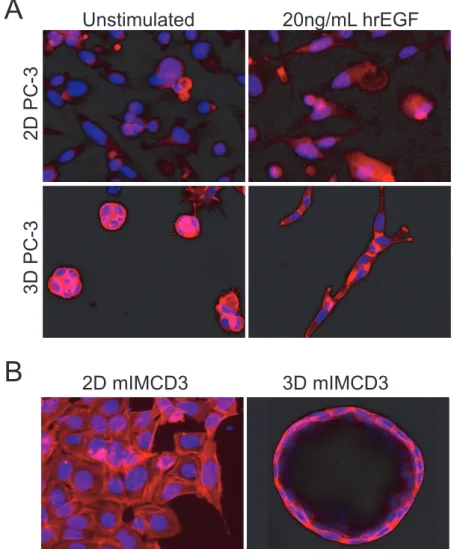
A
Unstimulated
20ng/mL hrEGF
2D PC-3
3D PC-3
B
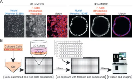
2D mIMCD3
3D mIMCD3
FIGURE 1 3D cell cultures provide a more physiologically relevant context for drug screening. A) Prostate Carcinoma (PC-3) cells cultured as 2D monolayer (top) or embedded in 3D hydrogels (bottom) display differential morphology and response to growth factors.75 Images in top panel obtained using wide-field BD pathway 855 with a
10x objective and images in bottom panel obtained using a Nikon Ti Eclipse confocal
microscope with 20x objective. B) mIMCD3 cells transduced with a short-hairpin
targeting Pkd1, deactivation of which is responsible for cyst growth in polycystic