In Vitro Evaluation of the Effect of Different Sandblasting Times on
the Bond Strength of Feldspathic Porcelain to Composite Resin
E. Amin Salehi1, H. Heshmat1, E. Moravej Salehi 2, MJ. Kharazifard3
1Assistant Professor, Department of Restorative Dentistry, School of Dentistry, Dental Branch Islamic Azad University. Tehran,
Iran
2Assistant Professor, Department of Restorative Dentistry, School of Dentistry, Shahid Beheshti University of Medical Sciences,
Tehran, Iran
3Research Member of Dental Research Center, Tehran University of Medical Sciences. Tehran, Iran
Corresponding author: E. Moravej Salehi, Assistant Professor, Department of Restorative Dentistry, School of Dentistry, Shahid Beheshti University of Medical Sciences, Tehran, Iran
e.msalehi@yahoo.com Received: 1 May 2012 Accepted: 16 Aug 2012
Abstract
Background and Aim:Intraoral repair of fractured porcelain is an acceptable method to avoid replacement and therefore saving time and cost. The purpose of this study was to determine the in-vitro shear bond strengths of composite resin to feldspathic porcelain after different durations of sandblasting and to compare the effect of sandblasting with that of hydrofluoric acid (HF).
Materials and Methods: In this in-vitro study, 40 porcelain disks were fabricated and randomly divided into 4 groups (n=10). Porcelain surface in group 1 was etched with 9.5% HF for 2 minutes. Groups 2, 3 and 4 were sandblasted with 50:m alumina particles for 5, 10 and 15 seconds, respectively. All specimens received the same silane agent, bonding agent and composite resin. The samples were subjected to 5000 thermal cycles and then underwent shear bond strength testing. The mean bond strength was analyzed with one-way ANOVA. The mode of failure was determined using stereomicroscope and scanning electron microscope. An additional porcelain sample was fabricated and prepared according to the aforementioned protocols in each group and its surface topography was observed by SEM.
Results: The mean bond strength was 15/28 (±3/64), 13/82(±4/03), 15/77(±3/94) and 16/54(±3/73) MPa in the 4 groups, respectively. There were no statistically significant differences among groups. The most common mode of failure was cohesive in porcelain. No statistically significant difference was found in SEM results of different durations of sandblasting.
Conclusion: The shear bond strength was not significantly different after various durations of sandblasting treatment. The bond strength after sandblasting was similar to that of HF. SEM showed that HF acid etching and sandblasting patterns were different.
Key Words: Sandblasting , Bond strength , Dental porcelain ,Composite resin Journal of Islamic Dental Association of IRAN (JIDAI) Spring 2013 ;25, (2)
Introduction
Ceramic restorations are becoming increasingly popular due to their optimal characteristics like favorable esthetics and biocompatibility [1-4]. Silica-based ceramics like feldspathic porcelain are used in metal-ceramic and all-ceramic veneer
restorations [5,6]. Excellent esthetic properties make these ceramics a good candidate for other ceramic restorations such as laminate veneers [6, 7]. However, the porcelain may fracture or chip in the oral cavity, during function as the result of factors such as occlusal forces, trauma, internal
defects and inappropriate design [7, 8]. Fracture is the most common cause of failure of various ceramic restorations [9]. Clinical studies have reported 5 to 10% prevalence rate for ceramic fractures in more than 10 years of clinical use [10]. Finding a standard method with optimal strength seems necessary for intraoral repair of porcelain fractures with composite resin, avoiding the replacement of restoration, sparing time and cost [11, 7-12] and bonding of orthodontic brackets to porcelain [2,13]. Resin to porcelain bond requires adequate porcelain surface treatment. At present, such bond is achieved through the application of micromechanical and chemical techniques [6,7, 12, 14]. Published studies recommend acid-etching or sandblasting with alumina particles for micromechanical retention [14] and application of silane agent for chemical bonding [14, 15].
Etching with hydrofluoric acid (HF) and subsequent silanization [6, 14] is a commonly used conventional surface treatment technique for increasing the bond strength to feldspathic porcelain. Fabianelli and Pollington in 2010 gave good reasons for maintaining the HF etching phase because HF is a very toxic chemical and a potentially serious health hazard. On the other hand, HF etching of silica-based ceramics produces insoluble hexafluorosilicate that can stay on the surface as a by-product and if not removed, interfere with the bond strength to resin [14, 15]. Application of silane agent after HF etching or air abrasion with alumina particles for ceramic surface treatment creates a good long-lasting bond [5, 16] which is stronger than the bond to the etched-only ceramic surface [5]. Silane agent improves bond strength by increasing the wettability [7, 17] and formation of covalence bond between ceramic and resin [7].
Intraoral sandblasting especially with 50-micron alumina particles is an easy effective method for repair of a fractured porcelain restoration and can be a suitable substitute for HF etching [12] by increasing the surface area and improving the micromechanical retention and bond strength [18]. Alumina particles create a clean and reactive
bonding surface in porcelain. Furthermore, the patient does not have to tolerate severe acid burns [12]. It should be noted that the efficacy of sandblasting is dependent on various factors like the size of particles, air pressure, duration of procedure, the selected angle, type of substrate, cleaning method, etc. [7, 19-20]. Xiong et al, in 2005 evaluated the effects of three factors involved in sandblasting (size of particles, pressure and time) on flexural strength of dental infiltrated Al2O3ceramics and reported the size of particles to
be the only effective factor in this regard [21]. Numerous studies have been conducted on the effect of different ceramic surface treatments i.e. the use of various types of burs, acid etching, sandblasting and laser on bond strength to resins yielding controversial results [22, 2, 6, 11, 14, 16-23]. However, a standard method for sandblasting of feldspathic porcelain and surface treatment for bonding to resin with intraoral sandblasting machine is yet to be found and various related parameters like the optimal duration, distance, pressure and angle need to be specified. To date, no study has compared the effect of different sandblasting durations on the bond strength of porcelain to composite resin and every study recommends a different time period. Therefore, considering the lack of adequate information on this subject, the present study was conducted aiming at finding the optimal duration of sandblasting with 50-micron alumina particles at constant pressure, distance and angulation and evaluating its effect on bond strength of composite resin to feldspathic porcelain.
Materials and Methods
At first, 40 porcelain disks (Ceramco 3, Dentsply Ceramco Co., Burlington, NJ) (with metal base from nickel titanium alloy) with 6 mm diameter and 3 mm thickness were fabricated in this in-vitro single blind experimental study. In order to match samples, the porcelain surfaces were ground on wet 400 and 600 grit silicon carbide discs (Mounted stones, American Dent-All Inc., Glendale, CA) for 15 seconds and were then rinsed and dried. Porcelain disks were randomly divided
into 4 groups of 10 each, coded and received the following surface treatments:
Group 1(controls): 9.5% HF gel (Porcelain Etchant Gel, Bisco Achaumburg, IL, USA) was applied on the surface of samples for 2 minutes. Samples were then rinsed with water and air dried for one minute. Group 2(cases): Surface of samples was sandblasted
by an intraoral sandblasting machine (Micro-sandblaster, Dento-Prop Ronvig, Denmark) with 50-micron alumina particles (Ronvig, Denmark) for 5 seconds at a constant distance of 5 mm, pres-sure of 3 bar and 90 degree angle in a circular mo-tion (a special jig was fabricated to meet the men-tioned criteria). After sandblasting, samples were rinsed with water for 1 minute and then air dried. Group 3: The exactly similar steps were repeated as in group 2. The only difference was duration of sandblasting for 10 seconds in this group.
Group 4: The exactly similar steps were repeated as in group 2. The only difference was duration of sandblasting for 15 seconds in this group.
A single layer of silane agent (Bis-Silane agent Parts A & B, Bisco, Schaumburg, IL, USA) was then applied on the surface of all samples with a microbrush for 1 minute and air dried for 30 seconds followed by the application of bonding resin (D/E Resin, Bisco, Schaumburg, IL, USA) and curing for 20 seconds with LED light cure unit (Starlight Pro, Mectron, Italy) with an intensity of 600 mW/cm2. Composite resin was then applied on
the adhesive area of samples in the form of 2-mm increments using clear Tygon plastic tubing with 3 mm diameter and 4 mm height. Each increment was cured for 40 seconds. Composite resin cylind-ers were light cured for an extra 120 seconds from all three dimensions at a 45 degree angulation to the surface of porcelain. The plastic tube was then cut with a sharp blade under stereomicroscope (Nikon SMZ800, Japan).
All samples were stored in distilled water at 37KC for 24 hours and were then subjected to 5000 thermal cycles in a thermocycler at 5-55KC with 30 seconds of dwell time and 10 seconds of transfer time from one bath to the other (Malek Teb, Iran). Samples were then mounted in self-polymerizing
acrylic resin molds (Acropars Co., Tehran, Iran), transferred to Universal Testing Machine (Zwick Roell Z050, Germany) and subjected to a load with a crosshead speed of 1 mm/min applied to the in-terface of composite resin/porcelain. The respec-tive shear bond strength was calculated and rec-orded in mega Pascals (MPa).
Mode of failure of samples was observed under stereomicroscope (Nikon SMZ800, Japan) with 40X magnification. Also, one sample from each group was randomly selected and its mode of fail-ure was evaluated with a scanning electron micro-scope (SEM) at 100X and 1000X magnifications. Modes of failure of samples were divided into the categories of cohesive, adhesive and cohesive/ ad-hesive (mixed). Coad-hesive failure occured in ceram-ic or composite resin. Also, an extra porcelain sample was prepared in each group according to the aforementioned preparation techniques (with-out the application of silane agent) and its surface topography was evaluated by SEM at 500X, 2000X and 4000X magnifications. For SEM analy-sis, first a 15 nm thick gold coat was applied on samples with sputter coater (K450X, EMITECH, England) and then they were evaluated under SEM (VEGA, TESCAN, Czech).
After ensuring the normal distribution of bond strength data, one way ANOVA was used for data analysis with a 95% confidence interval (M=0.05).
Results
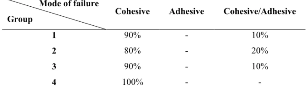
The mean shear bond strength of samples is dem-onstrated in Table 1. The highest mean bond strength was observed in group 4 (16.54±3.73 MPa). The lowest mean bond strength was de-tected in group 2 (13.82±4.03 MPa). No statistical-ly significant difference was detected between the understudy groups in this respect (p=0.455) (Table 2). By increasing the duration of sandblasting from 5 to 15 seconds, bond strength slightly increased but this increase was not statistically significant. The frequency of modes of failure observed with stereomicroscope in the studied groups is demon-strated in Table 3. All cohesive failures occurred in porcelain.
SEM results revealed significant differences in surface morphology of the porcelain etched with 9.5% HF for 2 minutes and the porcelain surface
sandblasted with 50 micron alumina particles; al-though in both methods the surface was porous as demonstrated in Figure 2.
Group Mean Standard deviation 95% CI for the mean Minimum Maximum Coefficient of change
Min Max
1 15/28 3/64 13/02 17/53 9/04 19/88 23 2 13/82 4/03 11/31 16/31 7/31 19/05 29 3 15/77 3/94 13/32 18/21 8/98 21/20 25 4 16/54 3/73 14/22 18/85 11/92 23/00 22
Source of variation Total sum of squares Degrees of freedom Mean squares F Level of significance
Inter-groups 39/416 3 13/139 0/891 0/455
Intra-groups 530/823 36 14/745
Total 570/239 39
Mode of failure
Group Cohesive Adhesive Cohesive/Adhesive
1 90% - 10%
2 80% - 20%
3 90% - 10%
4 100% - -
Table 1. Shear bond strength of samples in MPa
Table 2. Statistical comparison of groups using ANOVA
Table 3. Frequency (percentage) of modes of failure of samples in the studied groups
Figure 1. SEM analysis of the mode of failure of samples in groups 1 to 4 from right to left (1000X magnification)
Discussion
Nowadays, dentists try to repair chipped or fractured porcelain intraorally in order to avoid restoration replacement and spare time and cost [3, 7, 10, 11, 17]. The restorative material of choice for this purpose is composite resin due to its low cost and ease of application. The success of this repair depends on the presence of strong micromechanical and chemical bonds between the ceramic and composite resin, which requires adequate ceramic surface preparation and treatment [6, 17]. Various studies have evaluated different ceramic surface treatment regimens in order to achieve maximum bond strength [24]. The bond strengths achieved in various studies cannot be compared because the bond strength value for a specific material is widely affected by the type of substrate, preparation of sample, storage environment, and method of loading. Unfortunately, there is not much standard for laboratory investigations. Therefore, comparison of various study results must be done with great caution [25].
Shiu et al, in 2007 found the highest shear bond strength between resin cement and feldspathic porcelain after surface treatment with HF and sandblasting with alumina particles. Moderate bond strength was achieved in the group treated with the combination of HF etching and alumina sandblasting. Similar to our study, they applied silane agent to their samples but in contrast to ours, they did not perform any thermocycling or artificial aging [22].
Oral environment is different from in-vitro conditions [24]. Mechanical, thermal and chemical factors, presence of water and oral pH can significantly affect the bond strength between composite resin and ceramic [3, 6]. Studies have demonstrated that porcelain repair systems subjected to water storage or thermocycling result in lower bond strength [26, 23, 7-27]. Brose and Ruter stated that the water absorbed by composite resin causes hydrolysis and gradual dissolution of silane agent [7]. However, thermocycling is a more precise testing method for this purpose and
decreases bond strength more than water storage. Thus, thermocycling seems to be a logical method for screening of restorative materials in terms of their bond strength [28].
Several methods are available for evaluation of bond strength among which shear and tensile testing can be named [15, 23]. Adhesive interface is a highly stressed area which is not resistant to mechanical tests [7]. Shear bond strength is the most common method of assessment but mostly causes a cohesive type of failure in the substrate mass rather than fracture in the interface which results in complex stress distribution during the test and error in interpretation of data [25, 15, 7]. Analysis of the modes of failure in the present study (considering the evaluation of shear bond strength similar to some other studies) demonstrated the most common mode of failure to be cohesive within the porcelain [26] which may be rather attributed to the method of assessment which was shear bond strength testing.
Various researchers have demonstrated that 2 minutes of etching with 10% HF gel is the best method for increasing the bond strength of resin to feldspathic ceramic [3, 23]. Yadav et al, in 2010 reported the highest micro-shear bond strength values in feldspathic porcelain surface etched with hydrofluoric acid and coated with silane agent. Samples treated with airborne-particle abrasion with alumina and application of silane agent ranked second in this respect. The mode of failure was cohesive in porcelain similar to the present study result [2]. Thus, this method was selected for the control group in our study. In the present study, two-part silane was used since the atmospheric humidity is not optimal for the pre-hydrolyzed (one part) silane [15]. In a study by Khoroushi and Motamedi the bond strength achieved after Ceramco3 porcelain surface treatment with HF and silane was almost similar to that in the present study although samples were subjected to 5000 thermal cycles in the present study whereas only 1000 cycles were used in theirs [29].
On the other hand, there are several claims regarding the inefficacy of HF that suggest the
elimination of ceramic HF etching phase [15]. HF is a highly toxic chemical and a serious health hazard. It has adverse effects on soft tissue and its inhalation in the clinic by the dentist or patient is dangerous [15, 23]. HF results in formation of insoluble fluoride salt (hexafluorosilicate). This by-product remains on the surface and interferes with the bond strength to resin [23].
Sandblasting techniques are successful in improving the bond strength to gold, ceramic and amalgam surfaces [30]. Thus, a commonly recommended method, other than HF etching, is sandblasting with alumina particles that provides a clean and reactive porcelain surface for bonding [7, 10]. This method does not expose patients to severe acidic burns [12] and its efficacy depends on various factors like the size of particles, air pressure, duration of procedure, etc. [19, 17-20]. Alumina particles remove the weak ceramic phases and cause surface irregularities that increase surface area and improve micromechanical retention and bond strength [18].
Menezes et al, in their study in 2009 on glass matrix ceramics (IPS, Empress 2) reported the highest microshear bond strength in both groups of HF etching and sandblasting with 50-micron alu-mina particles from 4 mm distance. These two groups were not significantly different from each other. On SEM analysis, the most prominent mode of failure was ceramic cohesive type [27]. In the present study, the shear bond strength after sandblasting was not significantly different that the rate following HF etching.
In our study, SEM analysis showed that the feldspathic porcelain surface etched with 9.5% HF for 2 minutes had porosities from small and shallow to large and deep pores formed by the coalescence of small pores. The porosities had formed a three-dimensional network of canals and voids. Bottino et al, in 2008 and some others also observed this pattern [3, 24]. Borges et al, in 2003 compared this pattern to a honeycomb [31]. This surface topography is explained by the preferred reaction of HF with glass, Leucite and acid-sensitive phase of feldspathic porcelain and is
ideal for micromechanical retention [23, 27, 31]. Also, it improves wettability of silane due to the higher surface energy of the etched surface [24]. In the current study, 5 to 15 seconds of sandblasting did not cause a statistically significant difference in shear bond strength compared to 9.5% HF etching for 2 min. However, SEM showed that the porcelain surface morphology after sandblasting included same shape and same size porosities as in HF-treated samples but with a more homogenous depth and shallower pores which may indicate the fact that in addition to surface roughness, some other factors also affect the shear bond strength of ceramic to composite resin [3]. Also, it should be noted that there is a threshold for surface porosities that limits their impact on bond strength [3]. In the present study, the etching pattern and porosities observed under SEM were different following HF etching and sandblasting. This finding is in accordance with the SEM results of other studies although the type of ceramic used in our study was different from the ones used in other studies [3]. Furthermore, SEM analysis in the present study failed to find a significant difference in surface topography of samples by increasing the duration of sandblasting although it may be stated that number of porosities slightly increased. Lack of a significant difference in surface topography by increasing the duration of sandblasting from 5 to 15 seconds can, by some means, explain the lack of a significant difference in bond strength although several factors are involved in this respect. In the present study, no statistically significant difference was observed in bond strength by increasing the duration of sandblasting from 5 to 15 seconds although it was slightly improved. Therefore, by increasing the duration of sandblasting to more than 30 to 60 seconds bond strength may significantly be improve. However, we do know that increasing the duration of intraoral sandblasting for long periods of time is not feasible. On the other hand it causes distinct sharp margins in surface topography of the ceramic that may act as stress points and result in formation and propagation of cracks that can adversely affect
the fracture resistance of porcelain. Therefore, further investigations are required on this subject. Matsumuara et al. demonstrated that all systems used for chemical or mechanical retention need to have a minimum of 10 MPa bond strength in order to be qualified for application in a clinical setting [7]. The obtained bond strength in the current study after 5 to 15 seconds of sandblasting was similar to that of HF etching both being in an acceptable range. However, these results cannot be easily generalized to the clinical setting and other factors present in the oral environment such as mechanical and chemical loads have to be considered as well. Thus, future studies are required to be performed in clinical or conditions close to clinical settings. Other parameters of the intraoral sandblasting machine like optimal pressure and distance can also be evaluated. Furthermore, considering the increasing application of ceramics in esthetic restorations, these tests have to be carried out and the obtained results should be reported.
Conclusion
Sandblasting with 50-micron alumina particles for 5, 10 and 15 seconds from a constant distance of 5 mm and with 3 bar pressure and 90 degree angulation cannot significantly change the shear bond strength of feldspathic porcelain to composite resin. Also, the bond strength after the mentioned treatment was similar to that after the application of 9.5% HF for 2 min. SEM results demonstrated a different etching pattern on the surface morphology of samples after HF etching and sandblasting surface treatments although a porous surface was resulted in both methods. SEM analysis of samples subjected to different durations of sandblasting from 5 to 15 seconds failed to find a statistically significant difference between them.
References
1- Blum IR, Jagger DC, Wilson NH. Defective dental restorations: to repair or not to repair? Part 2: All-ceramics and porcelain fused to metal systems.Dent Update. 2011 Apr;38(3):150-2, 154-6, 158.
2- Yadav S, Upadhyay M, Borges GA, Roberts WE. Influence of ceramic (feldspathic) surface treatments on the micro-shear bond strength of composite resin. Angle Orthod. 2010 Jul; 80(4): 577-82.
3- Kukiattrakoon B, Thammasitboon K.The effect of different etching times of acidulated phosphate fluoride gel on the shear bond strength of high-leucite ceramics bonded to composite resin. J Prosthet Dent. 2007 Jul; 98(1):17-23.
4- Lee SY, Vang MS, Yang HS, Park SW, Park HO, Lim HP. Shear bond strength of composite resin to titanium according to various surface treatments.J Adv Prosthodont. 2009 Jul;1(2):68-74. Epub 2009 Jul 31.
5- Kim TH, Jivraj SA, Donovan TE.Selection of luting agents: part 2.J Calif Dent Assoc. 2006 Feb; 34(2):161-6.
6- Blatz MB, Sadan A, Kern M.Resin-ceramic bonding: a review of the literature. J Prosthet Dent. 2003 Mar;89(3):268-74.
7- de Melo RM, Valandro LF, Bottino MA. Microtensile bond strength of a repair composite to leucite-reinforced feldspathic ceramic. Braz Dent J. 2007;18(4):314-9.
8- Haselton DR, Diaz-Arnold AM, Dunne JT Jr.Shear bond strengths of 2 intraoral porcelain repair systems to porcelain or metal substrates. J Prosthet Dent. 2001 Nov;86(5):526-31.
9- Summitt JB, Robbins JW, Hilton TJ. Schwartz RS, Santos J. Fundamentals of Operative Dentistry: a contemporary approach. 3rd ed.
Quintessence, USA; 2006: 482.
10- Ozcan M. Fracture reasons in ceramic-fused-to-metal restorations. J Oral Rehabil. 2003 Mar; 30(3):265-9. Review.
11- Shahverdi S, Canay S, Sahin E, Bilge A. Effects of different surface treatment methods on the bond strength of composite resin to porcelain. J Oral Rehabil. 1998 Sep;25(9):699-705.
12- Ozcan M.Evaluation of alternative intra-oral repair techniques for fractured ceramic-fused-to-metal restorations. J Oral Rehabil. 2003 Feb; 30 (2):194-203.
13- Trakyali G, Malkondu O, KazazoRlu E, Arun T. Effects of different silanes and acid concentrations on bond strength of brackets to porcelain surfaces.Eur J Orthod. 2009 Aug; 31(4): 402-6. Epub 2009 Apr 1.
12
14- Pollington S, Fabianelli A, van Noort R. Microtensile bond strength of a resin cement to a novel fluorcanasite glass-ceramic following different surface treatments. Dent Mater. 2010 Sep; 26(9):864-72. Epub 2010 Jun 12.
15- Fabianelli A, Pollington S, Papacchini F, Goracci C, Cantoro A, Ferrari M, van Noort. The effect of different surface treatments on bond strength between leucite reinforced feldspathic ceramic and composite resin. - J Dent. 2010 Jan; 38(1):39-43.
16- Thurmond JW, Barkmeier WW, Wilwerding TM.Effect of porcelain surface treatments on bond strengths of composite resin bonded to porcelain. J Prosthet Dent. 1994 Oct;72(4):355-9.
17- Akyil MS, Yilmaz A, KaraalioRlu OF, DuymuS
ZY.Shear bond strength of repair composite resin to an acid-etched and a laser-irradiated feldspathic ceramic surface. Photomed Laser Surg. 2010 Aug; 28(4):539-45.
18- Della Bona A, Anusavice KJ. Microstructure, composition, and etching topography of dental ceramics. Int J Prosthodont. 2002 Mar-Apr; 15(2): 159-67.
19- Della Bona A, Borba M, Benetti P, Cecchetti D. Effect of surface treatments on the bond strength of a zirconia-reinforced ceramic to composite resin. Braz Oral Res. 2007 Jan-Mar; 21(1): 10-5.
20-Shiu P, De Souza-Zaroni WC, Eduardo Cde P, Youssef MN. Effect of feldspathic ceramic surface treatments on bond strength to resin cement. Photomed Laser Surg. 2007 Aug; 25(4):291-6. 21-Xiong F, Yu HA, Liao Y, Zhu Z, Zhou Z, Zhu M. Effect of erosion on strength of dental infiltrated Al2O3 ceramics. Sheng Wu Yi Xue Gong Cheng Xue Za Zhi. 2005Dec;22(6):1189-92, 1199.
22-Shiu P, De Souza-Zaroni WC, Eduardo Cde P, Youssef MN. Effect of feldspathic ceramic surface treatments on bond strength to resin cement. Photomed Laser Surg. 2007 Aug; 25(4):291-6. 23-Brentel AS, Ozcan M, Valandro LF, Alarça LG, Amaral R, Bottino MA. Microtensile bond strength of a resin cement to feldpathic ceramic after different etching and silanization regimens in dry and aged conditions. Dent Mater. 2007 Nov; 23(11):1323-31. Epub 2006 Dec 26.
24-Bottino MC, Ozcan M, Coelho PG, Valandro LF, Bressiani JC, Bressiani AH. Micro-morphological changes prior to adhesive bonding: high-alumina and glassy-matrix ceramics. Braz Oral Res. 2008 Apr-Jun;22(2):158-63.
25-Sakaguch R, Power J. Craig’s Restorative Dental Materials. 13th ed. United States: Mosby;
2012, 330, 97-98.
26- Yassini E, Tabari K. comparison of shear bond strength between composite resin and porcelain using different bonding systems. J Dentistry, Tehran Univ Med Sci., Tehran, Iran. 2005; 2(1):1-6.
27- Menezes F , Borges G , Valentino T, Oliveira M, Tussi C, Sobrinho L. Effect of surface treatment and storage on the bond strength of different ceramic systems. Braz J Oral Sci. 2009; 8 (3):119-123.
28- Mair L, Padipatvuthikul P.Variables related to materials and preparing for bond strength testing irrespective of the test protocol. Dent Mater. 2010 Feb;26(2):e17-23. Epub 2010 Jan 13.
29- Khoroushi M, Motamdi Sh. Shear bond strength of composite-resin to porcelain: Effect of thermocycling. J Dent, Teh Univ. Med. Sci., Tehran, Iran. 2007; 4(1):21-26
30-Cal-Neto JP, Castro S, Moura PM, Ribeiro D, Miguel JA. Influence of enamel sandblasting prior to etching on shear bond strength of indirectly bonded lingual appliances. Angle Orthod. 2011 Jan; 81(1):149-52.
31- Borges GA, Sophr AM, de Goes MF, Sobrinho LC, Chan DC. Effect of etching and airborne particle abrasion on the microstructure of different dental ceramics. J Prosthet Dent. 2003 May; 89 (5):479-88.