ACC/AHA GUIDELINE REVISION
ACC/AHA 2007 Guidelines for the Management of
Patients With Unstable Angina/Non–ST-Elevation
Myocardial Infarction
A Report of the American College of Cardiology/American Heart Association Task Force on Practice Guidelines (Writing Committee to Revise the 2002 Guidelines for the
Management of Patients With Unstable Angina/Non–ST-Elevation Myocardial Infarction)
Developed in Collaboration with the American College of Emergency Physicians, the Society for Cardiovascular Angiography and Interventions, and the Society of Thoracic Surgeons
Endorsed by the American Association of Cardiovascular and Pulmonary Rehabilitation and the Society for Academic Emergency Medicine
Writing Committee Members
Jeffrey L. Anderson, MD, FACC, FAHA,Chair
Cynthia D. Adams, RN, PHD, FAHA
Elliott M. Antman, MD, FACC, FAHA
Charles R. Bridges, SCD, MD, FACC, FAHA*
Robert M. Califf, MD, MACC
Donald E. Casey, JR, MD, MPH, MBA, FACP†
William E. Chavey II, MD, MS‡ Francis M. Fesmire, MD, FACEP§ Judith S. Hochman, MD, FACC, FAHA
Thomas N. Levin, MD, FACC, FSCAI储
A. Michael Lincoff, MD, FACC
Eric D. Peterson, MD, MPH, FACC, FAHA Pierre Theroux, MD, FACC, FAHA
Nanette Kass Wenger, MD, FACC, FAHA R. Scott Wright, MD, FACC, FAHA
*Society of Thoracic Surgeons Representative; †American College of Physicians Representative; ‡American Academy of Family Physicians Representative; §American College of Emergency Physicians Repre-sentative;储Society for Cardiovascular Angiography and Interventions Representative
TABLE OF CONTENTS
Preamble...e3
1. Introduction...e4
1.1. Organization of Committee and Evidence Review...e4
1.2. Purpose of These Guidelines...e6
1.3. Overview of the Acute Coronary Syndromes...e6
1.3.1. Definition of Terms...e6
1.3.2. Pathogenesis of UA/NSTEMI...e8
1.3.3. Presentations of UA and NSTEMI...e9
1.4. Management Before UA/NSTEMI and Onset of UA/NSTEMI...e9
1.4.1. Identification of Patients at Risk of UA/NSTEMI...e10
1.4.2. Interventions to Reduce Risk of UA/NSTEMI...e10
1.5. Onset of UA/NSTEMI...e11
1.5.1. Recognition of Symptoms by Patient...e11
1.5.2. Silent and Unrecognized Events...e11
2. Initial Evaluation and Management...e12
2.1. Clinical Assessment...e12
This document was approved by the American College of Cardiology Foundation Board of Trustees in June 2007 and by the American Heart Association Science Advisory and Coordinating Committee in June 2007.
When citing this document, the American College of Cardiology Foundation and the American Heart Association request that the following citation format be used: Anderson JL, Adams CD, Antman EM, Bridges CR, Califf RM, Casey DE Jr, Chavey WE II, Fesmire FM, Hochman JS, Levin TN, Lincoff AM, Peterson ED, Theroux P, Wenger NK, Wright RS. ACC/AHA 2007 guidelines for the management of patients with unstable angina/non–ST-elevation myocardial infarction: a report of the American College of Cardiology/American Heart Association Task Force on Practice Guidelines (Writing Committee to Revise the 2002 Guidelines for the Management of Patients With Unstable Angina/Non–ST-Elevation Myocardial Infarction): developed in collab-oration with the American College of Emergency Physicians, American College of Physicians, Society for Academic Emergency Medicine, Society for Cardiovascular
Angiography and Interventions, and Society of Thoracic Surgeons. J Am Coll Cardiol 2007;50:e1–157.
This article has been copublished in the August 7, 2007 issue ofCirculation. Copies: This document is available on the World Wide Web sites of the American College of Cardiology (www.acc.org), and the American Heart Association (www.americanheart.org). For copies of this document, please contact Elsevier Inc. Reprint Department, fax (212) 633-3820, e-mail reprints@elsevier.com.
2.1.1. Emergency Department or Outpatient Facility Presentation...e16
2.1.2. Questions to Be Addressed at the Initial Evaluation...e16
2.2. Early Risk Stratification...e16
2.2.1. Estimation of the Level of Risk...e18
2.2.2. Rationale for Risk Stratification...e18
2.2.3. History...e19
2.2.4. Anginal Symptoms and Anginal Equivalents...e19
2.2.5. Demographics and History in Diagnosis and Risk Stratification...e20
2.2.6. Estimation of Early Risk at Presentation...e21
2.2.6.1. ELECTROCARDIOGRAM...e23 2.2.6.2. PHYSICAL EXAMINATION...e24 2.2.7. Noncardiac Causes of Symptoms and Secondary
Causes of Myocardial Ischemia...e24
2.2.8. Cardiac Biomarkers of Necrosis and the
Redefinition of AMI...e25
2.2.8.1. CREATINE KINASE-MB...e25
2.2.8.2. CARDIAC TROPONINS...e26
2.2.8.2.1 CLINICAL USE...e26 2.2.8.2.1.1. Clinical Use of Marker
Change Scores ...e28 2.2.8.2.1.2. Bedside Testing for Cardiac
Markers...e29
2.2.8.3. MYOGLOBIN AND CK-MB SUBFORMS COMPARED WITH TROPONINS...e29
2.2.8.4. SUMMARY COMPARISON OF BIOMARKERS OF NECROSIS: SINGLY AND IN COMBINATION...e29
2.2.9. Other Markers and Multimarker Approaches...e29
2.2.9.1. ISCHEMIA...e29
2.2.9.2. COAGULATION...e30
2.2.9.3. PLATELETS...e30
2.2.9.4. INFLAMMATION...e30
2.2.9.5. B-TYPE NATRIURETIC PEPTIDES...e31
2.3. Immediate Management...e31
2.3.1. Chest Pain Units...e32
2.3.2. Discharge From ED or Chest Pain Unit...e33
3. Early Hospital Care...e34
3.1. Anti-Ischemic and Analgesic Therapy...e35
3.1.1. General Care...e38
3.1.2. Use of Anti-Ischemic Therapies...e39
3.1.2.1. NITRATES...e39
3.1.2.2. MORPHINE SULFATE...e40
3.1.2.3. BETA-ADRENERGIC BLOCKERS...e41
3.1.2.4. CALCIUM CHANNEL BLOCKERS...e43
3.1.2.5. INHIBITORS OF THE RENIN-ANGIOTENSIN-ALDOSTERONE SYSTEM...e44
3.1.2.6. OTHER ANTI-ISCHEMIC THERAPIES...e44
3.1.2.7. INTRA-AORTIC BALLOON PUMP COUNTERPULSATION...e45
3.1.2.8. ANALGESIC THERAPY...e45
3.2. Recommendations for Antiplatelet/Anticoagulant Therapy in Patients for Whom Diagnosis of
UA/NSTEMI Is Likely or Definite...e45
3.2.1. Antiplatelet Therapy Recommendations...e45
3.2.2. Anticoagulant Therapy Recommendations...e46
3.2.3. Additional Management Considerations for
Antiplatelet and Anticoagulant Therapy...e47
3.2.4. Antiplatelet Agents and Trials (Aspirin,
Ticlopidine, Clopidogrel)...e49
3.2.4.1. ASPIRIN...e49 3.2.4.2. ADENOSINE DIPHOSPHATE RECEPTOR ANTAGONISTS
AND OTHER ANTIPLATELET AGENTS...e51 3.2.5. Anticoagulant Agents and Trials...e55
3.2.5.1. UNFRACTIONATED HEPARIN...e56 3.2.5.2. LOW-MOLECULAR-WEIGHT HEPARIN...e57
3.2.5.3. LMWH VERSUS UFH...e58
3.2.5.3.1. EXTENDED THERAPY WITH LMWHS...e61 3.2.5.4. DIRECT THROMBIN INHIBITORS...e61
3.2.5.5. FACTOR XA INHIBITORS...e64
3.2.5.6. LONG-TERM ANTICOAGULATION...e65
3.2.6. Platelet GP IIb/IIIa Receptor Antagonists...e66
3.2.7. Fibrinolysis...e72
3.3. Initial Conservative Versus Initial Invasive
Strategies...e72
3.3.1. General Principles...e72
3.3.2. Rationale for the Initial Conservative Strategy...e73
3.3.3. Rationale for the Invasive Strategy...e73
3.3.4. Immediate Angiography...e73
3.3.5. Deferred Angiography...e73
3.3.6. Comparison of Early Invasive and Initial
Conservative Strategies...e73
3.3.7. Subgroups...e77
3.3.8. Care Objectives...e77
3.4. Risk Stratification Before Discharge...e79
3.4.1. Care Objectives...e80
3.4.2. Noninvasive Test Selection...e81
3.4.3. Selection for Coronary Angiography...e82
3.4.4. Patient Counseling...e82
4. Coronary Revascularization...e83
4.1. Recommendations for Revascularization With PCI and CABG in Patients With UA/NSTEMI...e83
4.1.1. Recommendations for PCI...e83
4.1.2. Recommendations for CABG...e83
4.2. General Principles...e84
4.3. Percutaneous Coronary Intervention...e85
4.3.1. Platelet Inhibitors and Percutaneous
Revascularization...e86
4.4. Surgical Revascularization...e87
4.5. Conclusions...e89
5. Late Hospital Care, Hospital Discharge, and Post-Hospital Discharge Care...e89
5.1. Medical Regimen and Use of Medications...e90
5.2. Long-Term Medical Therapy and Secondary
Prevention...e90
5.2.1. Antiplatelet Therapy...e91
5.2.2. Beta Blockers...e91
5.2.3. Inhibition of the Renin-Angiotensin-Aldosterone System...e91
5.2.4. Nitroglycerin...e92
5.2.5. Calcium Channel Blockers...e92
5.2.6. Warfarin Therapy...e92
5.2.7. Lipid Management...e92
5.2.8. Blood Pressure Control...e94
5.2.9. Diabetes Mellitus...e95
5.2.10. Smoking Cessation...e95
5.2.11. Weight Management...e95
5.2.12. Physical Activity...e96
5.2.13. Patient Education...e96
5.2.14. Influenza...e96
5.2.15. Depression...e96
5.2.16. Nonsteroidal Anti-Inflammatory Drugs...e96
5.2.17. Hormone Therapy...e97
5.2.18. Antioxidant Vitamins and Folic Acid...e98
5.3. Postdischarge Follow-Up...e98
5.4. Cardiac Rehabilitation...e99
5.6. Other Activities...e101
5.7. Patient Records and Other Information Systems....e102
6. Special Groups...e102
6.1. Women...e102
6.1.1. Profile of UA/NSTEMI in Women...e102
6.1.2. Management...e103
6.1.2.1. PHARMACOLOGICAL THERAPY...e103 6.1.2.2. CORONARY ARTERY REVASCULARIZATION...e103 6.1.2.3. INITIAL INVASIVE VERSUS INITIAL CONSERVATIVE
STRATEGY...e104 6.1.3. Stress Testing...e106
6.1.4. Conclusions...e106
6.2. Diabetes Mellitus...e106
6.2.1. Profile and Initial Management of Diabetic and Hyperglycemic Patients With UA/NSTEMI...e107
6.2.2. Coronary Revascularization...e108
6.2.3. Conclusions...e109
6.3. Post-CABG Patients...e109
6.3.1. Pathological Findings...e109
6.3.2. Clinical Findings and Approach...e109
6.3.3. Conclusions...e110
6.4. Older Adults...e110
6.4.1. Pharmacological Management...e111
6.4.2. Functional Studies...e111
6.4.3. Percutaneous Coronary Intervention in Older
Patients...e111
6.4.4. Contemporary Revascularization Strategies in Older Patients...e112
6.4.5. Conclusions...e112
6.5. Chronic Kidney Disease...e112
6.6. Cocaine and Methamphetamine Users...e113
6.6.1. Coronary Artery Spasm With Cocaine Use...e114
6.6.2. Treatment...e115
6.6.3. Methamphetamine Use and UA/NSTEMI...e115
6.7. Variant (Prinzmetal’s) Angina...e115
6.7.1. Clinical Picture...e116
6.7.2. Pathogenesis...e116
6.7.3. Diagnosis...e116
6.7.4. Treatment...e117
6.7.5. Prognosis...e117
6.8. Cardiovascular “Syndrome X”...e117
6.8.1. Definition and Clinical Picture...e117
6.8.2. Treatment...e118
6.9. Takotsubo Cardiomyopathy...e119
7. Conclusions and Future Directions...e119
Appendix 1 . . . .e121
Appendix 2 . . . .e126
Appendix 3 . . . .e131
References. . . .e133
Preamble
It is important that the medical profession play a significant role in critically evaluating the use of diagnostic procedures and therapies in the detection, management, or prevention of disease states. Rigorous and expert analysis of the available data documenting absolute and relative benefits and risks of those procedures and therapies can produce helpful guidelines that improve the effectiveness of care, optimize patient outcomes, and favorably affect the overall cost of care by focusing resources on the most effective strategies.
The American College of Cardiology Foundation (ACCF) and the American Heart Association (AHA) have jointly engaged in the production of such guidelines in the area of cardiovascular disease since 1980. The American College of
Cardiology (ACC)/AHA Task Force on Practice Guidelines, whose charge is to develop, update, or revise practice guidelines for important cardiovascular diseases and procedures, directs this effort. Writing committees are charged with the task of performing an assessment of the evidence and acting as an independent group of authors to develop, update, or revise written recommendations for clinical practice.
Experts in the subject under consideration have been selected from both organizations to examine subject-specific data and write guidelines. The process includes additional representatives from other medical practitioner and specialty groups when appropriate. Writing committees are specifi-cally charged to perform a formal literature review, weigh the strength of evidence for or against a particular treatment or procedure, and include estimates of expected health ACC/AHA
Task Force Members
Sidney C. Smith, JR, MD, FACC, FAHA,Chair
Alice K. Jacobs, MD, FACC, FAHA,Vice-Chair
Cynthia D. Adams, RN, PHD, FAHA
Jeffrey L. Anderson, MD, FACC, FAHA Elliott M. Antman, MD, FACC, FAHA¶ Jonathan L. Halperin, MD, FACC, FAHA Sharon A. Hunt, MD, FACC, FAHA Harlan M. Krumholz, MD, FACC, FAHA
Frederick G. Kushner, MD, FACC, FAHA Bruce W. Lytle, MD, FACC, FAHA Rick Nishimura, MD, FACC, FAHA Joseph P. Ornato, MD, FACC, FAHA** Richard L. Page, MD, FACC, FAHA Barbara Riegel, DNSC, RN, FAHA
outcomes where data exist. Patient-specific modifiers, co-morbidities, and issues of patient preference that might influence the choice of particular tests or therapies are considered, as well as frequency of follow-up and cost effectiveness. When available, information from studies on cost will be considered; however, review of data on efficacy and clinical outcomes will constitute the primary basis for preparing recommendations in these guidelines.
The ACC/AHA Task Force on Practice Guidelines makes every effort to avoid any actual, potential, or per-ceived conflict of interest that may arise as a result of an industry relationship or personal interest of a member of the Writing Committee. Specifically, all members of the Writ-ing Committee, as well as peer reviewers of the document, were asked to provide disclosure statements of all such relationships that may be perceived as real or potential conflicts of interest. Writing Committee members are also strongly encouraged to declare a previous relationship with industry that may be perceived as relevant to guideline development. If a Writing Committee member develops a new relationship with industry during their tenure, they are required to notify guideline staff in writing. The continued participation of the Writing Committee member will be reviewed. These statements are reviewed by the parent task force, reported orally to all members of the Writing Com-mittee at each meeting, and updated and reviewed by the Writing Committee as changes occur. Please refer to the methodology manual for ACC/AHA Guideline Writing Committees further description of relationships with indus-try policy, available on the ACC and AHA World Wide Web sites (http://www.acc.org/qualityandscience/clinical/ manual/manual%5Fi.htm and http://www.circ.ahajournals. org/manual/). See Appendix 1 for a list of Writing Committee member relationships with industry and Appendix 2 for a listing of peer reviewer relationships with industry that are pertinent to this guideline.
These practice guidelines are intended to assist health care providers in clinical decision making by describing a range of generally acceptable approaches for the diagnosis, management, and prevention of specific diseases or condi-tions. Clinical decision making should consider the quality and availability of expertise in the area where care is provided. These guidelines attempt to define practices that meet the needs of most patients in most circumstances. These guideline recommendations reflect a consensus of expert opinion after a thorough review of the available, current scientific evidence and are intended to improve patient care.
Patient adherence to prescribed and agreed upon medical regimens and lifestyles is an important aspect of treatment. Prescribed courses of treatment in accordance with these recommendations will only be effective if they are followed. Since lack of patient understanding and adherence may adversely affect treatment outcomes, physicians and other health care providers should make every effort to engage the
patient in active participation with prescribed medical reg-imens and lifestyles.
If these guidelines are used as the basis for regulatory/ payer decisions, the ultimate goal is quality of care and serving the patient’s best interests. The ultimate judgment regarding care of a particular patient must be made by the health care provider and patient in light of all the circum-stances presented by that patient. There are circumcircum-stances in which deviations from these guidelines are appropriate.
The guidelines will be reviewed annually by the ACC/ AHA Task Force on Practice Guidelines and will be considered current unless they are updated, revised, or sunsetted and withdrawn from distribution. The executive summary and recommendations are published in the August 7, 2007, issue of the Journal of the American College of Cardiology and August 7, 2007, issue of Circulation. The full- text guidelines are e-published in the same issue of the journals noted above, as well as posted on the ACC (www.acc.org) and AHA (www.americanheart.org) World Wide Web sites. Copies of the full text and the executive summary are available from both organizations.
Sidney C. Smith, Jr, MD, FACC, FAHA Chair, ACC/AHA Task Force on Practice Guidelines
1. Introduction
1.1. Organization of Committee and Evidence Review
The ACC/AHA Task Force on Practice Guidelines was formed to make recommendations regarding the diagnosis and treatment of patients with known or suspected cardiovascular disease (CVD). Coronary artery disease (CAD) is the leading cause of death in the United States. Unstable angina (UA) and the closely related condition of non–ST-segment elevation myocardial infarction (NSTEMI) are very common manifes-tations of this disease.
The committee members reviewed and compiled pub-lished reports through a series of computerized literature searches of the English-language literature since 2002 and a final manual search of selected articles. Details of the specific searches conducted for particular sections are pro-vided when appropriate. Detailed evidence tables were developed whenever necessary with the specific criteria outlined in the individual sections. The recommendations made were based primarily on these published data. The weight of the evidence was ranked highest (A) to lowest (C). The final recommendations for indications for a diag-nostic procedure, a particular therapy, or an intervention in patients with UA/NSTEMI summarize both clinical evi-dence and expert opinion.
Classification of Recommendations
how the grading system provides an estimate of the size of the treatment effect and an estimate of the certainty of the treatment effect.
A complete list of the thousands of publications on various aspects of this subject is beyond the scope of these guidelines; only selected references are included. The Committee consisted of acknowledged experts in general internal medicine representing the American College of Physicians (ACP), family medicine from the American Academy of Family Physicians (AAFP), emergency med-icine from the American College of Emergency Physi-cians (ACEP), thoracic surgery from the Society of Thoracic Surgeons (STS), interventional cardiology from the Society for Cardiovascular Angiography and Inter-ventions (SCAI), and general and critical care cardiology, as well as individuals with recognized expertise in more specialized areas, including noninvasive testing,
preven-tive cardiology, coronary intervention, and cardiovascular surgery. Both the academic and private practice sectors were represented. This document was reviewed by 2 outside reviewers nominated by each of the ACC and AHA and by 49 peer reviewers. These guidelines will be considered current unless the Task Force revises them or withdraws them from distribution.
These guidelines overlap several previously published ACC/ AHA practice guidelines, including the ACC/AHA Guide-lines for the Management of Patients With ST-Elevation
Myocardial Infarction (1), the ACC/AHA/SCAI 2005
Guideline Update for Percutaneous Coronary Intervention (2), the AHA/ACC Guidelines for Secondary Prevention for Patients With Coronary and Other Atherosclerotic Vascular Disease: 2006 Update (3), and the ACC/AHA 2002 Guide-line Update for the Management of Patients With Chronic Stable Angina (4).
Table 1. Applying Classification of Recommendations and Level of Evidence†
1.2. Purpose of These Guidelines
These guidelines address the diagnosis and management of patients with UA and the closely related condition of NSTEMI. These life-threatening disorders are a major cause of emergency medical care and hospitalization in the United States. In 2004, the National Center for Health Statistics reported 1,565,000 hospitalizations for primary or secondary diagnosis of an acute coronary syndrome (ACS), 669,000 for UA and 896,000 for myocardial infarction (MI) (5). The average age of a person having a first heart attack is 65.8 years for men and 70.4 years for women, and 43% of ACS patients of all ages are women. In 2003, there were 4,497,000 visits to US emergency departments (EDs) for primary diagnosis of CVD (5). The prevalence of this presentation of CVD ensures that many health care provid-ers who are not cardiovascular specialists will encounter patients with UA/NSTEMI in the course of the treatment of other diseases, especially in outpatient and ED settings. These guidelines are intended to assist both cardiovascular specialists and nonspecialists in the proper evaluation and management of patients with an acute onset of symptoms suggestive of these conditions. These clinical practice
guide-lines also provide recommendations and supporting evi-dence for the continued management of patients with these conditions in both inpatient and outpatient settings. The diagnostic and therapeutic strategies that are recommended are supported by the best available evidence and expert opinion. The application of these principles with carefully reasoned clinical judgment reduces but does not eliminate the risk of cardiac damage and death in patients who present with symptoms suggestive of UA/NSTEMI.
1.3. Overview of the Acute Coronary Syndromes
1.3.1. Definition of Terms
Unstable angina/NSTEMI constitutes a clinical syndrome subset of the ACS that is usually, but not always, caused by atherosclerotic CAD and is associated with an increased risk of cardiac death and subsequent MI. In the spectrum of ACS, UA/NSTEMI is defined by electrocardiographic (ECG) ST-segment depression or prominent T-wave in-version and/or positive biomarkers of necrosis (e.g., tropo-nin) in the absence of ST-segment elevation and in an appropriate clinical setting (chest discomfort or anginal equivalent) (Table 2, Fig. 1). The results of angiographic
Table 2. Guidelines for the Identification of ACS Patients by ED Registration Clerks or Triage Nurses
Registration/clerical staff
Patients with the following chief complaints require immediate assessment by the triage nurse and should be referred for further evaluation: •Chest pain, pressure, tightness, or heaviness; pain that radiates to neck, jaw, shoulders, back, or 1 or both arms
•Indigestion or “heartburn”; nausea and/or vomiting associated with chest discomfort •Persistent shortness of breath
•Weakness, dizziness, lightheadedness, loss of consciousness
Triage nurse
Patients with the following symptoms and signs require immediate assessment by the triage nurse for the initiation of the ACS protocol: •Chest pain or severe epigastric pain, nontraumatic in origin, with components typical of myocardial ischemia or MI:
XCentral/substernal compression or crushing chest pain
XPressure, tightness, heaviness, cramping, burning, aching sensation
XUnexplained indigestion, belching, epigastric pain
XRadiating pain in neck, jaw, shoulders, back, or 1 or both arms •Associated dyspnea
•Associated nausea and/or vomiting
•Associated diaphoresis
If these symptoms are present, obtain stat ECG.
Medical history
The triage nurse should take a brief, targeted, initial history with an assessment of current or past history of: •CABG, PCI, CAD, angina on effort, or MI
•NTG use to relieve chest discomfort
•Risk factors, including smoking, hyperlipidemia, hypertension, diabetes mellitus, family history, and cocaine or methamphetamine use •Regular and recent medication use
The brief history must not delay entry into the ACS protocol.
Special considerations
Women may present more frequently than men with atypical chest pain and symptoms. Diabetic patients may have atypical presentations due to autonomic dysfunction.
Elderly patients may have atypical symptoms such as generalized weakness, stroke, syncope, or a change in mental status.
Adapted from National Heart Attack Alert Program. Emergency Department: rapid identification and treatment of patients with acute myocardial infarction. Bethesda, MD: US Department of Health and Human Services. US Public Health Service. National Institutes of Health. National Heart, Lung and Blood Institute, September 1993. NIH Publication No. 93-3278 (6).
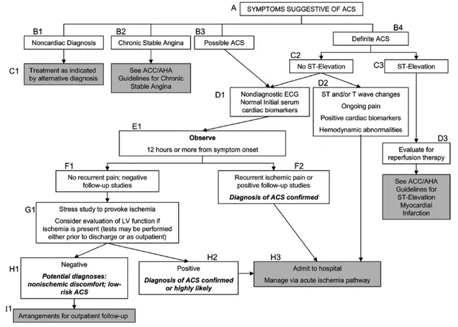
Figure 1. Acute Coronary Syndromes
and angioscopic studies suggest that UA/NSTEMI often results from the disruption or erosion of an atherosclerotic plaque and a subsequent cascade of pathological processes that decrease coronary blood flow. Most patients who die during UA/NSTEMI do so because of sudden death or the development (or recurrence) of acute MI. The efficient diagnosis and optimal management of these patients must derive from information readily available at the time of the initial clinical presentation. The clinical presentation of patients with a life-threatening ACS often overlaps that of patients subsequently found not to have CAD. Moreover, some forms of MI cannot always be differentiated from UA at the time of initial presentation.
“Acute coronary syndrome” has evolved as a useful operational term to refer to any constellation of clinical symptoms that are compatible with acute myocardial isch-emia (Fig. 1). It encompasses MI (ST-segment elevation and depression, Q wave and non-Q wave) and UA. These guidelines focus on 2 components of this syndrome: UA and NSTEMI. In practice, the term “possible ACS” is often assigned first by ancillary personnel, such as emergency medical technicians and triage nurses, early in the evaluation process. A guideline of the National Heart Attack Alert Program (6) summarizes the clinical information needed to make the diagnosis of possible ACS at the earliest phase of clinical evaluation (Table 2). The implication of this early diagnosis for clinical management is that a patient who is considered to have an ACS should be placed in an environ-ment with continuous ECG monitoring and defibrillation capability, where a 12-lead ECG can be obtained expeditiously and definitively interpreted, ideally within 10 min of arrival in the ED. The most urgent priority of early evaluation is to identify patients with ST-elevation MI (STEMI) who should be considered for immediate reperfusion therapy and to rec-ognize other potentially catastrophic causes of patient symp-toms, such as aortic dissection.
Patients diagnosed as having STEMI are excluded from management according to these guidelines and should be managed as indicated according to the ACC/AHA Guide-lines for the Management of Patients With ST-Elevation Myocardial Infarction (1,10). Similarly, management of electrocardiographic true posterior MI, which can masquer-ade as NSTEMI, is covered in the STEMI guidelines (1). The management of patients who experience periprocedural myocardial damage, as reflected in the release of biomarkers of necrosis, such as the MB isoenzyme of creatine kinase (CK-MB) or troponin, also is not considered here.
Patients with MI and with definite ischemic ECG changes for whom acute reperfusion therapy is not suitable should be diagnosed and managed as patients with UA. The residual group of patients with an initial diagnosis of ACS will include many patients who will ultimately be proven to have a noncardiac cause for the initial clinical presentation that was suggestive of ACS. Therefore, at the conclusion of the initial evaluation, which is frequently performed in the ED but sometimes occurs during the initial hours of inpatient
hospi-talization, each patient should have a provisional diagnosis of 1) ACS (Fig. 1), which in turn is classified as a) STEMI, a condition for which immediate reperfusion therapy (fibrinoly-sis or percutaneous coronary intervention [PCI]) should be considered, b) NSTEMI, or c) UA (definite, probable, or possible); 2) a non-ACS cardiovascular condition (e.g., acute pericarditis); 3) a noncardiac condition with another specific disease (e.g., chest pain secondary to esophageal spasm); or 4) a noncardiac condition that is undefined. In addition, the initial evaluation should be used to determine risk and to treat life-threatening events.
In these guidelines, UA and NSTEMI are considered to be closely related conditions whose pathogenesis and clinical presentations are similar but of differing severity; that is, they differ primarily in whether the ischemia is severe enough to cause sufficient myocardial damage to release detectable quan-tities of a marker of myocardial injury, most commonly troponin I (TnI), troponin T (TnT), or CK-MB. Once it has been established that no biomarker of myocardial necrosis has been released (based on 2 or more samples collected at least 6 h apart, with a reference limit of the 99th percentile of the normal population) (11), the patient with ACS may be considered to have experienced UA, whereas the diagnosis of NSTEMI is established if a biomarker has been released. Markers of myocardial injury can be detected in the blood-stream with a delay of up to several hours after the onset of ischemic chest pain, which then allows the differentiation between UA (i.e., no biomarkers in circulation; usually tran-sient, if any, ECG changes of ischemia) and NSTEMI (i.e., elevated biomarkers). Thus, at the time of presentation, pa-tients with UA and NSTEMI can be indistinguishable and therefore are considered together in these guidelines.
1.3.2. Pathogenesis of UA/NSTEMI
These conditions are characterized by an imbalance between myocardial oxygen supply and demand. They are not a specific disease, such as pneumococcal pneumonia, but rather a syndrome, analogous to hypertension. A relatively few nonexclusive causes are recognized (12) (Table 3).
The most common mechanisms involve an imbalance that is caused primarily by a reduction in oxygen supply to the myocardium, whereas with the fifth mechanism noted below, the imbalance is principally due to increased myo-cardial oxygen requirements, usually in the presence of a fixed, restricted oxygen supply:
cause this syndrome in the presence of an extensive collateral blood supply.
• The most common underlying molecular and cellular
pathophysiology of disrupted atherosclerotic plaque is arterial inflammation, caused by noninfectious (e.g., ox-idized lipids) and, possibly, infectious stimuli, which can lead to plaque expansion and destabilization, rupture or erosion, and thrombogenesis. Activated macrophages and T lymphocytes located at the shoulder of a plaque increase the expression of enzymes such as metallopro-teinases that cause thinning and disruption of the plaque, which in turn can lead to UA/NSTEMI.
• A less common cause is dynamic obstruction, which may be triggered by intense focal spasm of a segment of an epicardial coronary artery (Prinzmetal’s angina) (see Sec-tion 6.7). This local spasm is caused by hypercontractility of vascular smooth muscle and/or by endothelial dysfunc-tion. Large-vessel spasm can occur on top of obstructive or destabilized plaque, resulting in angina of “mixed” origin or UA/NSTEMI. Dynamic coronary obstruction can also be caused by diffuse microvascular dysfunction; for example, due to endothelial dysfunction or the ab-normal constriction of small intramural resistance vessels. Coronary spasm also is the presumed mechanism under-lying cocaine-induced UA/NSTEMI.
• A third cause of UA/NSTEMI is severe narrowing without spasm or thrombus. This occurs in some patients with progressive atherosclerosis or with restenosis after a PCI.
• A fourth cause of UA/NSTEMI is coronary artery
dissection (e.g., as a cause of ACS in peripartal women).
• The fifth mechanism is secondary UA, in which the
precipitating condition is extrinsic to the coronary arterial bed. Patients with secondary UA usually, but not always, have underlying coronary atherosclerotic narrowing that limits myocardial perfusion, and they often have chronic stable angina. Secondary UA is precipitated by conditions that 1) increase myocardial oxygen requirements, such as
fever, tachycardia, or thyrotoxicosis; 2) reduce coronary blood flow, such as hypotension; or 3) reduce myocardial oxygen delivery, such as anemia or hypoxemia.
These causes of UA/NSTEMI are not mutually exclusive.
1.3.3. Presentations of UA and NSTEMI
There are 3 principal presentations of UA: 1) rest angina (angina commencing when the patient is at rest), 2) new-onset (less than 2 months) severe angina, and 3) increasing angina (increasing in intensity, duration, and/or frequency) (Table 4) (14). Criteria for the diagnosis of UA are based on the duration and intensity of angina as graded according to the Canadian Cardiovascular Society classification (Table 5) (15). Non–ST-elevation MI generally presents as pro-longed, more intense rest angina or angina equivalent.
1.4. Management Before UA/NSTEMI and Onset of UA/NSTEMI
The ACS spectrum (UA/MI) has a variable but potentially serious prognosis. The major risk factors for development of coronary heart disease (CHD) and UA/NSTEMI are well established. Clinical trials have demonstrated that
modifi-Table 3. Causes of UA/NSTEMI*
Thrombus or thromboembolism, usually arising on disrupted or eroded plaque
●Occlusive thrombus, usually with collateral vessels†
●Subtotally occlusive thrombus on pre-existing plaque
●Distal microvascular thromboembolism from plaque-associated thrombus Thromboembolism from plaque erosion
●Non–plaque-associated coronary thromboembolism
Dynamic obstruction (coronary spasm‡ or vasoconstriction) of epicardial and/ or microvascular vessels
Progressive mechanical obstruction to coronary flow Coronary arterial inflammation
Secondary UA
Coronary artery dissection§
*These causes are not mutually exclusive; some patients have 2 or more causes. †DeWood MA, Stifter WF, Simpson CS, et al. Coronary arteriographic findings soon after non–Q-wave myocardial infarction. N Engl J Med 1986;315:417–23 (13). ‡May occur on top of an atherosclerotic plaque, producing missed-etiology angina or UA/NSTEMI. §Rare. Modified with permission from Braunwald E. Unstable angina: an etiologic approach to management. Circulation 1998;98:2219–22 (12).
UA⫽unstable angina; UA/NSTEMI⫽unstable angina/non–ST-elevation myocardial infarction.
Table 4. Three Principal Presentations of UA
Class Presentation
Rest angina* Angina occurring at rest and prolonged, usually greater than 20 min
New-onset angina New-onset angina of at least CCS class III severity Increasing angina Previously diagnosed angina that has become
distinctly more frequent, longer in duration, or lower in threshold (i.e., increased by 1 or more CCS class to at least CCS class III severity)
*Patients with non–ST-elevated myocardial infarction usually present with angina at rest. Adapted with permission from Braunwald E. Unstable angina: a classification. Circulation 1989;80:410 – 4 (14).
CCS⫽Canadian Cardiovascular Society classification; UA⫽unstable angina.
Table 5. Grading of Angina Pectoris According to CCS Classification
Class Description of Stage
I “Ordinary physical activity does not cause . . . angina,” such as walking or climbing stairs. Angina occurs with strenuous, rapid, or prolonged exertion at work or recreation.
II “Slight limitation of ordinary activity.” Angina occurs on walking or climbing stairs rapidly; walking uphill; walking or stair climbing after meals; in cold, in wind, or under emotional stress; or only during the few hours after awakening. Angina occurs on walking more than 2 blocks on the level and climbing more than 1 flight of ordinary stairs at a normal pace and under normal conditions.
III “Marked limitations of ordinary physical activity.” Angina occurs on walking
1 to 2 blocks on the level and climbing 1 flight of stairs under normal conditions and at a normal pace.
IV “Inability to carry on any physical activity without discomfort— anginal symptoms may be present at rest.”
Adapted with permission from Campeau L. Grading of angina pectoris (letter). Circulation 1976;54:522–3 (15).
cation of those risk factors can prevent the development of CHD (primary prevention) or reduce the risk of experienc-ing UA/NSTEMI in patients who have CHD (secondary prevention). All practitioners should emphasize prevention and refer patients to primary care providers for appropriate long-term preventive care. In addition to internists and family physicians, cardiologists have an important leader-ship role in primary (and secondary) prevention efforts.
1.4.1. Identification of Patients at Risk of UA/NSTEMI
CLASS I
1. Primary care providers should evaluate the presence and status of
control of major risk factors for CHD for all patients at regular
intervals (approximately every 3 to 5 years).(Level of Evidence: C)
2. Ten-year risk (National Cholesterol Education Program [NCEP]
global risk) of developing symptomatic CHD should be calculated
for all patients who have 2 or more major risk factors to assess
the need for primary prevention strategies (16,17). (Level of
Evidence: B)
3. Patients with established CHD should be identified for secondary
prevention efforts, and patients with a CHD risk equivalent (e.g.,
atherosclerosis in other vascular beds, diabetes mellitus, chronic
kidney disease, or 10-year risk greater than 20% as calculated by
Framingham equations) should receive equally intensive risk factor
intervention as those with clinically apparent CHD. (Level of
Evi-dence: A)
Major risk factors for developing CHD (i.e., smoking, family history, adverse lipid profiles, diabetes mellitus, and elevated blood pressure) have been established from large, long-term epidemiological studies (18,19). These risk fac-tors are predictive for most populations in the United States. Primary and secondary prevention interventions aimed at these risk factors are effective when used properly. They can also be costly in terms of primary care provider time, diversion of attention from other competing and important health care needs, and expense, and they may not be effective unless targeted at higher-risk patients (20). It is therefore important for primary care providers to make the identification of patients at risk, who are most likely to benefit from primary prevention, a routine part of everyone’s health care. The Third Report of the NCEP provides guidance on identifying such patients (18). Furthermore, the Writing Committee supports public health efforts to reach all adults at risk, not just those under the care of a primary care physician.
Patients with 2 or more risk factors who are at increased 10-year and lifetime risk will have the greatest benefit from primary prevention, but any individual with a single elevated risk factor is a candidate for primary prevention (19). Waiting until the patient develops multiple risk factors and increased 10-year risk contributes to the high prevalence of CHD in the United States (18,21). Such patients should have their risk specifically calculated by any of the several valid prog-nostic tools available in print (18,22), on the Internet (23), or for use on a personal computer or personal digital assistant (PDA) (18). Patients’ specific risk levels determine
the absolute risk reductions they can obtain from preventive interventions and guide selection and prioritization of those interventions. For example, target levels for lipid lowering and for antihypertensive therapy vary by patients’ baseline risk. A specific risk number can also serve as a powerful educational intervention to motivate lifestyle changes (24). The detection of subclinical atherosclerosis by noninva-sive imaging represents a new, evolving approach for refin-ing individual risk in asymptomatic individuals beyond traditional risk factor assessment alone. A recent AHA scientific statement indicates that it may be reasonable to measure atherosclerosis burden using electron-beam or mul-tidetector computed tomography (CT) in clinically selected intermediate-CAD-risk individuals (e.g., those with a 10% to 20% Framingham 10-year risk estimate) to refine clinical risk prediction and to select patients for aggressive target values for lipid-lowering therapies (Class IIb, Level of Evidence: B) (25).
1.4.2. Interventions to Reduce Risk of UA/NSTEMI
The benefits of prevention of UA/NSTEMI in patients with CHD are well documented and of large magnitude (3,21,26 –28). Patients with established CHD should be identified for secondary prevention efforts, and patients with a CHD risk equivalent should receive equally intensive risk factor intervention for high-risk primary prevention regard-less of sex (29). Patients with diabetes mellitus and periph-eral vascular disease have baseline risks of UA/NSTEMI similar to patients with known CHD, as do patients with multiple risk factors that predict a calculated risk of greater than 20% over 10 years as estimated by the Framingham equations (18). Such patients should be considered to have the risk equivalents of CHD, and they can be expected to have an absolute benefit similar to those with established CHD.
All patients who use tobacco should be encouraged to quit and should be provided with help in quitting at every opportunity (30). Recommendations by a clinician to avoid tobacco can have a meaningful impact on the rate of cessation of tobacco use. The most effective strategies for encouraging quitting are those that identify the patient’s level or stage of readiness and provide information, support, and, if necessary, pharmacotherapy targeted at the individ-ual’s readiness and specific needs (26,31). Pharmacotherapy may include nicotine replacement or withdrawal-relieving medication such as bupropion. Varenicline, a nicotine ace-tylcholine receptor partial antagonist, is a newly approved nonnicotine replacement therapy for tobacco avoidance (32–35). Many patients require several attempts before they succeed in quitting permanently (36,37). Additional discus-sion in this area can be found in other contemporary documents (e.g., the ACC/AHA 2002 Guideline Update for the Management of Patients With Chronic Stable Angina [4]).
low-cholesterol diets high in soluble (viscous) fiber and rich in vegetables, fruits, and whole grains. All patients also should be encouraged to be involved with a regular aerobic exercise program, including 30 to 60 min of moderate-intensity physical activity (such as brisk walking) on most and preferably all days of the week (3,38). For those who need to weigh less, an appropriate balance of increased physical activity (i.e., 60 to 90 min daily), caloric restriction, and formal behavioral programs is encouraged to achieve and maintain a body mass index between 18.5 and 24.9 kg/m2and a waist circumference of less than or equal to 35
inches in women and less than or equal to 40 inches in men. For those who need lipid lowering beyond lifestyle mea-sures, the statin drugs have the best outcome evidence supporting their use and should be the mainstay of phar-macological intervention (21). The appropriate levels for lipid management are dependent on baseline risk; the reader is referred to the NCEP report (http://www.nhlbi.nih.gov/ guidelines/cholesterol/index.htm) for details (17,18,39–41).
Primary prevention patients with high blood pressure should be treated according to the recommendations of the Seventh Joint National Committee on High Blood Pressure (JNC 7) (42,43). Specific treatment recommendations are based on the level of hypertension and the patient’s other risk factors. A diet low in salt and rich in vegetables, fruits, and low-fat dairy products should be encouraged for all hypertensive patients, as should a regular aerobic exercise program (44 – 47). Most patients will require more than 1 medication to achieve blood pressure control, and pharma-cotherapy should begin with known outcome-improving medications (primarily thiazide diuretics as first choice, with the addition of beta blockers, angiotensin-converting en-zyme [ACE] inhibitors, angiotensin receptor blockers, and/or long-acting calcium channel blockers) (42,48). Sys-tolic hypertension is a powerful predictor of adverse out-come, particularly among the elderly, and it should be treated even if diastolic pressures are normal (49).
Detection of hyperglycemic risk (e.g., metabolic syn-drome) and diabetes mellitus should be pursued as part of risk assessment. Lifestyle changes and pharmacotherapy are indicated in individuals with diabetes mellitus to achieve a glycosylated hemoglobin [HbA1c] level less than 7% but to avoid hypoglycemia (3,50,51).
Aspirin prophylaxis can uncommonly result in hemor-rhagic complications and should only be used in primary prevention when the level of risk justifies it. Patients whose 10-year risk of CHD is 10% or more are most likely to benefit, and 75 to 162 mg of aspirin (ASA) per day as primary prophylaxis should be discussed with such patients (29,38,52–55).
1.5. Onset of UA/NSTEMI
1.5.1. Recognition of Symptoms by Patient
Early recognition of symptoms of UA/NSTEMI by the patient or someone with the patient is the first step that
must occur before evaluation and life-saving treatment can be obtained. Although many laypersons are generally aware that chest pain is a presenting symptom of UA/NSTEMI, they are unaware of the other common symptoms, such as arm pain, lower jaw pain, shortness of breath (56), and diaphoresis (57) or anginal equivalents, such as dyspnea or extreme fatigue (56,58). The average patient with NSTEMI or prolonged rest UA (e.g., longer than 20 min) does not seek medical care for approximately 2 h after symptom onset, and this pattern appears unchanged over the last decade (58 – 60). A baseline analysis from the Rapid Early Action for Coronary Treatment (REACT) research pro-gram demonstrated longer delay times among non-Hispanic blacks, older patients, and Medicaid-only recipients and shorter delay times among Medicare recipients (compared with privately insured patients) and patients who came to the hospital by ambulance (58). In the majority of studies examined to date, women in both univariate- and multivariate-adjusted analyses (in which age and other potentially confounding variables have been controlled) exhibit more prolonged delay patterns than men (61).
A number of studies have provided insight into why patients delay in seeking early care for heart symptoms (62). Focus groups conducted for the REACT research program (63,64) revealed that patients commonly hold a preexisting expectation that a heart attack would present dramatically with severe, crushing chest pain, such that there would be no doubt that one was occurring. This was in contrast to their actual reported symptom experience of a gradual onset of discomfort involving midsternal chest pressure or tightness, with other associated symp-toms often increasing in intensity. The ambiguity of these symptoms, due to this disconnect between prior expectations and actual experience, resulted in uncer-tainty about the origin of symptoms and thus a “wait-and-see” posture by patients and those around them (62). Other reported reasons for delay were that patients thought the symptoms were self-limited and would go away or were not serious (65– 67); that they attributed symptoms to other preexisting chronic conditions, espe-cially among older adults with multiple chronic condi-tions (e.g., arthritis), or sometimes to a common illness such as influenza; that they were afraid of being embar-rassed if symptoms turned out to be a “false alarm”; that they were reluctant to trouble others (e.g., health care providers, Emergency Medical Services [EMS]) unless they were “really sick” (65– 67); that they held stereotypes of who is at risk for a heart attack; and that they lacked awareness of the importance of rapid action, knowledge of reperfusion treatment, or knowledge of the benefits of calling EMS/9-1-1 to ensure earlier treatment (62). Notably, women did not perceive themselves to be at risk (69).
1.5.2. Silent and Unrecognized Events
first to show that as many as half of all MIs may be clinically silent and unrecognized by the patient (71). Canto et al. (72) found that one third of the 434,877 patients with confirmed MI in the National Registry of Myocardial Infarction presented to the hospital with symptoms other than chest discomfort. Compared with MI patients with chest discomfort, MI patients without chest discomfort were more likely to be older, to be women, to have diabetes, and/or to have prior heart failure [HF]. Myocardial infarc-tion patients without chest discomfort delayed longer before they went to the hospital (mean 7.9 vs. 5.3 h) and were less likely to be diagnosed as having an MI when admitted (22.2% vs. 50.3%). They also were less likely to receive fibrinolysis or primary PCI, ASA, beta blockers, or heparin. Silent MI patients were 2.2 times more likely to die during the hospitalization (in-hospital mortality rate 23.3% vs. 9.3%). Unexplained dyspnea, even without angina, is a particularly worrisome symptom, with more than twice the risk of death than for typical angina in patients undergoing cardiovascular evaluation (56). Recently, the prognostic significance of dyspnea has been emphasized in patients undergoing cardiac evaluation. Self-reported dyspnea alone among 17,991 patients undergoing stress perfusion testing was an independent predictor of cardiac and total mortality
and increased the risk of sudden cardiac death 4-fold even in those with no prior history of CAD (56).
Health care providers should maintain a high index of suspicion for UA/NSTEMI when evaluating women, pa-tients with diabetes mellitus, older papa-tients, those with unexplained dyspnea (56), and those with a history of HF or stroke, as well as those patients who complain of chest discomfort but who have a permanent pacemaker that may confound recognition of UA/NSTEMI on their 12-lead ECG (73).
2. Initial Evaluation and Management
2.1. Clinical Assessment
Because symptoms are similar and the differentiation of UA/NSTEMI and STEMI requires medical evaluation, we will refer to prediagnostic clinical presentation as ACS,
defined as UA or MI (NSTEMI or STEMI) (Fig. 2).
RECOMMENDATIONS
CLASS I
1. Patients with symptoms that may represent ACS (Table 2) should
not be evaluated solely over the telephone but should be referred to a facility that allows evaluation by a physician and the recording of
Figure 2. Algorithm for Evaluation and Management of Patients Suspected of Having ACS
a 12-lead ECG and biomarker determination (e.g., an ED or other
acute care facility).(Level of Evidence: C)
2. Patients with symptoms of ACS (chest discomfort with or without
radiation to the arm[s], back, neck, jaw or epigastrium; shortness of
breath; weakness; diaphoresis; nausea; lightheadedness) should
be instructed to call 9-1-1 and should be transported to the hospital
by ambulance rather than by friends or relatives.(Level of Evidence: B)
3. Health care providers should actively address the following issues
regarding ACS with patients with or at risk for CHD and their
families or other responsible caregivers:
a. The patient’s heart attack risk;(Level of Evidence: C)
b. How to recognize symptoms of ACS;(Level of Evidence: C)
c. The advisability of calling 9-1-1 if symptoms are unimproved or
worsening after 5 min, despite feelings of uncertainty about the
symptoms and fear of potential embarrassment;(Level of
Evi-dence: C)
d. A plan for appropriate recognition and response to a potential
acute cardiac event, including the phone number to access EMS, generally 9-1-1 (74).(Level of Evidence: C)
4. Prehospital EMS providers should administer 162 to 325 mg of
ASA (chewed) to chest pain patients suspected of having ACS
unless contraindicated or already taken by the patient. Although some trials have used enteric-coated ASA for initial dosing, more
rapid buccal absorption occurs with non–enteric-coated
formula-tions.(Level of Evidence: C)
5. Health care providers should instruct patients with suspected ACS
for whom nitroglycerin [NTG] has been prescribed previously to take
not more than 1 dose of NTG sublingually in response to chest
discomfort/pain. If chest discomfort/pain is unimproved or is
wors-ening 5 min after 1 NTG dose has been taken, it is recommended that the patient or family member/friend/caregiver call 9-1-1
immediately to access EMS before taking additional NTG. In
pa-tients with chronic stable angina, if symptoms are significantly
improved by 1 dose of NTG, it is appropriate to instruct the patient
or family member/friend/caregiver to repeat NTG every 5 min for a
maximum of 3 doses and call 9-1-1 if symptoms have not resolved
completely.(Level of Evidence: C)
6. Patients with a suspected ACS with chest discomfort or other ischemic symptoms at rest for greater than 20 min, hemodynamic
instability, or recent syncope or presyncope should be referred
immediately to an ED. Other patients with suspected ACS who are
experiencing less severe symptoms and who have none of the
above high-risk features, including those who respond to an NTG
dose, may be seen initially in an ED or an outpatient facility able to
provide an acute evaluation.(Level of Evidence: C)
CLASS IIa
1. It is reasonable for health care providers and 9-1-1 dispatchers to
advise patients without a history of ASA allergy who have symptoms
of ACS to chew ASA (162 to 325 mg) while awaiting arrival of
prehospital EMS providers. Although some trials have used
enteric-coated ASA for initial dosing, more rapid buccal absorption occurs
with non–enteric-coated formulations.(Level of Evidence: B)
2. It is reasonable for health care providers and 9-1-1 dispatchers to
advise patients who tolerate NTG to repeat NTG every 5 min for a maximum of 3 doses while awaiting ambulance arrival.(Level of
Evidence: C)
3. It is reasonable that all prehospital EMS providers perform and
evaluate 12-lead ECGs in the field (if available) on chest pain
patients suspected of ACS to assist in triage decisions.
Electrocar-diographs with validated computer-generated interpretation
algo-rithms are recommended for this purpose.(Level of Evidence: B)
4. If the 12-lead ECG shows evidence of acute injury or ischemia, it is
reasonable that prehospital ACLS providers relay the ECG to a
predetermined medical control facility and/or receiving hospital.
(Level of Evidence: B)
Patients with suspected ACS must be evaluated rapidly. Decisions made on the basis of the initial evaluation have substantial clinical and economic consequences (75). The first triage decision is made by the patient, who must decide whether to access the health care system. Media campaigns such as “Act in Time,” sponsored by the National Heart, Lung, and Blood Institute (NHLBI), provide patient edu-cation regarding this triage decision (www.nhlbi.nih.gov/ actintime). The campaign urges both men and women who feel heart attack symptoms or observe the signs in others to wait no more than a few minutes, 5 min at most, before calling 9-1-1 (76,77). Campaign materials point out that patients can increase their chance of surviving a heart attack by learning the symptoms and filling out a survival plan. They also are advised to talk with their doctor about heart attacks and how to reduce their risk of having one. The patient materials include a free brochure about symptoms and recommended actions for survival, in English (78) and Spanish (79), as well as a free wallet card that can be filled in with emergency medical information (80). Materials geared directly to providers include a Patient Action Plan Tablet (81), which contains the heart attack warning symp-toms and steps for developing a survival plan, individualized with the patient’s name; a quick reference card for address-ing common patient questions about seekaddress-ing early treatment to survive a heart attack (82), including a PDA version (83); and a warning signs wall chart (84). These materials and others are available on the “Act in Time” Web page (www.nhlbi.nih.gov/health/public/heart/mi/core_bk.pdf) (77).
When the patient first makes contact with the medical care system, a critical decision must be made about where the evaluation will take place. The health care provider then must place the evaluation in the context of 2 critical questions: Are the symptoms a manifestation of an ACS? If so, what is the prognosis? The answers to these 2 questions lead logically to a series of decisions about where the patient will be best managed, what medications will be prescribed, and whether an angiographic evaluation will be required.
environ-ment for the level of care needed based on diagnostic criteria and an estimation of the underlying risk of specific negative outcomes.
Health practitioners frequently receive telephone calls from patients or family members/friends/caregivers who are concerned that their symptoms could reflect heart disease. Most such calls regarding chest discomfort of possible cardiac origin in patients without known CAD do not represent an emergency; rather, these patients usually seek reassurance that they do not have heart disease or that there is little risk due to their symptoms. Despite the frequent inclination to dismiss such symptoms over the telephone, health care providers, EMS dispatchers, and staff positioned to receive these calls should advise patients with possible accelerating angina or angina at rest that an evaluation cannot be performed solely via the telephone. This advice is essential because of the need for timely evaluation, including a physical examination, ECG, and appropriate blood tests to measure cardiac biomarkers.
Patients with known CAD—including those with chronic stable angina, recent MI, or prior intervention (i.e., coronary artery bypass graft surgery [CABG] or PCI)—who contact a physician or other appropriate member of the health care team because of worsening or recurrent symp-toms should be instructed to proceed rapidly to an ED, preferably one equipped to perform prompt reperfusion therapy. When the discomfort is moderate to severe or sustained, they should be instructed to access the EMS system directly by calling 9-1-1. Patients who have been evaluated recently and who are calling for advice regarding modification of medications as part of an ongoing treatment plan represent exceptions.
Even in the most urgent subgroup of patients who present with acute-onset chest pain, there usually is ade-quate time for transport to an environment in which they can be evaluated and treated (85). In a large study of consecutive patients with chest pain suspected to be of cardiac origin who were transported to the ED via ambu-lance, one third had a final diagnosis of MI, one third had a final diagnosis of UA, and one third had a final diagnosis of a noncardiac cause; 1.5% of these patients developed cardiopulmonary arrest before arrival at the hospital or in the ED (86).
Every community should have a written protocol that guides EMS system personnel in determining where to take patients with suspected or confirmed ACS. Active involve-ment of local health care providers, particularly cardiologists and emergency physicians, is needed to formulate local EMS destination protocols for these patients. In general, patients with suspected ACS should be taken to the nearest appropriate hospital; however, patients with known STEMI and/or cardiogenic shock should be sent as directly as possible to hospitals with interventional and surgical capa-bility (1).
The advent of highly effective, time-dependent treatment for ACS, coupled with the need to reduce health care costs,
adds further incentive for clinicians to get the right answer quickly and to reduce unnecessary admissions and length of hospital stay. Investigators have tried various diagnostic tools, such as clinical decision algorithms, cardiac biomar-kers, serial ECGs, echocardiography, myocardial perfusion imaging, and multidetector (e.g., 64-slice) coronary CT angiography (CCTA), in an attempt to avoid missing patients with MI or UA. The most successful strategies to emerge thus far are designed to identify MI patients and, when clinically appropriate, screen for UA and underlying CAD. Most strategies use a combination of cardiac biomar-kers, short-term observation, diagnostic imaging, and pro-vocative stress testing. An increasing number of high-quality centers now use structured protocols, checklists, or critical pathways to screen patients with suspected MI or UA (87–99). It does not appear to matter whether the institution designates itself a chest pain center; rather, it is the multifaceted, multidisciplinary, standardized, and struc-tured approach to the problem that appears to provide clinical, cost-effective benefit (100,101). One randomized trial has confirmed the safety, efficacy, and cost-effectiveness of the structured decision-making approach compared with standard, unstructured care (102).
Regardless of the approach used, all patients presenting to the ED with chest discomfort or other symptoms suggestive of MI or UA should be considered high-priority triage cases and should be evaluated and treated on the basis of a predetermined, institution-specific chest pain protocol. The protocol should include several diagnostic possibilities (Fig. 2) (103). The patient should be placed on a cardiac monitor immediately, with emergency resuscitation equipment, in-cluding a defibrillator, nearby. An ECG also should be performed immediately and evaluated by an experienced emergency medicine physician, with a goal of within 10 min of ED arrival. If STEMI is present, the decision as to whether the patient will be treated with fibrinolytic therapy or primary PCI should be made within the next 10 min (1). For cases in which the initial diagnosis and treatment plan are unclear to the emergency medicine physician or are not covered directly by an institutionally agreed-upon protocol, immediate cardiology consultation is advisable.
Several studies have confirmed that patients with ACS frequently do not call 9-1-1 and are not transported to the hospital by ambulance. A follow-up survey of chest pain patients presenting to participating EDs in 20 US commu-nities who were either released or admitted to the hospital with a confirmed coronary event revealed that the average proportion of patients who used EMS was 23%, with significant geographic difference (range 10% to 48%). Most patients were driven by someone else (60%) or drove themselves to the hospital (16%) (109). In the National Registry of Myocardial Infarction 2, just over half (53%) of all patients with MI were transported to the hospital by ambulance (105).
Even in areas of the country that have undertaken substantial public education campaigns about the warning signs of ACS and the need to activate the EMS system rapidly, either there were no increases in EMS use (58,110 – 113) or EMS use increased (as a secondary outcome measure) but was still suboptimal, with a 20% increase from a baseline of 33% in all 20 communities in the REACT study (63) and an increase from 27% to 41% in southern Minnesota after a community campaign (114). Given the importance of patients using EMS for possible acute cardiac symptoms, communities, including medical providers, EMS systems, health care insurers, hospitals, and policy makers at the state and local level, need to have agreed-upon
emer-gency protocols to ensure patients with possible heart attack symptoms will be able to access 9-1-1 without barriers, to secure their timely evaluation and treatment (115).
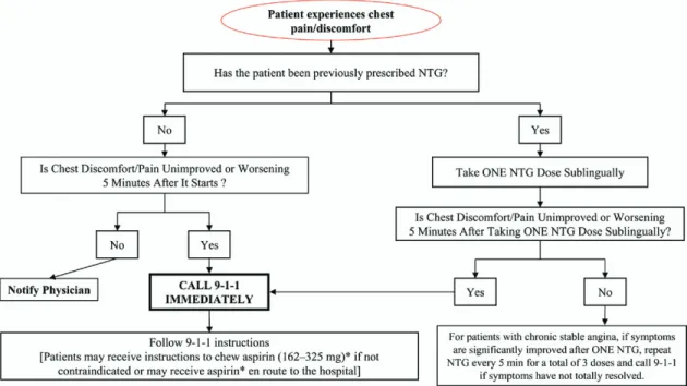
As part of making a plan with the patient for timely recognition and response to an acute event, providers should review instructions for taking NTG in response to chest discomfort/pain (Fig. 3). If a patient has previously been prescribed NTG, it is recommended that the patient be advised to take 1 NTG dose sublingually promptly for chest discomfort/pain. If symptoms are unimproved or worsening 5 min after 1 NTG dose has been taken, it also is recommended that the patient be instructed to call 9-1-1 immediately to access EMS. Although the traditional rec-ommendation is for patients to take 1 NTG dose sublin-gually, 5 min apart, for up to 3 doses before calling for emergency evaluation, this recommendation has been mod-ified by the UA/NSTEMI Writing Committee to encour-age earlier contacting of EMS by patients with symptoms suggestive of ACS. While awaiting ambulance arrival, patients tolerating NTG can be instructed by health care providers or 9-1-1 dispatchers to take additional NTG every 5 min up to 3 doses. Self-treatment with prescription medication, including nitrates, and with nonprescription medication (e.g., antacids) has been documented as a frequent cause of delay among patients with ACS, including those with a history of MI or angina (65,116). Both the rate
Figure 3. Patient (Advance) Instructions for NTG Use and EMS Contact in the Setting of Non–Trauma-Related Chest Discomfort/Pain
of use of these medications and the number of doses taken were positively correlated with delay time to hospital arrival (65).
Family members, close friends, caregivers, or advocates should be included in these discussions and enlisted as reinforcement for rapid action when the patient experiences symptoms of a possible ACS (74,117,118) (Fig. 3). For patients known to their providers to have frequent angina, physicians may consider a selected, more tailored message that takes into account the frequency and character of the patient’s angina and their typical time course of response to NTG. In many of these patients with chronic stable angina, if chest pain is significantly improved by 1 NTG, it is still appropriate to instruct the patient or family member/friend/ caregiver to repeat NTG every 5 min for a maximum of 3 doses and to call 9-1-1 if symptoms have not resolved completely. Avoidance of patient delay associated with self-medication and prolonged reevaluation of symptoms are paramount. An additional consideration in high-risk CHD patients is to train family members in CPR and/or to have home access to an automatic external defibrillator, now available commercially to the public.
The taking of aspirin by patients in response to acute symptoms has been reported to be associated with a delay in calling EMS (109). Patients should focus on calling 9-1-1, which activates the EMS system, where they may receive instructions from emergency medical dispatchers to chew aspirin (162 to 325 mg) while emergency personnel are en route, or emergency personnel can give an aspirin while transporting the patient to the hospital (119). Alternatively, patients may receive an aspirin as part of their early treatment once they arrive at the hospital if it has not been given in the prehospital setting (117).
Providers should target those patients at increased risk for ACS, focusing on patients with known CHD, peripheral vascular disease, or cerebral vascular disease, those with diabetes, and patients with a 10-year Framingham risk of CHD of more than 20% (120). They should stress that the chest discomfort will usually not be dramatic, such as is commonly misrepresented on television or in the movies as a “Hollywood heart attack.” Providers also should describe anginal equivalents and the commonly associated symptoms of ACS (e.g., shortness of breath, a cold sweat, nausea, or lightheadedness) in both men and women (56,106), as well as the increased frequency of atypical symptoms in elderly patients (72).
2.1.1. Emergency Department or Outpatient Facility Presentation
It is recommended that patients with a suspected ACS with chest discomfort or other ischemic symptoms at rest for more than 20 min, hemodynamic instability, or recent syncope or presyncope to be referred immediately to an ED or a specialized chest pain unit. For other patients with a suspected ACS who are experiencing less severe symptoms and are having none of the above high-risk features, the
recommendation is to be seen initially in an ED, a chest pain unit, or an appropriate outpatient facility. Outcomes data that firmly support these recommendations are not available; however, these recommendations are of practical importance because differing ACS presentations require differing levels of emergent medical interventions, such as fibrinolytics or emergency coronary angiography leading to PCI or surgery, or sophisticated diagnostic evaluation such as nuclear stress testing or CCTA. When symptoms have been unremitting for more than 20 min, the possibility of MI must be considered. Given the strong evidence for a relationship between delay in treatment and death (121– 123), an immediate assessment that includes a 12-lead ECG is essential. Patients who present with hemodynamic instability require an environment in which therapeutic interventions can be provided, and for those with presyn-cope or synpresyn-cope, the major concern is the risk of sudden death. Such patients should be encouraged to seek emer-gency transportation when it is available. Transport as a passenger in a private vehicle is an acceptable alternative only if the wait for an emergency vehicle would impose a delay of greater than 20 to 30 min.
2.1.2. Questions to Be Addressed at the Initial Evaluation
The initial evaluation should be used to provide information about the diagnosis and prognosis. The attempt should be made to simultaneously answer 2 questions:
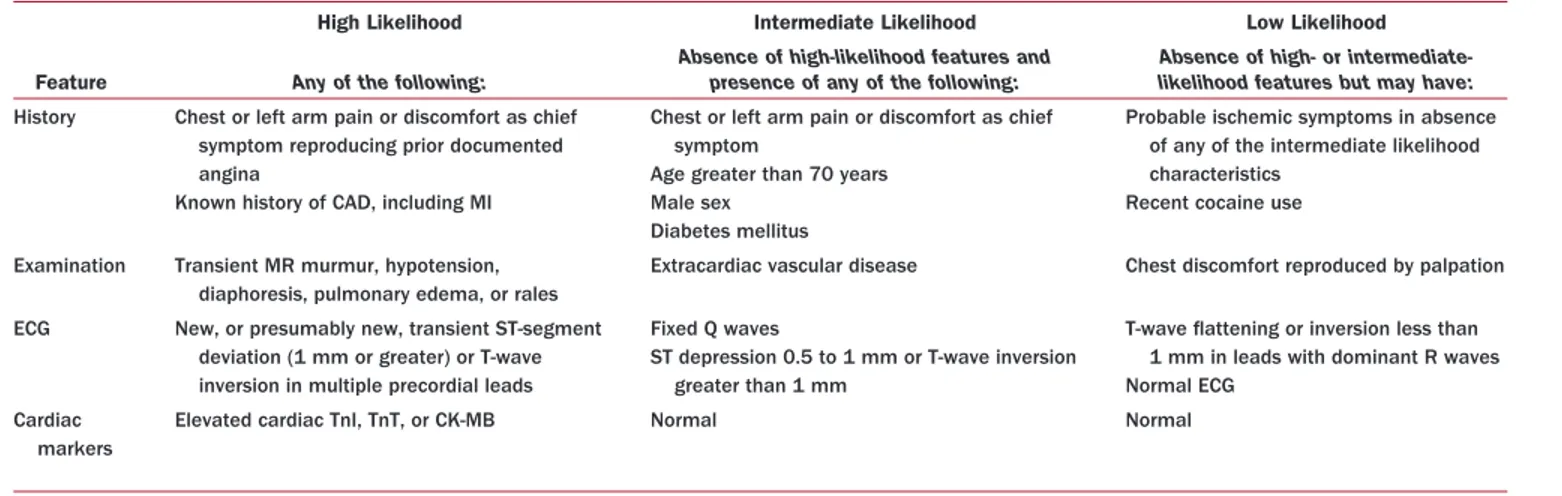
• What is the likelihood that the signs and symptoms represent ACS secondary to obstructive CAD (Table 6)?
• What is the likelihood of an adverse clinical outcome (Table 7)? Outcomes of concern include death, MI (or recurrent MI), stroke, HF, recurrent symptomatic isch-emia, and serious arrhythmia.
For the most part, the answers to these questions form a sequence of contingent probabilities. Thus, the likelihood that the signs and symptoms represent ACS is contingent on the likelihood that the patient has underlying CAD. Similarly, the prognosis is contingent on the likelihood that the symptoms represent acute ischemia. However, in patients with symptoms of possible ACS, traditional risk factors for CAD are less important than are symptoms, ECG findings, and cardiac biomarkers. Therefore, the presence or absence of these tradi-tional risk factors ordinarily should not be heavily weighed in determining whether an individual patient should be admitted or treated for ACS.
2.2. Early Risk Stratification
RECOMMENDATIONS FOR EARLY RISK STRATIFICATION
CLASS I
1. A rapid clinical determination of the likelihood risk of obstructive
CAD (i.e., high, intermediate, or low) should be made in all
patients with chest discomfort or other symptoms suggestive of
an ACS and considered in patient management. (Level of