In Vitro Toxicity Evaluation of Caffeine Imprinted Polymer (CAF-MIP) for
Decaffeination Method on Normal Chang Liver Cells
Fatimah Hashim
*, Faizatul Shimal Mehamod, and Naizatul Akmal Nawi
School of Fundamental Science, Universiti Malaysia Terengganu, 21030 Kuala Terengganu, Terengganu, Malaysia
*e-mail: [email protected]
Abstract
Over consuming of caffeine is one of the factors to a few health problems such as insomnia, hypertension and cardio-vascular disease. This preliminary study was conducted to evaluate the Caffeine-Imprinted Polymer (CAF-MIP) toxicity that was synthesized for a new alternative method for decaffeination. It is crucial to evaluate the toxicity of CAF-MIPas this product is potential to be used as complimentary with any drinks containing caffeine. In this study, the CAF-MIP toxicity potential was confirmed on Normal Chang Liver cell (NCLC) based on its IC50 value andacridine orange and
propidium iodide (AO/PI) staining for mode of cell death observation.Proliferation assay was also conducted after 24, 48 and 72 hours at 30 µg/ml on NCLC and it showed that CAF-MIP promote NCLC growth as shown by at various concentrationof CAF-MIPincrease the percentage of NCLC viability. Observation under light microscopes on NCLC incubated wit CAF-MIP and NIP showed the normal, viable cell morphology, cuboidal and monolayer cell morphology and this can be seen with green fluorescence when view under fluorescence microscope. In conclusion, from this study, it is proved that the CAF-MIP does not initiate toxicity effects on human liver cells, meanwhile induction of cell prolif-eration was observed.
Abstrak
Evaluasi Ketoksikan Caffeine Imprinted Polymer (CAF-MIP) Secara In Vitro untuk Proses Dekafeinasi pada Sel Normal Hati Chang. Konsumsi kafein yang berlebihan merupakan salah satu faktor penyebab beberapa masalah kesehatan seperti insomnia, tekanan darah tinggi atau hipertensi dan penyakit kardiovaskular. Kajian ini bertujuan untuk menguji kadar ketoksiksan Caffeine Imprinted Polymer (CAF-MIP) yang telah disintesis untuk membantu proses deka-feinasi pada tubuh. Evaluasi ketoksikan CAF-MIP penting dilakukan, karena produk ini berpotensi untuk dikonsumsi bersama minuman yang mengandung kafein. Dalam kajian ini, potensi ketoksikan CAF-MIP telah dianalisis dengan menggunakan sel normal hati Chang (NCLC) berdasarkan nilai IC50 dan pewarnaan sel oleh akridinoren dan propidium
iodide untuk menentukan nilai kematian sel. Analisis pergandaan sel juga telah dilakukan selama 24, 48, dan 72 jam pada konsentrasi 30 µg/mL terhadap NCLC. Hasil penelitian menunjukkan bahwa CAF-MIP menginduksi pertumbuhan NCLC yang bergantung pada konsentrasi CAF-MIP yang digunakan juga meningkatkan derajat viabilitas NCLC. Analisis di bawah mikroskop cahaya menunjukkan bahwa inkubasi NCLC bersama CAF-MIP dan NIP tidak mengubah bentuk sel seperti bentuk sel normal, morfologi sel yang viabel, berbentuk kuboid dan morfologi lapisan sel juga dapat dilihat dibawah mikroskopi floresen. Data penelitian ini telah menunjukkan CAF-MIP tidak menyebabkan ketoksikan pada sel-sel hati manusia, hasil ini juga menunjukkan bahwa sel tetap bisa bertambah.
Keywords: caffeine, toxicity, in vitro, liver cells, cytotoxicity
1.
Introduction
Caffeine (1,3,7- trimethylxanthine) is a purine alkaloid that can be found naturally in plants. It provide taste and flavor in coffee, tea, energy drink and other caffeine containing materials. Utility of this compound is due to its various benefits such as to prevent sleepiness, used in asthma treatment and able to inhibit the development of
It is also dangerous for pregnant woman as caffeine is slowly metabolized and it remain for a long period of time in our body.In this study, CAF-MIP was synthesized by precipitation polymerization technique that require caffeine (CAF) as the template, methacrylic acid (MAA) as the monomer, Divinylbenzene-80 (DVB-80) as the cross-linker, Azobisisobutyronitrile (AIBN) as the initiator and toluene/ACN as the solvent.
Non-Imprinted Polymer (NIP) was also used as a control to CAF-MIP, where NIP do not use caffeine as template while the rest of the components stay the same. There are four main existing methods for the decaffeination which are the Ethyl-Acetate method, Water method, Supercritical Carbon Dioxide and Liquid Carbon Dioxide methodas well as the Methylene Chloride method. All these four methods are widely used to produce decaffe-inated beverages especially water method. These four method shares the basic processing procedures. However, there are evidences stated that these methods need to be improved as there were negative effects and disadvantages to the consumers.
This study was done to evaluate the toxicity of the CAF-MIP and NIP on normal Chang liver cell for 72 hours at 0-30 µg/mL by MTT assay to determine the IC50 value.
Normal Chang liver cells proliferation test was also conducted by incubating the cells with CAF-MIPat the highest concentration, 30 µg/mL for 24, 48 and 72 hours.
Besides, acridine orange/propidium iodide (AO/PI) staining were performed to observe the morphological changes of the NCLC for both untreated and CAF-MIP, NIP- treated.
Hydrogen peroxide (H2O2) was used as a positive control
at 0-17.48 M concentration on NCLC to compare all experiments carried out in this study.
2.
Materials and Methods
Normal Chang Liver Cell culture. The normal Chang liver cells (NCLC) was obtained from Animal Tissue Culture Lab, Kampus Kota, Universiti Sultan Zainal Abidin. NCLC was cultured at 37 °C in 5% CO2 incubator
in a complete growth medium MEM (modified Eagle medium (D-MEM, Gibco/BRL, Carlsbad, CA, USA). Cells were maintained or sub-cultured every 3-5 days.
Caffeine-MIPs, NIP and H2O2 stock solution. One mg
Caffeine-MIP complex was dissolved in the 1000 µL MEM media. The final concentration for stock solution was 1000 µg/mL and 60 µ L/mL were placed in the 1.5 ml vial. Two fold serial dilutions were used to make different CAF-MIP concentrations ranging from 0-30 µg/mL as the treatments. The method was also applied to the Non-Imprinted Polymer (NIP) to make similar concentration with MIP as the control to
CAF-MIP. H2O2 was also prepared as a positive control in
this experiment.
Normal Chang Liver Cell viability treated with CAF-MIP, NIP and H2O2. In this experiment, Normal
Chang liver cells were placed in 96-well microplate at 2.5 x 10⁵ cells/mL for each well and kept in 5% CO2
incubator at 37 °C for 24 hours for cells attachment. Then, the medium was removed and replaced with medium ranging from 0 to 30 µg/mL ofCAF-MIP, NIP and H2O2 as a positive control. Each treatment was done
with 3-5 replicates and were incubated for 72 hours. After 72 hours of treatment, all medium were removed and replaced with 20 µL of MTT (5 mg/1 mL) in PBS solution into each well and again incubated for four hours.Following that, 80 µL of DMSO was added to dissolve the purple formazan crystal and the percentage of cells viability was calculated after absorbance of formazan crystal was read by the ELISA at 570 nm.
Normal Chang Liver Cell treated with CAF-MIP, NIP and H2O2Proliferation assayThe normal Chang
liver cell were placed in 96-well plate at 2.5 x 10⁵ cells concentration for each well and kept in 5% CO2
incubator for 24 hours for cell attachment. Then, the medium was removed and placed with medium containing 10, 20 and 30 µg/mL CAF-MIP with five replicates each and again kept in CO2 incubator for
another 24, 48 and 72 hours.MTT assay than were performed according to MTT method in section 2.3.
Observation of Normal Chang Liver cell culture under light microscope and fluorescence microscope treated with CAF-MIP and NIP. NCLC were seeded in 6-well plate at 2.5 x 10⁶ cells/mL and incubated at 37 °C in 5 % CO2. After 24 hours, the cultu re medium was
replaced with medium containing CAF-MIP, NIP and H2O2 at highest concentration tested in this study for
each compound and H2O2 and incubated for another 72
hours. CAF-MIP and NIP-treated and untreated NCLC images were viewed under phase contrast light microscope to detect any morphological changes on cells and the images were then captured and recorded. Next, for mode of cell death observation, the culture media were then gently separated and the remaining adhered cells were smoothly stained with AO/PI dye solution to detect the color changes. Lastly, stained cells were observed under a fluorescence microscope with blue filter and the images were captured and recorded.
3.
Results and Discussion
Cell viability by MTT assay. In this study, MTT assay was used to determine the IC50 value of CAF-MIP and
P
er
ce
n
ta
g
e
o
f
ce
ll
v
ia
b
il
it
y
(
%
)
Figure 1. Normal Chang Liver Cells Viability against CAF-MIP and NIP at Concentrations Ranging from 0- 30 µg/mL. High Cell viability Indicated that Both Compounds Wight not cause Toxicity towards NCLC when Percentage of Cell Via-bility were more than 100%
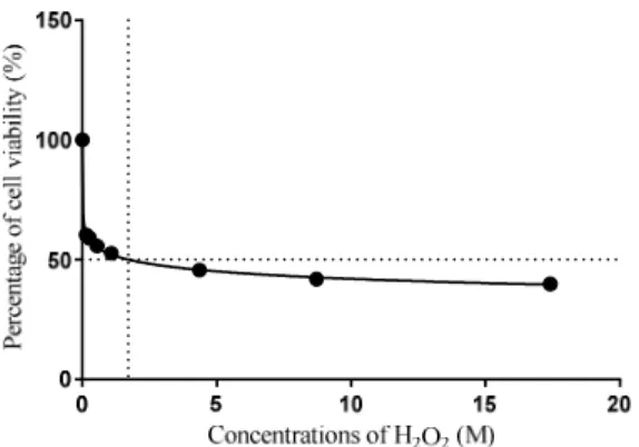
Figure 2. The Relationship between the Cell Viability against Hydrogen Peroxide. IC50 obtained from
the Graph was 2M
peroxide, H2O2 on normal Chang liver cells after 72
hours incubation time with all the test agents.
MTT assay is a quantitative colorimetric assay that is used to quantitate animal cell culture viability in vitro. Yellow tetrazolium salts, MTT were reduced into a purple formazan crystal in the mitochondria of living cells that contain mitochondrial dehydrogenase enzyme. Solubilization buffer, the DMSO were added to dissolve the purple formazan crystal. The higher absorbance reading by the spectrophotometer indicated that the purple crystal formazan is higher as well as the number of living or viable cells increase. CAF-MIP and NIP were prepared with a 2-fold serial dilution to make a concentration from 0 to 30 µg/mL.
Results showed that CAF-MIP and NIP were not toxic as the percentage of normal Chang liver cells is mostly increasing. The result is dependable as the MTT assay is said to be simple, rapid, inexpensive and repeatable even though it is time-consuming. A study done by
Sargant & Taylor [4] on the determination of the chemo sensitivity of an acute myeloid leukemia by using MTT assay showed that the result has no significant difference and it stated that the MTT assay is reproducible and repeatable for cytotoxicity research. In this study, Hydrogen peroxide, H2O2 was used as a positive control
to CAF-MIP and NIP in MTT assay, proliferation assay and AO/PI staining.
The concentration (M, molar) used starting from 0, 0.136, 0.27, 0.544, 1.08, 2.17, 4.35, 8.71, and 17.41 which prepared by 2-fold serial dilution. Based on the figure 2, at overall, the percentage of cell viability was decreasing from 0 to 17.41. The IC50 value for H2O2 was
measured at concentration 2.0M in which only small concentration of this apoptosis agent is able to reduce the cell viability by 50%. It showed that the H2O2
possessed a toxicity effect on normal Chang liver cells as it decreased the percentage of cell viability by 50%. IC50 values represent the compound concentration that
reduced the cell viability to 50% of those in the untreated control wells.
From the graph, it is clearly showed that the percentage of cells viability treated with CAF-MIP and NIP were increased more than 100% after 72 hours treatment. However, the percentage of cell viability were fluctuated at certain concentration for both CAF-MIP and NIP. This may suggested that the normal cell stop dividing and proliferates due to cell crowding, growth factor depletion, accumulation of waste products and drop in pH. Basically, the normal Chang liver cells are monolayer and anchorage dependent cells that are adhere to bottom of culture flask. After sometimes when the cells over proliferates, the space is limited in which it will caused cell death. The high proliferations rates of cell exhausted the medium faster than lower concentrations of cells would[5].The normal pH for normal Chang liver cells culture medium are between 7.0-7.4. A drop in pH was indicated by the changing of MEM media from orange-red to cloudy light yellow. Most cells stop growing as the pH falls from Ph 7.0 to pH 6.5 and start to lose viability between pH 6.5 and pH 6.0. Besides, cells incubated after sometimes, the growth factor, FBS that was added into MEM media will be depleted and this can cause cell death. It is also a source of minerals, lipids, and hormones [5]. One of the product of cell metabolism is waste and the accumulation of waste is toxic to cell that can alsocause cell death. There is no IC50 value determination for CAF-MIP and NIPas they
did not show any toxicity effects on normal Chang liver cells.
in food analysis using MISPE were prepared via non-covalent imprinting by bulk polymerization which is the same technique use to synthesize the CAF-MIP. MIP in food analysis were widely used and it proven to be safe [6]. However, there are no report on the in vitro study of this version of MIP as they are not being use internally, and it does not interferes with the human metabolism. Thus, it is suggested that CAF-MIP that was synthesized using the same components and techniques may be safe to be use which is consequently to our preliminary result.
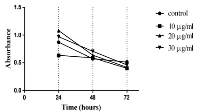
Normal Chang Liver Cell Proliferation Assay after treatment with CAF-MIP. Cell proliferation assay is an alternative way to determine the cell viability or number of cell dividing at designated times. In this study, normal Chang liver cells were incubated with CAF-MIP for 24, 48 and 72 hours at 30 µg/mL in 5% CO2 incubator at 37 ºC.
The treatments were done with 3-5 replicates for eachconcentration and compound tested, CAF-MIP. Yellow MTT tetrazolium salt was used in this assay to assess the metabolic activity of the NCLC. Figure3 shows the result of proliferation assay for 24, 48 and 72 hours incubation time with different concentration of the compounds.
From the graph, percentage of cell viability for the untreated was increased from 0 to 24 hours. However, there was significance decreased in cell viability from 24 to 72 hours. Normal Chang liver cells are an anchorage dependent in which it needs a surface to attach. At 24 hours, the cells still have enough space to divide and carry out their metabolic activities as resulted in the increasing percentage of cell viability up to 133.62%. The decreasing in the percentage of cell viability after 24 until 72 hours is suggested due to limited space and the accumulation of their metabolic wastes.
Meanwhile, percentage of normal Chang liver cells viability that was treated with CAF-MIP showed an increased from 0 to 48 hours and decreased at 72 hours. Here, it is suggested that CAF-MIP that have template caffeine may play an important role as a proliferative agent as we can see that the percentage of the cell viability was increased at 48 hours. CAF-MIP contain caffeine as template even it has been removed while in NIP, they do not contain caffeine as template as well as for untreated. However, at 72 hours, the percentage of cell viability was decreased. Most normal cells need to spread out on a substrate to proliferate, and inadequate spreading due to poor adhesion orovercrowding will inhibit proliferation. Takuwa et al. [7] reported that the cell growth and proliferation of normal vertebrate cells depend on the physiological concentration of extra-cellular calcium. Calcium influx is required at various stages of the cell cycle, but little is known about the nature
Figure 3. The Relationships between the NCLC Viability against Times (hour) at 30 µg/mL.
of the calcium channels involved in this process in non-excitable cells such as liver cells.
Therefore, it is suggested that the normal Chang liver cells may possess the same calcium channels involved as in liver cells as it is well-known that the rate of proliferation of the normal cells is slower than the cancer cells even it still divide continuously [8]. In fact, it is reported that the hepatocytes cells from adult liver are quiescent and blocked in the G0 phase of the cell
cycle. Low levels of cell proliferation occur in the adult liver to replace the small percentage of the hepatocyte cell population undergoing apoptosis [9]. Thus, based on this proliferation assay, it is clearly showed that CAF-MIP do not possess toxicity effects on normal Chang liver cells.
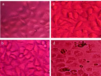
Morphological changes observation on Normal Chang liver cell treated with CAF-MIP and NIP AO/PI staining. For the microscopic observation of NCLC changes after incubated with CAF-MIP and NIP, bright field images were taken using light microscope using phase contrast filter to detect the morphological changes on the NCLC. It is important to detect their morphological changes as it can be used to determine and indicate the cells viability. Moraco et al., [10] stated that the cells viability can be detected by observing any changes in the cells shape and size. Figure 4 shows the image of normal Chang liver cells observed after incubated with CAF-MIP, NIP and H2O2 for 72 hours at
highest concentrations.
Figure 4. Normal Chang Liver Cells under Light Micro-scope Observations. For (a) Untreated, (b) treated with CAF-MIP, and (c) treated with NIP, Nor-mal Chang Liver Cells are Intact, Confluent and Still in their Cuboidal Shape with some Visible Nuclei while (d) a Positive Control, NCLC treated with H2O2 that showed Swelling of Cells and
Precipitation of Cytoplasmic Content as well as Apoptotic Bodies. Magnification was 400 x
But, after the polymerization process, the caffeine as a template was removed [11] while synthesis of NIP does not use caffeine as template at all. So, basically both CAF-MIP and NIP does not contain caffeine as their component; however, CAF-MIP has a template of caffeine and it is suggested that the template of caffeine may have a residue of caffeine and it is able to promote the cell growth.
For NIP that was used as a control to CAF-MIP, they also do not show toxicity effects on normal Chang liver cells. Thus, the components used to synthesized the CAF-MIP were safe and do not imply any toxicity effects on normal Chang liver cell.
While in normal Chang liver cells treated with H2O2 as a
positive control in this study, Figure 4(d) showed the cells were not confluence as in (a), (b) and (c). Besides, the cells lose their cuboidal shape as well as loose between neighboring cells. The membrane blebbing, with nuclear chromatin condensation, DNA fragmenta tion and the formation of small apoptotic bodies cell. The formation of membrane bound vesicles does not affect a group of contagious cells showed that the cell undergo apoptosis. From the images, the normal Chang liver cells are apoptosis death and it is accordingly to a report said that H2O2 as an apoptosis agent [12] which is
in contrast to cells treated with CAF-MIP and NIP.
Fluorescence microscopic observationfor mode of cell death observation on Normal Chang liver cell treated with CAF-MIP and NIP. Alternative viability
assays using the fluorescent dyes acridine orange (AO) and propidium iodide (PI) have been developed for the simultaneous visualization of both viable and nonviable cells. In this study, AO/PI staining was used to detect the mode of the cell death of the normal Chang liver cells when treated with CAF-MIP, NIP and H2O2. Figure 5 shows
the image of untreated and treated normal Chang liver cells under AO/PI staining at highest concentrations.
The combination of the two dyes were used to distinguish the mode of cell death either apoptosis or necrosis based on changes and color emitted by the nuclear structure under fluorescence microscope. Howe ver, based on the result of MTT assay, both cells treated with CAF-MIP and NIP are viable which indicate that both compound used are not toxic to cells. Thus, AO/PI staining was used to confirm the MTT assay results. AO is a membrane-permeable, cationic dye that binds to nucleic acids of viable cells and that at low concen trations causes a green fluorescence. PI is impermeable to intact membranes but readily penetrates the mem branes of nonviable cells and binds to DNA or RNA, causing orange fluorescence [13]. When AO and PI are used simultaneously, viable cells fluoresce green and nonviable cells fluoresce orange under fluorescence microscopy were observed. As show in the Figure 5 (a-c), all three image which are untreated cells, treated with CAF-MIP and treated with NIP, the cells are green fluorescence with an intact nucleus, show that the cells is viable caused by the binding of the AO to the nucleic [13].
In addition, nucleus of all the cells in all untreated, CAF-MIP and NIP-treated were intact, round or oval in shape, without any condensation and membrane blebbing.
Figure 5. Normal Chang liver Cells under Fluorescence Microscope (a) Untreated Cells, (b) Treated with CAF-MIP, (c) Treated with NIP that showed Nuclei is Intact While (d) Treated with H2O2, shows Cells with Yellow Nuclei-apoptotic
and Orange-red Fluorescence-necrotic cells. Magnification was 400 x
c a
b d
a
b
c
Cytoplasm was normal with intact cell membrane and cytoplasm disintegration was not observed.
Thus, it clearly shows both compounds especially CAF-MIP may not imply any toxicity effects on normal Chang liver cells.
As discussed before, the normal Chang liver cells that were treated with CAF-MIP and NIP were viable that indicated there are no toxicity effects. In fact, it is suggested that the CAF-MIP play an important role as an agent for cell growth as it might have the residue of caffeine. Caffeine contain few active compounds such as anti-inflammatory and polyphenol as an antioxidants that fight against free radical that can be found naturally in human body that may cause damage to human cells at higher concentration [14]. As comparison, the normal Chang liver cell treated with CAF-MIP and NIP were green fluorescence, intact nuclei and no membrane blebbing indicated that the cells were viable. In contrast, the normal Chang liver cells treated with H2O2 are
red-orange fluorescence with loss of membrane integrity and disintegration of nuclei indicated a necrosis death while the cell with yellow fluorescence nuclei and membrane blabbing with a formation of small apoptotic bodies indicated apoptosis death. Thus, CAF-MIP do not cause toxicity effects on normal Chang liver cells in contrast to H2O2 that is well-known as an apoptosis
inducing agent.
H2O2 is a strong oxidizing agent that could disrupt many
aspects of a microbe-acting on proteins, lipids, DNA and leading to cell death [15]. At a certain concentration, H2O2 acted as an apoptosis inducing agents and inducer
of DNA strand breaks and can cause cytotoxic effect on cells. This peroxide was also reported to cause DNA damage, chromosomal alterations and gene mutation by generating hydroxyl radicals close to the DNA molecule. Many previous studied on various cell culture in vitro showed that H2O2 cause the cell to undergo apoptosis
type of cell death [16,17]. Based on Figure 5(d), there were two types of normal Chang liver cells showed which are apoptosis and necrosis type of cells. It is clearly showed that the cells are dead as the cell shape is changes and shrink in which comparable to study done by Moracoet al. [10] on the mode of cell death. They reported that the shape of dead cells will change due to the membrane integrity interruption and alteration in both apoptosis and necrosis. The cells with yellow nucleus showed that it is apoptosis with a membrane blebbing, condensation of nuclear chromatin, DNA fragmentation and the formation of apoptotic bodies. The apoptotic cell does not affect the neighboring cell and no inflammatory reaction could occur to neighboring cells. It was reported that, for a biochemical features, the apoptosis death is energy (ATP) dependent with an activation of caspase cascade [18]. Previous study on the toxicity effect of goniothalamin on smooth vascular
muscle cell also showed that the cell undergo apoptosis as the nucleus is in yellow fluorescence and the blabbing of membrane with a formation of apoptotic bodies. The result is in comparison with the normal Chang liver cell when treated with H2O2 in which it
indicated an apoptotic cell [19]. On the other hand, cells with orange red fluorescence showed that the cell undergo necrotic type of cell death in which the cells shapes were changed, loss of membrane integrity, swelling of cytoplasm and mitochondria, and a total lysis with no formation of vesicle.
These changes may resulted from ATP depletion and the failure of plasma membrane ion pumps to maintain a stable osmotic gradient [10]. Necrosis type of cell death will effects the contagious cells and caused inflammatory reaction. Meanwhile, during necrosis type of cell death, no energy requirement and no activation of caspase cascade. As mentioned before, H2O2 is an apoptosis
inducing agents however result showed that the Chang livercells also undergo necrosis death. It is suggested that due to the massive damage to the mitochondria, and ATP depletion, the cell undergo necrosis death [17]. Apoptosis as highly regulated processes, characterized by specific morphological and biochemical properties which was induced by H2O2.
4.
Conclusion
In conclusion, both CAF-MIP and NIP did not reduce NCLC growth effects on human cells while promoting its proliferation. Not toxicity was observed as demon-strated by all experiments performed in this study. Therefore, CAF-MIP is suggested to be safe as a new alternative method for decaffeination in which it can reduce few health problem caused by over consuming of caffeine.
References
[1] B. Mariam, R.J. Carnachan, N.R. Cameron, S.A. Przyborski, J. Anatomy, 211/4 (2007) 567. [2] T. Mossman, J. Immunol. Methods, 65 (1983) 55 [3] S.A. Chintalwar, B. Rajkapoor, P.D. Ghode, Int.
J. Pharma. Bio. Sci. 3 (2012)155.
[4] J.M. Sargent, C.G. Taylor, Br. J. Cancer, 60 (1989) 206.
[5] Culture of Animal Cells-Basic Techniques, Roche Diagnostics GmbH, Mannheim, Germany, 2012, p.16. [6] S.S. Saini, A. Kaur, Minireview: Adv. Nanopart.
2 (2013) 60.
[7] N. Takuwa, Y. Fukui, Y. Takuwa, Mol. Cell. Biol. 19/2 (1999) 1346.
[9] A. Zimmermann, Med. Sci. Monit. 8 (2002) 53. [10] A.H. Moraco, H. Kornfeld, Semin. Immunol. 26/6
(2014) 497. http://dx.doi.org/10.1016/j.smim. [11] G. Vasapollo, R.D. Sole, L. Mergola, M.R. Lazzoi,
A. Scardino, S. Scorrano, G. Mele, Int. J. Molecu-lar Sci. 12 (2011) 5908.
[12] A. Zunino, P. Degan, T. Vigo, A. Abbondandolo, Mutagenesis, 16 (2001) 28
[13] K. Mascotti, J. McCullough, S.R. Burger, Trans-fussion, 40 (2000) 693.
[14] G.J. Troup, D.R. Hutton, J. Dobbie, J.R. Pilbrow, C.R. Hunter, B.R. Smith, B.J. Bryant, Med. J. Aus-tralia, 148/10 (1988) 537.
[15] J.K. Willcox, S.L. Ash, G.L. Catignani, Crit. Rev. Food Sci. Nutr., 44/4 (2004) 275.
[16] C. Cerella, S. Coppola, V. Maresca, M. De Nicola, F. Radogna, L. Ghibelli, Ann. N.Y. Acad. Sci. 1171 (2009) 559.
[17] H. Fatimah, A. Abdul-Manaf, M.A. Nakisah, Ma-lays. J. Microsc. 9 (2013) 133.
[18] A.I. Baba, Sci. Med. Vet. 12 (2009) 1.