R E S E A R C H
Open Access
Stable transgenic C9orf72 zebrafish model
key aspects of the ALS/FTD phenotype and
reveal novel pathological features
Matthew P. Shaw
1, Adrian Higginbottom
1, Alexander McGown
1, Lydia M. Castelli
1, Evlyn James
1,
Guillaume M. Hautbergue
1, Pamela J. Shaw
1†and Tennore M. Ramesh
1,2*†Abstract
A hexanucleotide repeat expansion (HRE) within the chromosome 9 open reading frame 72 (C9orf72) gene is the most prevalent cause of amyotrophic lateral sclerosis/fronto-temporal dementia (ALS/FTD). Current evidence suggests HREs induce neurodegeneration through accumulation of RNA foci and/or dipeptide repeat proteins (DPR). C9orf72 patients are known to have transactive response DNA binding protein 43 kDa (TDP-43) proteinopathy, but whether there is further cross over between C9orf72 pathology and the pathology of other ALS sub-types has yet to be revealed.
To address this, we generated and characterised two zebrafish lines expressingC9orf72HREs. We also characterised pathology in humanC9orf72-ALS cases. In addition, we utilised a reporter construct that expresses DsRed under the control of a heat shock promoter, to screen for potential therapeutic compounds.
Both zebrafish lines showed accumulation of RNA foci and DPR. Our C9-ALS/FTD zebrafish model is the first to recapitulate the motor deficits, cognitive impairment, muscle atrophy, motor neuron loss and mortality in early adulthood observed in human C9orf72-ALS/FTD. Furthermore, we identified that in zebrafish, human cell lines and human post-mortem tissue, C9orf72 expansions activate the heat shock response (HSR). Additionally, HSR activation correlated with disease progression in our C9-ALS/FTD zebrafish model. Lastly, we identified that the compound ivermectin, as well as riluzole, reduced HSR activation in both C9-ALS/FTD and SOD1 zebrafish models.
Thus, our C9-ALS/FTD zebrafish model is a stable transgenic model which recapitulates key features of human C9orf72-ALS/FTD, and represents a powerful drug-discovery tool.
Keywords: Amyotrophic lateral sclerosis, C9orf72, SOD1, TDP-43, Zebrafish, Drug-screening
Introduction
Amyotrophic lateral sclerosis (ALS) is a neurodegenerative disorder characterised by motor neuron loss, leading to progressive muscle weakness and eventual death, primar-ily due to respiratory failure. Approximately 10% of ALS is inherited in an autosomal dominant fashion, this is known as familial-ALS (fALS). The remaining 90% of ALS cases are caused by complex genetic and environmental
interactions which are currently not well understood, this is known as sporadic-ALS (sALS). Mutations in multiple genetic loci have been identified as causes of ALS
includ-ing theSOD1andTARDBPloci. See Amyotrophic Lateral
Sclerosis Online Genetics Database for comprehensive
information (http://alsod.iop.kcl.ac.uk/). The most
com-mon known genetic cause of ALS and frontotemporal de-mentia (FTD) is a hexanucleotide expansion within the first intron of the C9orf72gene [11, 32]. Carriers of the C9orf72 hexanucleotide expansion may show symptoms of ALS or FTD exclusively, but can also present with symptoms of both diseases concurrently.
Concerning pathology inC9orf72patients, there are three major, non-mutually exclusive routes of toxicity which have * Correspondence:[email protected]
†Pamela J. Shaw and Tennore Ramesh contributed equally to this work. 1Sheffield Institute for Translational Neuroscience, University of Sheffield,
385a Glossop Road, Sheffield S10 2HQ, UK
2The Bateson Centre, Firth Court, The University of Sheffield, Western Bank,
Sheffield S10 2TN, UK
© The Author(s). 2018Open AccessThis article is distributed under the terms of the Creative Commons Attribution 4.0 International License (http://creativecommons.org/licenses/by/4.0/), which permits unrestricted use, distribution, and reproduction in any medium, provided you give appropriate credit to the original author(s) and the source, provide a link to the Creative Commons license, and indicate if changes were made. The Creative Commons Public Domain Dedication waiver (http://creativecommons.org/publicdomain/zero/1.0/) applies to the data made available in this article, unless otherwise stated. Shawet al. Acta Neuropathologica Communications (2018) 6:125
been proposed to arise from the C9orf72 expansion: 1) Sense and antisense RNA foci which sequester RNA bind-ing proteins causbind-ing dysregulation of RNA processbind-ing
[7, 11]. 2) Dipeptide repeat proteins (DPRs) produced
via non-canonical repeat associated non-ATG (RAN) translation, form insoluble aggregates in the nucleus
and cytoplasm [42].3)Hexanucleotide expansion
medi-ated haploinsufficiency may cause dysregulation of en-dogenous C9orf72 pathways such as autophagy [12,39].
To date, several models have been generated to help
dissect out the mechanisms of C9orf72expansion
medi-ated toxicity. Most drosophila and zebrafish models sup-port an RNA/DPR mediated gain of toxic function hypothesis [18,26,28,37]. In addition, transgenic mouse models have been generated containing the human
patient C9orf72 gene (complete with G4C2 expansion
and flanking regions). Two transgenic mouse models demonstrate the reduced survival, neuronal loss and
motor deficits observed in human C9-ALS/FTD [14,20].
However, a further two independently generatedC9orf72
transgenic models showed no signs of neuronal loss or reduced survival. [29,30]. This highlights the wide
vari-ability observed in C9orf72 expansion in vivo models.
C9orf72 knockdown in the zebrafish causes mild motor
defects [6]. However, early reports from C9orf72
knock-out zebrafish do not recapitulate the knockdown motor
phenotypes ([34]; Schmid, Hruscha, Haass, unpublished).
Additionally, four independently generated C9orf72
knockout mice did not demonstrate any neurodegenera-tive phenotype [1,13,17,35].
Whilst mouse models are a useful tool for
understand-ing the pathobiology of C9orf72-related ALS, they are
not amenable to high throughput drug screening. Gen-etic modifier screens have been carried out in drosoph-ila, but their CNS is much simpler compared to the human CNS and findings in this invertebrate model are less likely to translate to the clinic [3,15]. Zebrafish are vertebrates with a more complex CNS, and therefore represent a practical compromise for assessing the effi-cacy of therapeutic compounds.
Here we present a novel transgenic zebrafish model
which stably expresses C9orf72 expansions. These
zeb-rafish recapitulate the behavioural deficits, cognitive ab-normalities, motor decline and early mortality observed
in C9-ALS patients. Additionally we show that C9orf72
expansions activate the heat shock response in human cell lines, post-mortem ALS tissue and our model
zeb-rafish. Using theseC9orf72zebrafish and our previously
reported SOD1 zebrafish in tandem [31], we show that
riluzole and a newly identified compound, ivermectin,
are able to reduce cellular stress in both C9orf72 and
SOD1 in vivo models. We therefore propose that our
C9orf72 zebrafish model effectively bridges the gap between drosophila and mouse models by providing an
efficient tool for high-throughput in vivo drug screen-ing assays.
Materials and methods
Generating and maintenance of transgenic zebrafish Zebrafish embryos were injected with a DNA construct containing 89 C9orf72 hexanucleotide repeats driven by a zebrafish ubiquitin promotor (Fig. 1a, Additional file 1). Creation and identification of transgenic zebrafish was
performed as previously described [31] and maintained
using established practices [40].
In situ hybridisation and immunofluorescence
In situ hybridization of paraffin embedded tissue
sec-tions to detect CCCCGG (C4G2) foci was performed on
5dpf embryos using methods described previously [8].
For immunofluorescence staining, paraffin embedded tissue was dewaxed, antigen retrieved and stained as previously described [9].
Western blotting
Ethical approval for use of human cerebellum samples was obtained by the Sheffield Brain Tissue Bank Manage-ment Board, and approval to release tissue under REC 08/ MRE00/103 was granted. Human cerebellum samples and adult zebrafish tissue, brain, spinal cord and whole zebra-fish embryos were snap frozen in liquid nitrogen and processed for western blotting. Laemmli buffer was added
in the ratio of 10 μl:1 mg of tissue and sonicated.
SDS-PAGE and immunoblotting were performed as previously
described [39]. Antibodies used were Rb-anti-PR (gift
from Dieter Edbauer), Rb-anti-Dsred (Clontech 632,496), Ms-anti-tubulin (Abcam). Species specific HRP conju-gated secondary antibodies were used and imaged by chemiluminescence using G-Box.
Embryonic behaviour
For spontaneous locomotor activity, 5 dpf zebrafish were placed into individual wells of a 96well plate and habituated in the dark for 10 min before a light stimulus was turned on. 10 min of light was followed by 10 min dark and repeated once more. Recordings were carried out using ZebraBox software (ViewPoint Behaviour Technologies), movement thresholds used were slow (x < 5 mm/sec), intermediate (5 < x < 15 mm/sec) and fast (x > 15 mm/sec).
centre of the well was visible, the % of zebrafish present in the centre of the well was then blind counted for every image and the average per well was calculated.
Adult locomotor behaviour
Zebrafish swimming ability was tested using a swim tun-nel with an intial flow-rate of 2 L/min, increasing in 2 L/ min increments every 5 min until the maximum flow rate of 11.6 L/min was achieved. Data were analysed as previ-ously described [31]. 5 min post-testing, the spontaneous swimming behaviour of the fish was measured for 30 min using a camera linked to ZebraLab software (ViewPoint Behaviour Technologies). Speed thresholds used were slow (x < 60 mm/sec), intermediate (60 < x < 120 mm/sec) and fast (x > 120 mm/sec).
Motor neuron counts and myotome measurements Spinal motor neurons were counted from paraffin em-bedded adult zebrafish segments cut anterior to the
pelvic fin, sectioned at 10 μm and stained with
haema-toxylin and eosin. Cells with a soma size >75μm2
and
within 25,000μm2
proximity of the central canal were designated as motor neurons. Three sections/per animal
were analysed by two independent blinded investigators and averaged. The areas of individual myotomes were measured by a blinded investigator from 6 images per animal. All muscle images were obtained from the ep-axial muscle region just lateral to the dorsal spinal bone. Any myotome which was incomplete due to being par-tially out of frame was not included in the analysis.
Cell culture and transfections
Cells were maintained in a 37 °C incubator with 5% CO2.
HEK293T cells were cultured in Dulbecco’s Modified
Eagle Medium (Sigma) supplemented with 10% foetal
bo-vine serum (FBS) (Gibco) and 5 U ml−1Penstrep (Lonza).
Neuro-2a(N2A) (ATCC) cells were cultured in Dulbecco’s
Modified Eagle Medium (Sigma) supplemented with 10%
FBS (Gibco), 5 U ml−1Penstrep (Lonza) and 5 mM
so-dium pyruvate.
HEK293T and N2A cells were transfected with 700 ng of
plasmid using 3.5 μg PEI/ml media and one tenth media
volume of OptiMEM in a 24 well format. Approximately, 50,000 HEK293T cells were seeded / well and 75,000 N2A cells were seeded per well of the 24 well plate. Proteins were extracted 72 h post-transfection. Cells were washed in
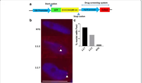
Fig. 1C9orf72model zebrafish display RNA foci in the nucleus. (a) Schematic representation of the transgene inserted into 2.2 zebrafish. A zebrafish ubiquitin promotor drives GFP-DPR expression. Anhsp70promotor then drives DsRed production as a read out of cellular stress. (b) In situ hybridisation of paraffin embedded sections of 10dpf 2.2–7 zebrafish using a Cy3-conjugated (red) GC probe (GGGGCC)X4 showed that RNA foci are present in the nuclei of muscle cells. Arrow heads denote RNA foci. Scale bar = 10μm. (c) Quantification of RNA foci showed Immunofluorescence labelling of adult zebrafish muscle tissue showed that poly-GP DPR protein localises to the nucleus in 2.2–2 and 2.2–7 transgenic zebrafish
ice cold phosphate buffered saline (PBS) and subsequently lysed in ice cold lysis buffer (50 mM Hepes pH 7.5, 150 mM NaCl, 10% glycerol, 0.5% Triton X-100, 1 mM EDTA, 1 mM DTT, protease inhibitor cocktail (Sigma)) for
10 min on ice. Extracts were then centrifuged at 17,000g
for 5 min at 4 °C. Extracts were quantified using Bradford Reagent (BioRAD), resolved by SDS-PAGE, electroblotted onto nitrocellulose membrane and probed to the relevant primary antibodies.
Heat shock cell stress drug screening assay
At 2 dpf, transgenic zebrafish were placed into a 96 well
plate in 200μl of drug or DMSO containing E3 zebrafish
media. At 5 dpf zebrafish were sonicated in the well for 10 s each and then centrifuged in a plate spinner at
3000 rpm for 10 min. From each well, 20 μl of
super-natant was transferred into a 385 well plate, and the DsRed levels in each individual lysate were quantified using a FLUOstar Omega fluorescence plate reader (BMG labtech).
Quantification and statistical analysis
Data were analysed by one way ANOVA with Tukey’s post
hoc test or two way ANOVA with Sidak’s post hoc test for
multiple comparisons, t-test or Kaplan Meier analysis as in-dicated in the appropriate figure legend. Significance is denoted as *P< 0.05, **P< 0.01, ***P< 0.001 and ****P< 0.0001. Individual myotome size data were counted into bins
with a 0.5mm2 size range. The frequency distribution of
each genotype was then compared using a chi-squared test for trend.
Results
Generation of transgenic zebrafish
To better understand ALS/FTD pathogenesis and screen
potential therapeutic agents, we generated aC9orf72
zeb-rafish model. At the single cell stage zebzeb-rafish embryos were injected with a DNA construct containing 89 C9orf72 hexanucleotide repeats (Fig.1a, Additional file1). Of the 3 zebrafish lines generated, one was extremely toxic, resulting in death within 7 days of fertilisation (dpf). Therefore, only the 2 remaining lines were maintained to breeding age and established for further characterisation. These two transgenic zebrafish lines which were
estab-lished to adulthood will henceforth be known as line 2.2–
2 and line 2.2–7, or collectively as 2.2-zebrafish lines. Both 2.2-zebrafish lines give rise to 1:1 ratios of transgenic:NTG offspring when outbred, suggesting a single site of trans-gene insertion.
C9orf72zebrafish lines express RNA foci and DPR
The hallmark features ofC9orf72pathology are expression
of RNA foci and DPR species. Using in situ hybridisation and immunofluorescence, we identified expression of
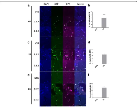
RNA foci and DPR species in both 2.2-zebrafish lines. Antisense RNA foci (CCCCGG, the same orientation with respect to the construct) can be detected in the nuclei of muscle cells in both 2.2-zebrafish lines (Fig.1b), no more than one focus is observed per nucleus, and no cytoplas-mic foci were detected. 50% (11/22) of nuclei in 2.2.7 line showed RNA nuclear foci while fewer foci (30%, 6/20) were observed in 2.2.2 line. Non-transgenics showed 4% (1/25) foci like staining but failed to show colocalisation in the nuclei (Fig. 1c). It is presumed, that the single focus observed in the NTG zebrafish was due to non-specific binding of the in situ probe. To determine whether repeat RNA was translated into DPR proteins, antibodies specific to antisense DPR species poly-GP, PA and PR were used. All three antisense DPR species were detected in the nuclei of muscle cells from both 2.2-zebrafish lines
(Fig. 2a,c,e) with over 50% of the nuclei expressing the
DPRs (Fig.2b,d,f).
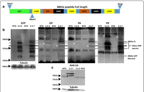
C9orf72zebrafish produce multiple distinct DPR species The various DPR species are known to have differential toxicity, with arginine rich species being considered the most toxic. To investigate whether there is a relationship between molecular weight (MW) and species toxicity, western blotting was performed on zebrafish lysates. The transgene construct expressed in both 2.2-zebrafish lines causes the production of GFP tagged DPR proteins via canonical ATG (start codon) dependent translation. The full length GFP fusion protein is predicted to be 48 kDa
while any truncation 3′of any C4G2 repeats would
re-sult in the production of GFP alone (28KDa). The
sche-matic of the GFP-DPR fusion protein is shown (Fig.3a).
GFP tagged DPRs are produced from C4G2 transcripts
and can be detected at 5 dpf (Fig. 3b, left most panel).
Interestingly, multiple GFP bands are detected in both
the 2.2–2 and 2.2–7 zebrafish lines, and these bands
were often unique to one zebrafish line or the other, and were consistent over > 10 clutches. The differential
ex-pression of DPRs between 2.2–2 and 2.2–7 zebrafish
also holds true when probing for the DPR proteins
dir-ectly (Fig. 3b, 3 right panels). The full length GFP-DPR
fusion at 48 kDa was expressed but at low levels
(Fig. 3b, 48 kDa band). Probing antibodies against DPR
proteins also revealed that some DPR bands detected did not co-localise with any of the ATG-dependent translation bands detected using the GFP antibody, suggesting that these bands are likely to be produced via non-canonical
RAN translation (Fig. 3b, three right panels marked
with asterix).
In addition to the poly-PA, PR and GP DPRs produced from the (C4G2) RNA transcripts, we were also able to
de-tect poly(GA) DPR produced from the (G4C2) RNA
zebrafish line (Fig. 3c). The detection of poly(GA) indi-cates that bidirectional transcription of the GC rich region is occurring in the presence of our transgene. As the tran-scription of the RNA transcript containing the (G4C2)
ex-pansion is not driven by a conventional promotor region, this strongly indicates that poly(GA) protein is indeed pro-duced via RAN translation.
In human ALS, C9orf72 associated toxicity occurs
primarily in cells of the CNS, and so it is essential to ascertain whether DPR species are also produced
within the CNS of this C9orf72 model zebrafish. In
adult brain and spinal cord of both 2.2-zebrafish lines, GFP-tagged DPR species and DPR species which were not immunoreactive with GFP antibodies (RAN
trans-lation bands), could be detected (Fig. 4). This suggests
that both ATG-dependent translation and RAN trans-lation of DPR species occurs within the CNS of the 2.2-zebrafish.
In summary, both 2.2-zebrafish lines exhibit DPR species generated by conventional ATG-dependent translation and RAN translation in the muscle and the CNS. In addition, the 2.2–7 zebrafish line shows bi-directional transcription,
producing DPR species from both G4C2 and C4G2 RNA
transcripts. In both 2.2-zebrafish lines, the band pattern of DPRs detected largely remains constant from 5 dpf until adulthood, although higher MW (>50KDa) poly(PR) posi-tive bands are more abundant in adult tissue. Of all the DPR species examined here, poly(PR) generally has the highest propensity to form high MW RAN-translation mediated bands.
Fig. 2C9orf72 model zebrafish display DPR expression in the nucleus. (a,b) Immunofluorescence labelling of adult zebrafish muscle tissue showed that poly-GP DPR protein localises to the nucleus in 2.2–2 and 2.2–7 transgenic zebrafish. (c.d) Immunofluorescence labelling of adult zebrafish muscle tissue showed that poly-PA DPR protein localises to the nucleus in 2.2–2 and 2.2–7 transgenic zebrafish. (e,f) Immunofluorescence labelling of adult zebrafish muscle tissue showed that poly-PA DPR protein localises to the nucleus in 2.2–2 and 2.2–7 transgenic zebrafish. For all DPR images nuclei are stained with Hoechst (blue), GFP is stained with GFP antibody (green) and DPR proteins are stained with the relevant DPR antibody (purple), white arrow heads denote DPR positive staining. Scale bar = 25μm for all DPR images
Early mortality, altered swimming behaviour and reduced weight gain in transgenicC9orf72zebrafish
Neither 2.2-zebrafish lines showed any overt
morpho-logical abnormalities during embryonic development (0–
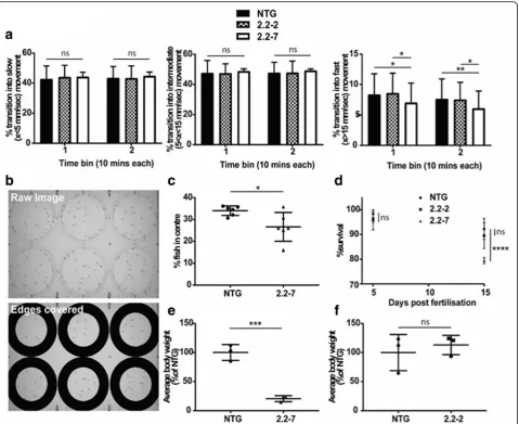
5 dpf ). At 5 dpf zebrafish begin to express a wider reper-toire of behaviours, including more frequent swimming and independent feeding. For this reason, rigorous evaluation was performed on 5 dpf zebrafish to test for underlying motor and behavioural deficits. In order to test the spontaneous locomotor activity of embryonic zebrafish, we monitored 5 dpf zebrafish in 96 well plates using the Viewpoint behaviour monitoring setup. No sig-nificant difference was observed between the groups in the proportion of times transitioning occurred into slow
or medium movements (Fig. 5a). However, a significant
reduction in the proportion of transitions into fast
movement was detected in 2.2–7 zebrafish, when
com-pared to either NTG or 2.2–2 zebrafish (Fig.5a). AsC9orf72expansions in human ALS cause a spectrum of both motor and cognitive deficits, we examined whether normal zebrafish behaviour was affected in
2.2–7 zebrafish at 5 dpf. Centre avoidance behaviour
assays are a validated means of measuring willingness
to explore in zebrafish [33], and are comparable to the
open field test performed in mice. It was determined that 2.2–7 zebrafish were significantly less likely to ven-ture into the centre of the well when compared to their
NTG clutchmates (Fig.5b+c).
To determine if the early embryonic expression of RNA foci and DPR impacted upon the viability of the C9orf72 zebrafish, we carried out early (1–15 dpf )
sur-vival analysis. Heterozygous 2.2–2 zebrafish do not show
any change in survival within 15 dpf as compared to NTG zebrafish (data from NTG clutchmates of all
geno-types are pooled; Fig. 5d). However, heterozygous 2.2–7
zebrafish did show a significant decrease in survival
within 15 dpf as compared to NTG zebrafish (Fig. 5d),
but not in comparison to 2.2–2 zebrafish.
It was noted that during early development, the 2.2–7
zebrafish appeared smaller than their NTG clutchmates. At 30 dpf there was a significant decrease in total body
weight of 2.2–7 zebrafish compared to their NTG
clutchmates (Fig. 5e). However, 2.2–2 zebrafish did not show a significant difference in body weight as compared to their own clutchmates at the same age (Fig.5f ).
In summary, 2.2–7 zebrafish but not 2.2–2 zebrafish,
show significant reduction in survival at 15 dpf, and
reduction in bodyweight at 30 dpf. At 5 dpf, 2.2–7
zeb-rafish also show defects in swimming activity and dis-played signs of atypical behaviour. Behaviour of the
phenotypically more severe 2.2–7 zebrafish was also
studied through adulthood.
C9orf72zebrafish display adult onset ALS-like behavioural phenotypes
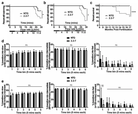
To assess the neuro-muscular integrity of the 2.2–7 trans-genic zebrafish, swimming endurance was tested using a swim tunnel, the aquatic equivalent to a treadmill [31]. At 9 months of age more 2.2–7 transgenic zebrafish failed to maintain swimming at the maximum flow rate as
com-pared with their NTG clutchmates (Fig. 6a). Despite
de-creased body mass during early development, body mass and body size were not significantly different between adult transgenic and NTG groups from 9 months of age
(Additional file2: Figure S1). Spontaneous swimming was
observed immediately following swim tunnel testing, but no difference was observed between the two groups
(Fig. 6d). The swim tunnel test was repeated with the
same cohort of zebrafish at 12 months of age, and the ability to swim at maximum speed continued to decrease in the 2.2–7 zebrafish (Fig.6b). Interestingly, at 12 months
2.2–7 zebrafish now showed defects in spontaneous
swim-ming behaviour following the swim tunnel testing. 12-month-old zebrafish showed an increase in the propor-tion of times transipropor-tioned into slow speed movements and a concomitant decrease in the proportion of times transi-tioned into fast speed movement, as compared to NTG clutchmates at the first-time point following swim tunnel testing (Fig.6e).
Adult survival was also monitored from 8 months post-fertilisation onwards. By 17 months post-post-fertilisation, sur-vival rates of the 2.2–7 transgenic zebrafish were signifi-cantly reduced in comparison to their NTG clutchmates
(44% vs 100% survival respectively; Fig. 6c). A zebrafish
was defined as having reached end-stage once it had lost the ability to maintain normal swimming (showing signs of paralysis) to the extent where it was no longer able to
obtain food. End-stage 2.2–7 zebrafish displayed severe
wasting in the body muscle region and had very poor locomotor skills (Additional file 3: video 1, no NTG zebra-fish displayed this wasting phenotype.
Muscle atrophy and motor neuron loss inC9orf72 zebrafish
Progressive muscle atrophy is observed in all ALS
pa-tients. Similarly, end-stage 2.2–7 zebrafish muscle
dis-played widespread severe atrophy, muscle fibres were disorganised, and a large increase in nuclei was observed
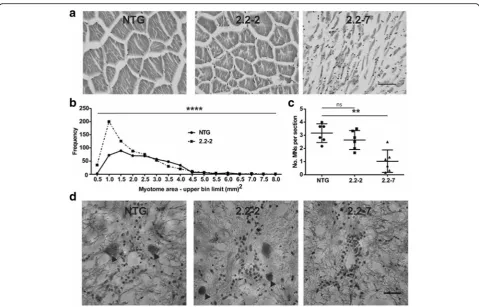
(Fig. 7a). The muscle of 2.2–2 zebrafish displayed more
subtle changes, with myotomes being significantly smaller
Fig. 4C9orf72model zebrafish produce multiple species of DPRs in the adult CNS. (a) Anti-sense DPRs (predominately ATG-driven) detected in adult brain lysates. (b) Anti-sense DPRs (predominately ATG-driven) detected in adult spinal cord lysates. Asterix (*) denotes protein bands which are proposed to have been produced via RAN-translation
and more numerous as compared to NTG muscle (Fig.7a +b). We did not quantify end-stage 2.2–7 zebrafish muscle fibre size, as their myofibres were too disorganised to dis-cern individual myotomes.
In ALS patients, the underlying molecular pathology ultimately leads to motor neuron death. Similarly, sig-nificant loss of ventral horn motor neurons was
ob-served in end-stage 2.2–7 zebrafish as compared with
NTG controls (Fig. 7c+d). A small, non-significant
reduction in motor neurons was observed in 2.2–2
zeb-rafish as compared with NTG controls.
Heat shock stress response is activated byC9orf72 expansions
Heat shock proteins are upregulated in response to the presence of aberrant cellular proteins [4,10]. We hypothe-sised that the low-complexity structure of DPR proteins might drive activation of the heat shock response (HSR).
To test this, we transfected both HEK 293 T and N2A
cells with C9orf72 expansion containing pure HRE and
interrupted HRE constructs. The repeats were expressed
in tandem with a hsp70promotor driving a DsRed gene,
as a readout of heat shock response activation. As DsRed is more stable than hsp70, it allows more sensitive
detec-tion of small but chronic HSR activadetec-tion [24]. In both
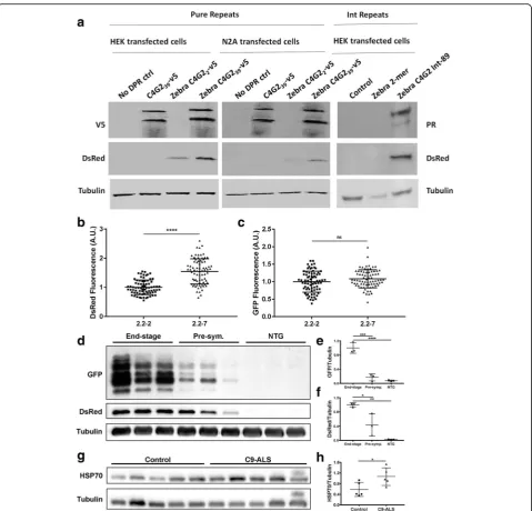
HEK and N2A cells, cells transfected with 39 C4G2pure
repeats (left two panels) or 89 interrupted repeats (right sided panel) showed strong RAN-translated V5-tagged DPR or ATG driven PR-tagged DPR production and
markedly higher DsRed production (Fig.8a). In contrast,
cells transfected with only 2 C4G2 repeats displayed no
RAN-translated DPRs and less or undectable DsRed
pro-duction. As expected cells transfected with 39 C4G2
re-peats but no hsp70:DsRed heat shock readout, produced
abundant RAN-translated DPRs but no DsRed protein. To assess differences in HSR activation in the more
phenotypically severe 2.2–7 zebrafish vs the less severe
2.2–2 zebrafish, we screened 5 dpf zebrafish for DsRed
(produced viahsp70promotor activation). The more severe
2.2–7 zebrafish showed significantly increased DsRed
Fig. 6C9orf72model zebrafish show adult onset swimming endurance deficits and reduced survival. (a) At nine months old, 2.2–7 transgenic zebrafish failed to continue swimming at earlier time points than their NTG clutch mates.N= 13 fish per genotype. (b) Also at twelve months old, 2.2–7 transgenic zebrafish failed to continue swimming at earlier time points than their NTG clutch mates. N = 13 fish per genotype. (c) Adult transgenic 2.2–7 zebrafish have reduced survival between 8 and 17 months in comparison to their NTG clutch mates which are housed in the same tank.N= 17 2.2–7 and 27 NTG at 8 months. (d) After being removed from the swim tunnel, 9 month old fish did not show any significant difference in proportion of transitions into slow (left), intermediate (middle) or fast (right) movements.N= 12 fish per genotype. (e) After being removed from the swim tunnel, 12 month old 2.2–7 zebrafish showed a significant increase in the proportion of transitions into slow movements and a corresponding significant decrease in the proportion of transitions into fast movement. There was no change in transition into intermediate movement. N = 13 fish per genotype. All data are shown as mean +/−standard deviation; *P < 0.05, **P < 0.01, ***P < 0.001 and ****P < 0.0001
fluorescence in comparison to 2.2–2 zebrafish at 5dpf (Fig.
8b). Importantly, GFP fluorescence (from GFP-tagged
DPRs) was not significantly different between 2.2–7 and
2.2–2 zebrafish (Fig.8c).
To assess how HSR activation changes as phenotypic severity increases, we examined GFP and DsRed
produc-tion in adult zebrafish brains, from 3 end-stage 2.2–7
zebrafish (ages 15, 15 and 19 months), 3 pre-symptomatic
2.2–7 zebrafish (all aged 7 months) and 3 NTG zebrafish
(age matched to end-stage). Pre-symptomatic was defined as fish which did not show any overt swimming or muscle abnormalities. GFP tagged DPRs were increased in the brains of end-stage zebrafish in comparison to the brains of pre-symptomatic zebrafish (Fig.8d+e). Similarly, DsRed also increased in the brains of end-stage zebrafish in comparison to the brains of pre-symptomatic
zebra-fish (Fig. 8d+f ), thus suggesting an association
be-tween DPR production and HSR induction.
Finally, we examined whether HSR activation could occur in the presence of the DPR proteins in cerebellar
post-mortem tissue from C9orf72 ALS patients.
Cerebellum tissue was selected to study the effect of DPRs on HSR, as previous reports indicate cerebellum tissue consistently shows a high DPR load [2,9,21,22]. Firstly, we confirmed that DPR species are expressed in the cerebellum of these C9-ALS patients (Additional
file 4: Figure S2. Next, HSP70 protein levels in human
cerebellum were assessed using western blotting. C9-ALS patients had significantly higher cerebellar levels of HSP70 as compared with
non-neurological-disease controls (Fig. 8g+h). Taken together, our data
demonstrate that C9orf72 expansions activate the heat
shock response.
Both C9orf72 and SOD1 ALS zebrafish models
ex-press a hsp70 promotor which drives DsRed protein
production. Cell stress from a variety of insults
in-creases the drive on thehsp70promotor, and
upregula-tion of the HSP70 protein has been reported in neurodegenerative disorders such as multiple sclerosis and, in the present study, ALS [19, 23, 27]. Therefore, in our ALS zebrafish models, the abundance of DsRed produced via hsp70 promotor activation is used as a
readout of cellular stress. Drugs which reduce cellular
stress, and thereby reduce hsp70 promotor mediated
DsRed production can be identified by treating zebra-fish with the drug from 2 to 5 dpf, and then measuring
DsRed levels in a fluorescence plate reader [25]. To
date, thousands of compounds have been tested using
this drug screening paradigm in SOD1-ALS zebrafish
models (current authors, data not shown). Ivermectin is a compound which was identified as one of the most
ef-ficacious drugs in theSOD1 zebrafish screen. In SOD1
Fig. 8Heat shock stress response activation is induced byC9orf72expansions. (a) In lysates from both HEK 293 T and N2A cell lines, DsRed levels are higher in cells transfected with 39 C4G2 pure repeats (Left and middle panel) or 89 C4G2 interrupted repeats (Right panel) compared with those transfected with only 2 C4G2 repeats. (b) At 5dpf, 2.2–7 zebrafish show significantly higher DsRed fluorescence than 2.2–2 zebrafish.N= 75 2.2–2 and 76 2.2–7 individual zebrafish. (c) At 5dpf, GFP fluorescence is not significantly different between 2.2–7 and 2.2–2 zebrafish. N = 75 2.2–2 and 76 2.2–7 individual zebrafish. (d) In end-stage 2.2–7 zebrafish brains, levels of GFP tagged DPR and DsRed proteins are increased compared with pre-symptomatic 2.2–7 and NTG. (e) Quantification of GFP tagged DPR protein normalised to tubulin in adult zebrafish brains. N = 3 adult brains per condition. (f) Quantification of DsRed protein normalised to tubulin in adult zebrafish brains. N = 3 adult brains per condition. (g) In human cerebellum samples, HSP70 protein levels are higher in C9-ALS patients as compared to non-neurological-disease controls.N= 5 samples per group. (h) Quantification of HSP70 protein levels normalised to tubulin in human cerebellum. All data are shown as mean +/−standard deviation; *P< 0.05, **P< 0.01, ***P< 0.001 and ****P< 0.0001
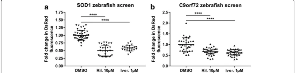
zebrafish ivermectin treatment reduced the level of HSR activation (as measured by DsRed fluorescence) to a similar degree as riluzole (the only disease modifying
treatment currently prescribed for ALS; Fig. 9a). Thus,
in C9orf72zebrafish ivermectin treatment also resulted in a significant reduction of HSR activation, and
com-pared with the SOD1 zebrafish screen, the efficacy of
ivermectin was comparable to that of riluzole (Fig.9b).
Therefore, these data suggest that cross over between SOD1 and C9orf72 pathology may allow for a single treatment to be efficacious in both disease forms.
Discussion
We have generatedC9orf72-related ALS model zebrafish
which stably express interrupted C4G2 expansions and
exhibit RNA foci and DPR pathology. These zebrafish accurately recapitulate key aspects of the behavioural, cognitive, motor defects and reduced survival associated with C9-ALS/FTD. Additionally, these zebrafish have been utilised to identify that poly(PR) DPRs form higher
molecular weight species. Furthermore, these C9orf72
zebrafish were used in conjunction with human cell lines
and human post-mortem tissue to identify that C9orf72
expansions activate the HSR. Finally, we identified that ivermectin treatment reduces cell stress HSR activation
in bothSOD1 and C9orf72 zebrafish models. The novel
aspects of the C9orf72 zebrafish model we have
gener-ated here are compared and contrasted to otherC9orf72
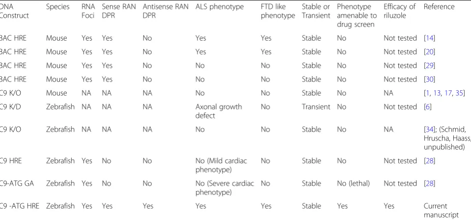
in vivo models in Table1.
The zebrafish model presented here lends support to a gain of function as the toxic mechanism underlying C9orf72 ALS/FTD. Our data are consistent with several other studies in animal models showing toxicity mediated by RNA foci and DPRs [5,20,26,38], including two
inde-pendently generated C9orf72 zebrafish models [18, 28].
Furthermore, our data are consistent with four
independ-ently generated C9orf72 knock-out mice and one
knockout zebrafish model, none of which display any motor or neurodegenerative changes, arguing against
hap-loinsufficiency as a major contributor to C9orf72 ALS/
FTD [1, 13, 17, 35], (Schmid, Hruscha, Haass,
unpub-lished). In contrast, decreased C9orf72 transcript levels
have been reported in the CNS of G4C2expansion bearing
patients, and morpholino mediated knockdown of C9orf72transcripts have been linked with motor deficits
in zebrafish [6, 11]. However, morpholinos notoriously
have off-target effects and may fail to mimic the
pheno-types observed in stable knockout mutant zebrafish [16].
Thus, the current body of evidence is heavily weighted to-wards RNA foci/DPR mediated gain of function toxicity in C9orf72expansion pathobiology.
Western blotting of zebrafish lysates revealed that mul-tiple lengths of GFP-tagged DPRs are produced (including the predicted 48KDa full length peptide) producing a lad-dered appearance. Both sense and antisense DPR were de-tected and were produced by both conventional and RAN-translation. Detection of species of varying MW has also been reported during RAN-translation of CAG
repeats [42], and during RAN-translation of GGGGCC in
C9-ALS patients [43]. More RAN-translation mediated
bands were detected in 2.2–7 zebrafish compared to 2.2–
2. Interestingly, poly(PR) species were detected at higher MWs than other DPR species, and it will be important to investigate whether the tendency of poly(PR)s to form high MW species is related to the potent in vivo toxicity. This suggests that RAN-translation blocking agents aimed specifically at inhibiting HMW poly(PR) formation may be an important therapeutic avenue to pursue.
The more severe 2.2–7 zebrafish line showed embryonic
onset motor defects and evidence of cognitive abnormal-ities, thus suggesting that DPR/RNA foci pathology is ad-versely affecting not only the motor unit, but also cognitive function; consistent with the spectrum of ALS/
FTD inC9orf72patients. Assessment of centre avoidance
behaviour indicated that 2.2–7 zebrafish showed an
un-willingness to explore, similar to C9orf72 mice assayed
with the open field paradigm [20]. Early mortality is also
observed in the more severe 2.2–7 zebrafish, indicating
that motor and cognitive defects detectable at the embry-onic stage later become severe enough to impact upon
survival. Reduction in body weight was observed in 2.2–7
zebrafish at the larval stage, however this later recovered by adulthood, suggesting that the reduction was due to re-tardation of the growth process rather than tissue degen-eration. Indeed, it is possible that slowed growth during early development of the 2.2–7 line may be due to the ob-served motor defects reducing access to food.
Swim tunnel performance of the 2.2–7 zebrafish was
significantly poorer than that of their NTG clutchmates at both 9 and 12 months. Swim tunnel performance is mainly indicative of the neuromuscular integrity of zeb-rafish body muscle, however cardiovascular involvement cannot be ruled out. Small differences in spontaneous swimming behaviour observed at 9 months became sig-nificantly different at 12 months, indicating progression of phenotypic severity. Disease progression was also
con-firmed when the same 2.2–7 swim tunnel tested
zebra-fish displayed clear signs of muscular atrophy and became unable to swim, necessitating culling. None of the NTG clutchmates showed this progressive atrophic
phenotype. By 17 months of age over 50% of the 2.2–7
zebrafish required to be culled, however most of the remaining zebrafish appeared healthy. This indicates a
heterogeneity in progression of phenotype in the 2.2–7
zebrafish, and suggests that genetic, epigenetic or other
factors may modulate the disease phenotype. Indeed, this
phenomenon may explain why the 2.2–2 zebrafish
model present a less severe phenotype. Similar variability in phenotypic severity has previously been reported in
BAC mice expressing theC9orf72gene [20].
Abnormal muscle histology was observed in both 2.2–
2 and 2.2–7 zebrafish. Generally muscle fibres were
smaller and more numerous in the transgenic zebrafish, consistent with atrophy and attempted regeneration.
Sig-nificant motor neuron loss was also observed in 2.2–7
zebrafish and a trend in the same direction was observed in the 2.2–2 zebrafish. At this point it is not possible to determine whether the degeneration of the neuromuscu-lar unit was neurogenic or myogenic in origin, and given that it is now known that DPR may transmit from cell to cell there may well be a contribution to toxicity from both tissues [41].
Previous transient RNA-injection zebrafish models
sug-gest that G4C2 RNA is sufficient to cause activation of
apoptosis and motor axonopathy [18,36]. It is important to note that transient RNA-injection models express RNA in much higher concentrations than would be observed in stable animal models, therefore the observed pathology is less likely to be reflective of pathology under physiological conditions. The RNA-injection zebrafish were not charac-terised longitudinally as the transgene is only expressed transiently (typically for 1–3 days). Additionally, an inde-pendently generated stable zebrafish model has previously
shown that 80 X (G4C2) RNA or poly(GA) DPR
expres-sion leads to pericardial oedema related toxicity at 4 dpf, but no neurological or motor phenotype was reported at Table 1Comparison of StableC9orf72stable mouse and zebrafish mutants showing the distinct phenotype and utility of each model
DNA Construct
Species RNA Foci
Sense RAN DPR
Antisense RAN DPR
ALS phenotype FTD like phenotype
Stable or Transient
Phenotype amenable to drug screen
Efficacy of riluzole
Reference
BAC HRE Mouse Yes Yes No Yes Yes Stable No Not tested [14]
BAC HRE Mouse Yes Yes No Yes Yes Stable No Not tested [20]
BAC HRE Mouse Yes Yes No No No Stable No Not tested [29]
BAC HRE Mouse Yes Yes No No No Stable No Not tested [30]
C9 K/O Mouse NA NA NA No No Stable No NA [1,13,17,35]
C9 K/D Zebrafish NA NA NA Axonal growth
defect
No Transient No Not tested [6]
C9 K/O Zebrafish NA NA NA No No Stable No NA [34]; (Schmid,
Hruscha, Haass, unpublished)
C9 HRE Zebrafish Yes No No No (Mild cardiac
phenotype)
No Stable No Not tested [28]
C9-ATG GA Zebrafish Yes No No No (Severe cardiac
phenotype)
No Stable No (lethal) Not tested [28]
C9 -ATG HRE Zebrafish Yes Yes Yes Yes Yes Stable Yes Yes Current
manuscript
NANot applicable,K/OKnockout,K/DKnockdown, Grey boxes represents the features that represent similarity to human ALS/FTD or utility in the high throughput screening of novel therapeutics
any time point [28]. In contrast, over a comparable time period (5 dpf ), the zebrafish presented here showed both motor and cognitive dysfunction. Additionally, our zebra-fish model survived to adulthood and displayed adult-on-set motor defects which eventually lead to motor neuron loss and death, thus recapitulating key features of human ALS/FTD over multiple time points. If model organisms are to be reliable in terms of the mechanistic insights or the therapeutic targets they generate, then they must re-flect disease features accurately. Future models should in-clude as many disease relevant features as possible until
the exact mechanisms of C9orf72 expansion toxicity are
better understood.
HSP70 protein levels were found to be increased in C9-ALS patient cerebellar tissue. Consistent with previ-ous reports, these cerebellum samples were found to have a substantial DPR load, thus DPRs may mediate cerebellar HSR activation [2,9,21,22]. Activation of the HSR as measured by DsRed protein expression under
the control of the hsp70 promotor, was found to be
higher in cells transfected with 39 C4G2 repeats
com-pared to cells transfected with only 2 C4G2repeats, thus
indicating that C9orf72 expansions of a pathological
length are required for activation of thehsp70promotor.
Additionally, activation of the HSR as measured by
DsRed protein expression, was higher in 2.2–7 zebrafish
compared with 2.2–2 zebrafish. However, in the same
fish GFP fluorescence was not significantly different, in-dicating that the total amount of DPR in each of the 2.2-zebrafish lines is equivalent. The reason for a greater
activation of HSR in 2.2–7 could be due to the
differen-tial pattern of DPR expression between the two zebrafish lines. Variability in transgene copy number is unlikely to underlie the difference in DsRed production between the
2.2–7 and 2.2–2 zebrafish, as GFP levels between the
two are not significantly different. DsRed and GFP tagged DPRs also progressively increased in the brains of end-stage zebrafish, indicating that DsRed production positively correlates with both DPR production and dis-ease severity.
Furthermore,C9orf72andSOD1ALS zebrafish models
were both validated as good quality drug screening models by demonstrating reduced cell stress HSR activa-tion following treatment with riluzole. More importantly,
SOD1 zebrafish identified the compound ivermectin as
reducing cell stress HSR activation, and this finding was
then mirrored in C9orf72 zebrafish, further suggesting
that there is cross over between SOD1 and C9orf72
pathology.
Conclusion
The stable transgenic C9orf72 zebrafish model we have
generated exhibits RAN-translation of DPRs, motor
neuron loss, muscle atrophy, motor impairment,
cognitive abnormalities and reduced adult survival. Thus, our zebrafish model accurately recapitulates the more complex aspects of human C9-ALS/FTD pathobi-ology, which is essential for studying the underlying mechanisms of ALS/FTD. In addition to all previous in vivo models of any species, our zebrafish model offers the unique benefit of being validated for screening of
therapeutic compounds. Using this C9orf72 zebrafish
model we have identified novel insights into the patho-genesis of C9-ALS/FTD. Specifically, we identified that poly(PR) DPRs are RAN-translated into higher molecu-lar weight species compared to other DPRs, which may explain the greater in vivo toxicity of this DPR species. Blocking formation of HMW poly(PR) proteins may therefore represent a novel therapeutic avenue. Add-itionally, we identified that the heat shock response is
activated by C9orf72 expansions, indicating that protein
chaperone machinery may modify the disease course through a role in attempted preservation of protein homeostasis. Finally, by tandem drug screening with sod1and C9orf72zebrafish we identified that ivermectin may hold therapeutic potential in both of these forms of ALS. Rapid drug screening and validation of hits in zeb-rafish models of multiple ALS disease genes will be a powerful drug-discovery tool going forward.
Additional files
Additional file 1:Sequence of transgene injected to create C9-HRE transgenic zebrafish. (PDF 120 kb)
Additional file 2:Figure S1.Body mass and body length were not significantly different at the time of swim tunnel testing. (a) At 9 months old, body mass of the 2.2–7 and NTG zebrafish tested in the swim tunnel was not significantly different.N= 12 zebrafish per genotype. (b) At 9 months old, body length of the 2.2–7 and NTG zebrafish tested in the swim tunnel was not significantly different. N = 12 zebrafish per genotype. (c) At 12 months old, body mass of the 2.2–7 and NTG zebrafish tested in the swim tunnel was not significantly different.N= 13 zebrafish per genotype. (d) At 12 months old, body length of the 2.2–7 and NTG zebrafish tested in the swim tunnel was not significantly different. N = 13 zebrafish per genotype. All measurements were carried out ~ 40 min after removal from the swim tunnel (5 min rest, 30 min spontaneous behaviour recording and another 5 min of rest). (TIF 360 kb)
Additional file 3: End-stage 2.2–7 zebrafish video. (MP4 15919 kb)
Acknowledgements
We thank Dr. Henry Roehl for providing the plasmid for Ubi cloning, and the staff at the Bateson Centre Zebrafish Facility for the maintenance of zebrafish. This work was supported by a Motor Neurone Disease Association (MNDA) Prize Studentship grant (Ramesh/Shaw/Oct14/875-792). PJS is supported as an NIHR Senior Investigator and by the Sheffield NIHR Biomedical Research Centre for Translational Neuroscience.
Declarations
Raw images and data of the figures and data represented in this manuscript is stored on hard-drives for permanent storage and additionally also stored on the cloud. These data will be available on request.
Authors’contributions
TR initiated and designed the project, TR, PJS and AH supervised and advised on the project, MPS, AH, AM, LMC, EJ, GMH and TR performed studies and analysed the data. MS wrote the manuscript and TR, AH and PJS edited the manuscript. All authors read and approved the final manuscript.
Ethical approval
All applicable international, national, and/or institutional guidelines for the care and use of animals were followed.
Competing interest
The authors declare that they have no competing interest.
Publisher’s Note
Springer Nature remains neutral with regard to jurisdictional claims in published maps and institutional affiliations.
Received: 1 November 2018 Accepted: 3 November 2018
References
1. Atanasio A, Decman V, White D, Ramos M, Ikiz B, Lee HC, Siao CJ, Brydges S, LaRosa E, Bai Y, Fury W, Burfeind P, Zamfirova R, Warshaw G, Orengo J, Oyejide A, Fralish M, Auerbach W, Poueymirou W, Freudenberg J, Gong G, Zambrowicz B, Valenzuela D, Yancopoulos G, Murphy A, Thurston G, Lai KM (2016) C9orf72 ablation causes immune dysregulation characterized by leukocyte expansion, autoantibody production, and glomerulonephropathy in mice. Sci Rep 6:23204.https://doi.org/10.1038/srep23204
2. Baborie A, Griffiths TD, Jaros E, Perry R, McKeith IG, Burn DJ, Masuda-Suzukake M, Hasegawa M, Rollinson S, Pickering-Brown S, Robinson AC, Davidson YS, Mann DM (2015) Accumulation of dipeptide repeat proteins predates that of TDP-43 in frontotemporal lobar degeneration associated with hexanucleotide repeat expansions in C9ORF72 gene. Neuropathol Appl Neurobiol 41:601–612.https://doi.org/10.1111/nan.12178
3. Boeynaems S, Bogaert E, Michiels E, Gijselinck I, Sieben A, Jovicic A, De Baets G, Scheveneels W, Steyaert J, Cuijt I, Verstrepen KJ, Callaerts P, Rousseau F, Schymkowitz J, Cruts M, Van Broeckhoven C, Van Damme P, Gitler AD, Robberecht W, Van Den Bosch L (2016) Drosophila screen connects nuclear transport genes to DPR pathology in c9ALS/FTD. Sci Rep 6:20877.https://doi.org/10.1038/srep20877
4. Bukau B, Weissman J, Horwich A (2006) Molecular chaperones and protein quality control. Cell 125:443–451.https://doi.org/10.1016/j.cell.2006.04.014
5. Chew J, Gendron TF, Prudencio M, Sasaguri H, Zhang YJ, Castanedes-Casey M, Lee CW, Jansen-West K, Kurti A, Murray ME, Bieniek KF, Bauer PO, Whitelaw EC, Rousseau L, Stankowski JN, Stetler C, Daughrity LM, Perkerson EA, Desaro P, Johnston A, Overstreet K, Edbauer D, Rademakers R, Boylan KB, Dickson DW, Fryer JD, Petrucelli L (2015) Neurodegeneration. C9ORF72 repeat expansions in mice cause TDP-43 pathology, neuronal loss, and behavioral deficits. Science (New York, NY) 348:1151–1154. doi:https://doi. org/10.1126/science.aaa9344
6. Ciura S, Lattante S, Le Ber I, Latouche M, Tostivint H, Brice A, Kabashi E (2013) Loss of function of C9orf72 causes motor deficits in a zebrafish model of amyotrophic lateral sclerosis. Ann Neurol 74:180–187.https://doi. org/10.1002/ana.23946
7. Cooper-Knock J, Bury JJ, Heath PR, Wyles M, Higginbottom A, Gelsthorpe C, Highley JR, Hautbergue G, Rattray M, Kirby J, Shaw PJ (2015) C9ORF72 GGGGCC expanded repeats produce splicing dysregulation which correlates
with disease severity in amyotrophic lateral sclerosis. PLoS One 10:e0127376.
https://doi.org/10.1371/journal.pone.0127376
8. Cooper-Knock J, Walsh MJ, Higginbottom A, Robin Highley J, Dickman MJ, Edbauer D, Ince PG, Wharton SB, Wilson SA, Kirby J, Hautbergue GM, Shaw PJ (2014) Sequestration of multiple RNA recognition motif-containing proteins by C9orf72 repeat expansions. Brain : a journal of neurology 137: 2040–2051.https://doi.org/10.1093/brain/awu120
9. Davidson Y, Robinson AC, Liu X, Wu D, Troakes C, Rollinson S, Masuda-Suzukake M, Suzuki G, Nonaka T, Shi J, Tian J, Hamdalla H, Ealing J, Richardson A, Jones M, Pickering-Brown S, Snowden JS, Hasegawa M, Mann DM (2016) Neurodegeneration in frontotemporal lobar degeneration and motor neurone disease associated with expansions in C9orf72 is linked to TDP-43 pathology and not associated with aggregated forms of dipeptide repeat proteins. Neuropathol Appl Neurobiol 42:242–254.https://doi.org/10. 1111/nan.12292
10. Dedmon MM, Christodoulou J, Wilson MR, Dobson CM (2005) Heat shock protein 70 inhibits alpha-synuclein fibril formation via preferential binding to prefibrillar species. J Biol Chem 280:14733–14740.https://doi. org/10.1074/jbc.M413024200
11. DeJesus-Hernandez M, Mackenzie IR, Boeve BF, Boxer AL, Baker M, Rutherford NJ, Nicholson AM, Finch NA, Flynn H, Adamson J, Kouri N, Wojtas A, Sengdy P, Hsiung GY, Karydas A, Seeley WW, Josephs KA, Coppola G, Geschwind DH, Wszolek ZK, Feldman H, Knopman DS, Petersen RC, Miller BL, Dickson DW, Boylan KB, Graff-Radford NR, Rademakers R (2011) Expanded GGGGCC hexanucleotide repeat in noncoding region of C9ORF72 causes chromosome 9p-linked FTD and ALS. Neuron 72:245–256.https://doi.org/10.1016/j.neuron.2011.09.011
12. Gijselinck I, Van Langenhove T, van der Zee J, Sleegers K, Philtjens S, Kleinberger G, Janssens J, Bettens K, Van Cauwenberghe C, Pereson S, Engelborghs S, Sieben A, De Jonghe P, Vandenberghe R, Santens P, De Bleecker J, Maes G, Baumer V, Dillen L, Joris G, Cuijt I, Corsmit E, Elinck E, Van Dongen J, Vermeulen S, Van den Broeck M, Vaerenberg C, Mattheijssens M, Peeters K, Robberecht W, Cras P, Martin JJ, De Deyn PP, Cruts M, Van Broeckhoven C (2012) A C9orf72 promoter repeat expansion in a Flanders-Belgian cohort with disorders of the frontotemporal lobar degeneration-amyotrophic lateral sclerosis spectrum: a gene identification study. The Lancet Neurology 11:54–65.https://doi.org/ 10.1016/s1474-4422(11)70261-7
13. Ji Y, Ugolino J, Brady NR, Hamacher-Brady A, Wang J (2017) Systemic deregulation of autophagy upon loss of ALS- and FTD-linked C9orf72. Autophagy 0.https://doi.org/10.1080/15548627.2017.1299312
14. Jiang J, Zhu Q, Gendron TF, Saberi S, McAlonis-Downes M, Seelman A, Stauffer JE, Jafar-Nejad P, Drenner K, Schulte D, Chun S, Sun S, Ling SC, Myers B, Engelhardt J, Katz M, Baughn M, Platoshyn O, Marsala M, Watt A, Heyser CJ, Ard MC, De Muynck L, Daughrity LM, Swing DA, Tessarollo L, Jung CJ, Delpoux A, Utzschneider DT, Hedrick SM, de Jong PJ, Edbauer D, Van Damme P, Petrucelli L, Shaw CE, Bennett CF, Da Cruz S, Ravits J, Rigo F, Cleveland DW, Lagier-Tourenne C (2016) Gain of toxicity from ALS/FTD-linked repeat expansions in C9ORF72 is alleviated by antisense oligonucleotides targeting GGGGCC-containing RNAs. Neuron 90:535–550.
https://doi.org/10.1016/j.neuron.2016.04.006
15. Jovicic A, Mertens J, Boeynaems S, Bogaert E, Chai N, Yamada SB, Paul JW 3rd, Sun S, Herdy JR, Bieri G, Kramer NJ, Gage FH, Van Den Bosch L, Robberecht W, Gitler AD (2015) Modifiers of C9orf72 dipeptide repeat toxicity connect nucleocytoplasmic transport defects to FTD/ALS. Nat Neurosci 18:1226–1229.https://doi.org/10.1038/nn.4085
16. Kok FO, Shin M, Ni CW, Gupta A, Grosse AS, van Impel A, Kirchmaier BC, Peterson-Maduro J, Kourkoulis G, Male I, DeSantis DF, Sheppard-Tindell S, Ebarasi L, Betsholtz C, Schulte-Merker S, Wolfe SA, Lawson ND (2015) Reverse genetic screening reveals poor correlation between morpholino-induced and mutant phenotypes in zebrafish. Dev Cell 32:97–108.https:// doi.org/10.1016/j.devcel.2014.11.018
17. Koppers M, Blokhuis AM, Westeneng HJ, Terpstra ML, Zundel CA, Vieira de Sa R, Schellevis RD, Waite AJ, Blake DJ, Veldink JH, van den Berg LH, Pasterkamp RJ (2015) C9orf72 ablation in mice does not cause motor neuron degeneration or motor deficits. Ann Neurol 78:426–438.https://doi. org/10.1002/ana.24453
18. Lee YB, Chen HJ, Peres JN, Gomez-Deza J, Attig J, Stalekar M, Troakes C, Nishimura AL, Scotter EL, Vance C, Adachi Y, Sardone V, Miller JW, Smith BN, Gallo JM, Ule J, Hirth F, Rogelj B, Houart C, Shaw CE (2013) Hexanucleotide repeats in ALS/FTD form length-dependent RNA foci, sequester RNA
binding proteins, and are neurotoxic. Cell Rep 5:1178–1186.https://doi.org/ 10.1016/j.celrep.2013.10.049
19. Lee YJ, Corry PM (1998) Metabolic oxidative stress-induced HSP70 gene expression is mediated through SAPK pathway. Role of Bcl-2 and c-Jun NH2-terminal kinase. J Biol Chem 273:29857–29863
20. Liu Y, Pattamatta A, Zu T, Reid T, Bardhi O, Borchelt DR, Yachnis AT, Ranum LP (2016) C9orf72 BAC mouse model with motor deficits and
neurodegenerative features of ALS/FTD. Neuron 90:521–534.https://doi.org/ 10.1016/j.neuron.2016.04.005
21. Mackenzie IR, Arzberger T, Kremmer E, Troost D, Lorenzl S, Mori K, Weng SM, Haass C, Kretzschmar HA, Edbauer D, Neumann M (2013) Dipeptide repeat protein pathology in C9ORF72 mutation cases: clinico-pathological correlations. Acta Neuropathol 126:859–879. https://doi.org/10.1007/s00401-013-1181-y
22. Mann DM, Rollinson S, Robinson A, Bennion Callister J, Thompson JC, Snowden JS, Gendron T, Petrucelli L, Masuda-Suzukake M, Hasegawa M, Davidson Y, Pickering-Brown S (2013) Dipeptide repeat proteins are present in the p62 positive inclusions in patients with frontotemporal lobar degeneration and motor neurone disease associated with expansions in C9ORF72. Acta Neuropathol Commun 1:68. https://doi.org/10.1186/2051-5960-1-68
23. Mansilla MJ, Comabella M, Rio J, Castillo J, Castillo M, Martin R, Montalban X, Espejo C (2014) Up-regulation of inducible heat shock protein-70 expression in multiple sclerosis patients. Autoimmunity 47:127–133.https://doi.org/10. 3109/08916934.2013.866104
24. McGown A, McDearmid JR, Panagiotaki N, Tong H, Al Mashhadi S, Redhead N, Lyon AN, Beattie CE, Shaw PJ, Ramesh TM (2013) Early interneuron dysfunction in ALS: insights from a mutant sod1 zebrafish model. Ann Neurol 73:246–258.https://doi.org/10.1002/ana.23780
25. McGown A, Shaw DP, Ramesh T (2016) ZNStress: a high-throughput drug screening protocol for identification of compounds modulating neuronal stress in the transgenic mutant sod1G93R zebrafish model of amyotrophic lateral sclerosis. Mol Neurodegener 11:56. https://doi.org/10.1186/s13024-016-0122-3
26. Mizielinska S, Gronke S, Niccoli T, Ridler CE, Clayton EL, Devoy A, Moens T, Norona FE, Woollacott IOC, Pietrzyk J, Cleverley K, Nicoll AJ, Pickering-Brown S, Dols J, Cabecinha M, Hendrich O, Fratta P, Fisher EMC, Partridge L, Isaacs AM (2014) C9orf72 repeat expansions cause neurodegeneration in drosophila through arginine-rich proteins. Science (New York, NY) 345:1192–1194.https://doi.org/10.1126/science.1256800
27. Mosser DD, Caron AW, Bourget L, Denis-Larose C, Massie B (1997) Role of the human heat shock protein hsp70 in protection against stress-induced apoptosis. Mol Cell Biol 17:5317–5327
28. Ohki Y, Wenninger-Weinzierl A, Hruscha A, Asakawa K, Kawakami K, Haass C, Edbauer D, Schmid B (2017) Glycine-alanine dipeptide repeat protein contributes to toxicity in a zebrafish model of C9orf72 associated neurodegeneration. Mol Neurodegener 12:6.https://doi.org/10.1186/ s13024-016-0146-8
29. O'Rourke JG, Bogdanik L, Muhammad AK, Gendron TF, Kim KJ, Austin A, Cady J, Liu EY, Zarrow J, Grant S, Ho R, Bell S, Carmona S, Simpkinson M, Lall D, Wu K, Daughrity L, Dickson DW, Harms MB, Petrucelli L, Lee EB, Lutz CM, Baloh RH (2015) C9orf72 BAC transgenic mice display typical pathologic features of ALS/FTD. Neuron 88:892–901.https://doi.org/10.1016/j.neuron. 2015.10.027
30. Peters OM, Cabrera GT, Tran H, Gendron TF, McKeon JE, Metterville J, Weiss A, Wightman N, Salameh J, Kim J, Sun H, Boylan KB, Dickson D, Kennedy Z, Lin Z, Zhang YJ, Daughrity L, Jung C, Gao FB, Sapp PC, Horvitz HR, Bosco DA, Brown SP, de Jong P, Petrucelli L, Mueller C, Brown RH, Jr. (2015) Human C9ORF72 Hexanucleotide expansion reproduces RNA foci and dipeptide repeat proteins but not neurodegeneration in BAC transgenic mice. Neuron 88:902–909. doi:https://doi.org/10.1016/j.neuron.2015.11.018
31. Ramesh T, Lyon AN, Pineda RH, Wang C, Janssen PM, Canan BD, Burghes AH, Beattie CE (2010) A genetic model of amyotrophic lateral sclerosis in zebrafish displays phenotypic hallmarks of motoneuron disease. Dis Model Mech 3:652–662.https://doi.org/10.1242/dmm.005538
32. Renton AE, Majounie E, Waite A, Simon-Sanchez J, Rollinson S, Gibbs JR, Schymick JC, Laaksovirta H, van Swieten JC, Myllykangas L, Kalimo H, Paetau A, Abramzon Y, Remes AM, Kaganovich A, Scholz SW, Duckworth J, Ding J, Harmer DW, Hernandez DG, Johnson JO, Mok K, Ryten M, Trabzuni D, Guerreiro RJ, Orrell RW, Neal J, Murray A, Pearson J, Jansen IE, Sondervan D, Seelaar H, Blake D, Young K, Halliwell N, Callister JB, Toulson G, Richardson
A, Gerhard A, Snowden J, Mann D, Neary D, Nalls MA, Peuralinna T, Jansson L, Isoviita VM, Kaivorinne AL, Holtta-Vuori M, Ikonen E, Sulkava R, Benatar M, Wuu J, Chio A, Restagno G, Borghero G, Sabatelli M, Heckerman D, Rogaeva E, Zinman L, Rothstein JD, Sendtner M, Drepper C, Eichler EE, Alkan C, Abdullaev Z, Pack SD, Dutra A, Pak E, Hardy J, Singleton A, Williams NM, Heutink P, Pickering-Brown S, Morris HR, Tienari PJ, Traynor BJ (2011) A hexanucleotide repeat expansion in C9ORF72 is the cause of chromosome 9p21-linked ALS-FTD. Neuron 72:257–268.https://doi.org/ 10.1016/j.neuron.2011.09.010
33. Schnorr SJ, Steenbergen PJ, Richardson MK, Champagne DL (2012) Measuring thigmotaxis in larval zebrafish. Behav Brain Res 228:367–374.
https://doi.org/10.1016/j.bbr.2011.12.016
34. Stepto A1, Gallo JM, Shaw CE, Hirth F (2014) Modelling C9ORF72 hexanucleotide repeat expansion in amyotrophic lateral sclerosis and frontotemporal dementia. Acta Neuropathol. 127(3):377–89. 35. Sudria-Lopez E, Koppers M, de Wit M, van der Meer C, Westeneng HJ,
Zundel CA, Youssef SA, Harkema L, de Bruin A, Veldink JH, van den Berg LH, Pasterkamp RJ (2016) Full ablation of C9orf72 in mice causes immune system-related pathology and neoplastic events but no motor neuron defects. Acta Neuropathol.https://doi.org/10.1007/s00401-016-1581-x
36. Swinnen B, Bento-Abreu A, Gendron TF, Boeynaems S, Bogaert E, Nuyts R, Timmers M, Scheveneels W, Hersmus N, Wang J, Mizielinska S, Isaacs AM, Petrucelli L, Lemmens R, Van Damme P, Van Den Bosch L, Robberecht W (2018) A zebrafish model for C9orf72 ALS reveals RNA toxicity as a pathogenic mechanism. Acta Neuropathol.https://doi.org/10.1007/ s00401-017-1796-5
37. Tran H, Almeida S, Moore J, Gendron TF, Chalasani U, Lu Y, Du X, Nickerson JA, Petrucelli L, Weng Z, Gao FB (2015) Differential toxicity of nuclear RNA foci versus dipeptide repeat proteins in a drosophila model of C9ORF72 FTD/ALS. Neuron 87:1207–1214.https://doi.org/10.1016/j.neuron.2015.09.015
38. Walker C, Herranz-Martin S, Karyka E, Liao C, Lewis K, Elsayed W, Lukashchuk V, Chiang SC, Ray S, Mulcahy PJ, Jurga M, Tsagakis I, Iannitti T, Chandran J, Coldicott I, De Vos KJ, Hassan MK, Higginbottom A, Shaw PJ, Hautbergue GM, Azzouz M, El-Khamisy SF (2017) C9orf72 expansion disrupts ATM-mediated chromosomal break repair. Nat Neurosci 20:1225–1235.https:// doi.org/10.1038/nn.4604
39. Webster CP, Smith EF, Bauer CS, Moller A, Hautbergue GM, Ferraiuolo L, Myszczynska MA, Higginbottom A, Walsh MJ, Whitworth AJ, Kaspar BK, Meyer K, Shaw PJ, Grierson AJ, De Vos KJ (2016) The C9orf72 protein interacts with Rab1a and the ULK1 complex to regulate initiation of autophagy. EMBO J 35:1656–1676.https://doi.org/10.15252/embj.201694401
40. Westerfield M (2000) The zebrafish book. In: A guide for the laboratory use of zebrafish (Danio rerio), 4th edn. Univ. of Oregon Press, Eugene 41. Westergard T, Jensen BK, Wen X, Cai J, Kropf E, Iacovitti L, Pasinelli P, Trotti
D (2016) Cell-to-cell transmission of dipeptide repeat proteins linked to C9orf72-ALS/FTD. Cell Rep 17:645–652.https://doi.org/10.1016/j.celrep. 2016.09.032
42. Zu T, Gibbens B, Doty NS, Gomes-Pereira M, Huguet A, Stone MD, Margolis J, Peterson M, Markowski TW, Ingram MA, Nan Z, Forster C, Low WC, Schoser B, Somia NV, Clark HB, Schmechel S, Bitterman PB, Gourdon G, Swanson MS, Moseley M, Ranum LP (2011) Non-ATG-initiated translation directed by microsatellite expansions. Proc Natl Acad Sci U S A 108:260–265.https://doi.org/10.1073/pnas.1013343108