ScholarWorks@UNO
ScholarWorks@UNO
University of New Orleans Theses and
Dissertations Dissertations and Theses
12-17-2004
Biomimetics and Host-Guest Chemistry
Biomimetics and Host-Guest Chemistry
Jiachang Gong
University of New Orleans
Follow this and additional works at: https://scholarworks.uno.edu/td
Recommended Citation Recommended Citation
Gong, Jiachang, "Biomimetics and Host-Guest Chemistry" (2004). University of New Orleans Theses and Dissertations. 211.
https://scholarworks.uno.edu/td/211
This Dissertation is protected by copyright and/or related rights. It has been brought to you by ScholarWorks@UNO with permission from the rights-holder(s). You are free to use this Dissertation in any way that is permitted by the copyright and related rights legislation that applies to your use. For other uses you need to obtain permission from the rights-holder(s) directly, unless additional rights are indicated by a Creative Commons license in the record and/ or on the work itself.
A Dissertation
Submitted to the Graduate Faculty of the
University of New Orleans
in partial fulfillment of the
requirement for the degree of
Doctor of Philosophy
in
The Department of Chemistry
by
Jiachang Gong
B.S., Ningbo University, 1993
M.S., Chinese Academy of Sciences, 1996
ii
ACKNOWLEDGEMENTS
I would like to express my greatest gratitude to my advisor, Professor Bruce C.
Gibb, for his patient guidance, support and encouragement throughout the course of this
work. My gratitude also goes to the members of my research advisory committee,
Professor Mark L. Trudell, Professor Steven P. Nolan, Professor Paul Hanson and
Professor Branko S. Jursic. They have given me helpful discussions and fresh ideas.
I would like to acknowledge all the past and present members in our group,
especially to Corrine Gibb, who has generously assisted me in the physical studies and
analytical instruments. This work would have been impossible for me to finish without
their contributions. I also want to thank Professor John B. Wiley for his help on my
presentation preparation, Professor Richard B. Cole and Dr. Chau-weu Chou for mass
spectroscopy analysis, and Professor Matthew A. Tarr for ICP analysis.
Great appreciation also goes to my dear wife, Li Chen, for her love, patience and
iii
TABLE OF CONTENTS
LIST OF TABLES... vi
LIST OF FIGURES... vii
LIST OF SCHEMES...xiii
ABBREVIATIONS... xiv
ABSTRACT... xv
I. Introduction ... 1
1.1 Supramolecular Chemistry ... 1
1.2 Molecular Recognition and Host-Guest Chemistry ... 2
1.3 Zinc Enzymes ... 3
1.4 Carbonic Anhydrase... 5
1.5 Carbonic Anhydrase Mimics... 7
1.6 Cation Binding Receptors ... 15
1.6.1 Crown Ethers... 15
1.6.2 Cryptands ... 19
1.6.3 Calixarences ... 21
1.7 Anion Receptors ... 24
1.7.1 Positively Charged Anion Receptors ... 24
1.7.2 Neutral Amide Based Receptors ... 26
iv
1.8 Dipotic Receptors ... 33
1.8.1 Simultaneous Complexation of Inorganic Ion Pairs ... 33
1.8.2 Simultaneous Complexation of Organic Pairs ... 38
1.8.2.1 Carboxylate Salts and Zwitterionic Amino Acids... 38
1.8.2.2 Tetraalkylammonium Salts ... 40
1.9 Enantioselective Receptors ... 42
1.9.1 Through Ammonium Cation Binding ... 43
1.9.2 Through Carboxylate Binding... 46
1.9.3 Recognition of Zwitterionic Amino Acids... 49
II. Toward the Mimicry of Carbonic Anhydrase... 52
2.1 Synthesis and Binding Studies of First Generation Ligands... 55
2.2 Synthesis and Binding Studies of Second Generation Ligands ... 60
2.3 Synthesis and Binding Studies of Third Generation Ligands ... 63
2.4 Kinetics Studies: Towards the Hydrolysis of p-nitrophenyl Acetate and Phosphate Esters... 68
III. Investigation of the Ditopic Properties of Novel Tris(pyridyl)macrocycles... 70
v
3.3 Anion Binding Studies ... 81
3.4 Primary Ammonium Salt Binding Studies... 85
IV. Enantioselective Recognition of Amino Acid Derivatives by chiral ditopic macrocycles... 89
4.1 Macrocycle Synthesis... 91
4.2 Enantioselective Binding Studies of Macrocycles ... 93
4.3 Investigation of the Ditopic Properties of Macrocycles... 97
V. Conclusions... 99
VI. Experimental Section... 100
6.1 General ... 100
6.2 Synthesized Compounds ... 100
6.3 Zinc Binding Studies of Tris(2-pyridyl)methanol Derivatives ... 124
6.4 Kinetics Studies of Zinc Complexes of Tris(2-pyridyl)methanol Derivatives in D2O ... 125
6.5 Kinetics of p-nitrophenyl Acetate Hydrolysis... 128
6.6 COSY and NOESY 1H NMR of Macrocycle 105... 131
6.7 IR studies... 134
6.8 1H NMR Titration Experiments of Macrocycle 105 ... 135
6.9 Job’s Plot of Macrocycle 105... 142
6.10 1H NMR Titration Experiment of Macrocycles 124 and 125... 143
VII. References... 156
vi
Table 1.1 Binding constants of ligands 33, 34 with alkali cations ... 23
Table 2.1 Equilibrium distribution for 1:1 mixtures of Zn2+ and the ‘first generation’ ligands ... 59
Table 2.2 Equilibrium distribution for 1:1 mixtures of Zn2+ and the ‘second generation’ ligands ... 62
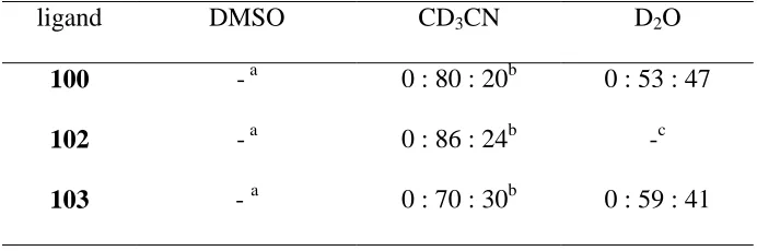
Table 2.3 Equilibrium distribution for 1:1 mixtures of Zn2+ and the ‘third generation’ ligands ... 64
Table 3.1 The yield of the cyclization reaction at different concentration of 119... 77
Table 3.2 Associate constants for macrocycle 105 with anions in CDCl3 ... 84
Table 3.3 Associate constants for macrocycle 105 with ammonium salts in CDCl3 ... 87
Table 4.1 Associate constants for macrocycles 124 and 125 with nitrate salt of amino acid methyl esters ... 95
Table 4.2 Associate constants for macrocycles 124 and 125 with nitrate salt of methionine and threinine methyl esters ... 97
vii
LIST OF FIGURES
Figure 1.1 Common structure feature of zinc enzymes ... 4
Figure 1.2 Cartoon of the active site of CAII... 6
Figure 1.3 The mechanism of hydration of CO2 by CAII ... 7
Figure 1.4 Facial binding of a tripodal ligand ... 8
Figure 1.5 Structures of common tri-podal ligands... 9
Figure 1.6 Reaction between the zinc hydroxide complex of 5 (R, R’ = t-Butyl) with CO2 ... 10
Figure 1.7 Synthesis of the novel biomimetic calix[6]arene-based zinc complexes... 11
Figure 1.8 Formation of [ML2]2+ complex of tris(pyridyl)methanol ligand ... 12
Figure 1.9 Catalytic mechanism of complex 13... 14
Figure 1.10 Synthesis of bis[2-(O-hydrxyphenoxy)ether] ether 14 and dibenzo 18-crown-6 15 ... 15
Figure 1.11 Binding of K+ by dibenzo-18-crown-6 15... 16
Figure 1.12 Templated synthesis of crown ether 16... 16
Figure 1.13 Metal complexation of N-substituted crown ethers ... 18
Figure 1.14 Complexation of Cl- by protonated forms of ligands 37 and 38... 25
Figure 1.15 The synthesis of bipyrrole based macrocycles 46, 47 ... 30
Figure 1.16 Simultaneous complexation of NaCl by calix[4]arene 60... 37
Figure 1.17 Simultaneous complexation of NaCl by ligand 62... 38
Figure 1.18 Simultaneous complexation of NaCl by ligand 63. ... 39
Figure 1.19 Complexation of p-nitrobenzoate with ligand 64... 39
Figure 1.20 Complexation of phenylalanine with ligand 65... 40
viii
Figure 2.1 Deprotonation of bound water ... 53
Figure 2.2 Formation of [ZnL2]2+ sandwich complexes ... 54
Figure 2.3 Formation of tetrahedral zinc complex with tris(pyrazolyl)borate 5 ... 55
Figure 2.4 1H NMR of a 1:1 mixture (1 mM) of ligand 84 and zinc perchlorate in DMSO ... 58
Figure 2.5 1H NMR COSY spectrum of a 1:1 mixture (1 mM) of ligand 84 and zinc perchlorate in DMSO. ... 59
Figure 2.6 1H NMR EXSY spectrum of a 1:1 mixture (1 mM) of ligand 84 and zinc perchlorate in DMSO ... 60
Figure 2.7 Schematic represent of two major causes of weak binding ... 60
Figure 2.8 Formation of oxygen-bridged zinc complex ... 63
Figure 2.9 ESmass spectrum of a 1:1 mixture (1 mM) of ligand 91 and zinc perchlorate in H2O... 63
Figure 2.10 Change of the percentages of [ZnL]2+ in aqueous solution as a function of time ... 66
Figure 3.1 Schematic represent of counteranion affect on cation binding ... 71
Figure 3.2 The structures of macrocycle 105 and the guest molecule ... 73
Figure 3.3 The structures of coupling reagents ... 78
Figure 3.4 Noesy signals of receptor 105... 81
Figure 3.5 Proposed binding model of receptor 105 with anions ... 82
Figure 3.6 Binding isotherm for the complexation of macrocycle 105 and TBA-Cl... 83
Figure 3.7 The job’s plot of macrocycle 105 with phenylalanine HBr salt ... 83
Figure 3.8 The structure of the guest molecule 123... 85
Figure 3.9 Binding isotherm for the complexation of macrocycle 105 and phenylalanine HNO3 salt ... 86
Figure 3.10 The Job’s plot of macrocycle 105 with phenylalanine HBr salt ... 86
ix
Figure 4.2 The structures of the guest molecule 133... 93
Figure 4.3 Binding isotherm for the complexation of macrocycle 124 and S-phenylalanine HNO3 salt... 94
Figure 6.1 1H NMR of a 1:1 mixture of ligand 91 and zinc perchlorate in D2O ... 126
Figure 6.2 Change of the percentages of [ZnL]2+ and [ZnL2]2+ of ligand 91 as a function of time ... 127
Figure 6.3 Change of the percentages of [ZnL]2+ and [ZnL2]2+ of ligand 100 as a function of time ... 128
Figure 6.4 Change of the percentages of [ZnL]2+ and [ZnL2]2+ of ligand 103 as a function of time ... 128
Figure 6.5 Change of the percentages of [ZnL]2+ and [ZnL2]2+ of ligand 104 as a function of time ... 129
Figure 6.6 The kinetics of p-nitrophenyl acetate hydrolysis in the presence of 1 mM zinc complex of ligand 100 at 298 K and pH = 8.4 ... 131
Figure 6.7 The kinetics of p-nitrophenyl acetate hydrolysis in the presence of 2 mM zinc complex of ligand 100 at 298 K and pH = 8.4 ... 132
Figure 6.8 The kinetics of p-nitrophenyl acetate hydrolysis in absence complexes at 298 K and pH = 8.4 ... 132
Figure 6.9 The structure of the macrocycle 105... 133
Figure 6.10 1H NMR of macrocycle 105 in CDCl3... 133
Figure 6.11 1H NMR COSY spectrum of macrocycle 105 in CDCl3... 134
Figure 6.12 1H NMR of macrocycle 105 in DMSO ... 134
Figure 6.13 1H NMR NOESY spectrum of macrocycle 105 in DMSO... 135
Figure 6.14 NH stretching region of the FT-IR spectra of macrocycle 105 in CDCl3.. 136
Figure 6.15 Binding isotherm for the complexation of macrocycle 105 and TBA-Br 138 Figure 6.16 Binding isotherm for the complexation of macrocycle 105 and TBA-F .. 138
x
Figure 6.19 Binding isotherm for the complexation of macrocycle 105 and
TBA-TFA ... 140
Figure 6.20 Binding isotherm for the complexation of macrocycle 105 and TBA-TsO ... 140
Figure 6.21 Binding isotherm for the complexation of macrocycle 105 and phenylalanine HNO3 salt ... 141
Figure 6.22 Binding isotherm for the complexation of macrocycle 105 and phenylalanine HCl salt ... 141
Figure 6.23 Binding isotherm for the complexation of macrocycle 105 and phenylalanine TFA salt ... 142
Figure 6.24 Binding isotherm for the complexation of macrocycle 105 and phenylalanine HBr salt ... 142
Figure 6.25 Binding isotherm for the complexation of macrocycle 105 and phenylalanine TsOH salt ... 143
Figure 6.26 Binding isotherm for the complexation of macrocycle 105 and phenylalanine HI salt ... 143
Figure 6.27 The job’s plot of macrocycle 105 with phenylalanine HBr salt ... 144
Figure 6.28 The job’s plot of macrocycle 105 with TBA-B ... 144
Figure 6.29 The structure of macrocycle 123 and 124... 145
Figure 6.30 Binding isotherm for the complexation of macrocycle 124 and S-phenylglycine HNO3 salt ... 146
Figure 6.31 Binding isotherm for the complexation of macrocycle 124 and R-phenylglycine HNO3 salt ... 146
Figure 6.32 Binding isotherm for the complexation of macrocycle 124 and S-phenylalanine HNO3 salt ... 147
Figure 6.33 Binding isotherm for the complexation of macrocycle 124 and R-phenylalanine HNO3 salt ... 147
xi
Figure 6.36 Binding isotherm for the complexation of macrocycle 124 and S-alanine HNO3 salt ... 149
Figure 6.37 Binding isotherm for the complexation of macrocycle 124 and R-alanine HNO3 salt ... 149
Figure 6.38 Binding isotherm for the complexation of macrocycle 125 and S-phenylalanine HNO3 salt ... 150
Figure 6.39 Binding isotherm for the complexation of macrocycle 125 and R-phenylalanine HNO3 salt ... 150
Figure 6.40 Binding isotherm for the complexation of macrocycle 125 and S-phenylglycine HNO3 salt ... 151
Figure 6.41 Binding isotherm for the complexation of macrocycle 125 and R-phenylglycine HNO3 salt ... 151
Figure 6.42 Binding isotherm for the complexation of macrocycle 125 and S-alanine HNO3 salt ... 152
Figure 6.43 Binding isotherm for the complexation of macrocycle 125 and R-alanine HNO3 salt ... 152
Figure 6.44 Binding isotherm for the complexation of macrocycle 125 and S-valine HNO3 salt ... 153
Figure 6.45 Binding isotherm for the complexation of macrocycle 125 and R-valine HNO3 salt ... 153
Figure 6.46 Binding isotherm for the complexation of macrocycle 124 and S-methionine HNO3 salt ... 154
Figure 6.47 Binding isotherm for the complexation of macrocycle 124 and R-methionine HNO3 salt ... 154
Figure 6.48 Binding isotherm for the complexation of macrocycle 124 and S-threonine HNO3 salt ... 155
Figure 6.49 Binding isotherm for the complexation of macrocycle 124 and R-threonine HNO3 salt ... 155
xii
Figure 6.52 Binding isotherm for the complexation of macrocycle 125 and S-threonine HNO3 salt ... 157
xiii
LIST OF SCHEMES
Scheme 2.1 Synthesis of Tris-(2-nicotinic acid)methanol methyl ether 85 ... 56
Scheme 2.2 The synthesis of ligands 86-88... 57
Scheme 2.3 The synthesis of ligands 91-93... 61
Scheme 2.4 The synthesis of ligands 100, 102 and 103... 65
Scheme 2.5 The synthesis of ligand 104... 67
Scheme 2.6 Catalytic hydrolysis of esters by CA... 68
Scheme 3.1 The synthesis of macrocycle 105... 75
Scheme 3.2 The synthesis of macrocycle 105 (continued)... 76
Scheme 3.3 Side reaction from synthesizing 110... 76
Scheme 3.4 Formation of guaridinated by-products... 78
Scheme 3.5 Optimized synthesis of diacid 112... 80
xiv
ABBREVIATIONS
BOP Benzotriazole-1-yl-oxy-tris-(dimethylamino)-phosphoniumhexa-fluorophosphate
n-BuLi n-Butyllithium
DMF N,N’-Dimethylformamide
DMSO Dimethylsulfoxide
DPPA Diphenylphosphoryl azide
HOBt N-hydroxylbenzotriazole
HBTU 2-(1H-benzotriazole-1-yl)-1, 1, 3, 3-tetramethyluronium hexaflorophosphate
MOE Ethoxymethyl
MOM Methoxymethyl
PyBOP Benzotriazole-1-yl-oxy-tris-pyrrolidino-phosphonium hexafluorophosphate
rt Room temperature
THF Tetrahydrofuran
TLC Thin layer chromatography
xv
ABSTRACT
In an effort to produce the tetrahedrally coordinated, catalytically active zinc center,
three families of tris(2-pyridyl)methanol derivatives were synthesized and characterized.
Zinc binding studies revealed that the binding behaviors of the ligands depended on the
steric and electronic properties of the substituents on the pyridyl rings, as well as the
functional group on the tertiary alcohol.
A novel tris-pyridyl macrocyclic receptor was synthesized. The receptor possesses
both hydrogen bond donors and acceptors. NMR titration experiments revealed that the
receptor simultaneously bound both ammonium cation and the counter anion. The
counter anion significantly influences the association between the receptor and the
ammonium cation.
Chiral ditopic macrocycles, which enantioselectively bind chiral ammonium cations,
have also been synthesized. Their enantioselective binding properties, as well as the
ditopic recognition properties were investigated.
I.
Introduction
1.1 Supramolecular Chemistry
Chemistry can be divided into two broad areas: molecular and supramolecular.1 Traditional chemistry, or molecular chemistry, deals mainly with the synthesis of
complex molecules. Its power arises through the manipulation of the covalent bonds
between atoms. However, most biological processes do not involve the making or
breaking of covalent bonds, but rather non-covalent, intermolecular interactions. These
non-covalent, intermolecular interactions are the basis of supramolecular chemistry.
As defined by Jean-Marie Lehn, supramolecular chemistry is “chemistry beyond the
molecule”.2 The field of supramolecular chemistry started with the selective binding of alkali metals by artificial receptors: crown ethers and cryptands, and then moved to
consider intermolecular interactions between neutral species and anions. In the past
several decades, supramolecular chemistry has undergone enormous development. It is
one of the most popular and fastest growing areas of chemistry now. This highly
interdisciplinary field of science has brought about wide-ranging collaborations between
1.2 Molecular Recognition and Host-Guest Chemistry
Molecular recognition is defined by the energy and the information involved in the
binding and selection of substrate by a given receptor or host molecule.3 The term “host” describes the ability of a molecule to bind another one with preference over all others,
and with greater strength than is commonly found in unspecific molecular interactions.
The chemical nature of the “guest” to be specifically bound mutually complements the
host. These include geometry and electronics.4 In general, molecular recognition relies on the factor that there is a molecular interaction between host and guest, leading to an
assembly of two or more species into a well-defined structure. These non-covalent
interactions could be: ion-ion, ion-dipole, dipole-dipole, hydrogen bonding, cation-π, π-π
stacking, van der waals forces, close packing in the solid-state, the hydrophobic effect or
the combination of those interactions.
Generally, molecular recognition is restricted to certain conditions. Changing one or
more condition may lead to no binding or reduced selectivity. For example, a compound
qualifying as a host for one particular guest species in one solvent may completely fail to
bind the same guest under different solvent conditions.
The design of a synthetic host requires a careful consideration of the nature of the
guest. This leads to conclusions about the general properties of the new host system.
There must be a good match or complementary between host and guest: Lewis acids must
match Lewis bases; hydrogen bond donors must match acceptors. Another very
important concept in host design is pre-organization. A pre-organized ligand means there
is less unfavorable conformational rearrangement to take place in order to adopt the
rigid host design, it is widely accepted that a rigid host having all of its anchor groups
preorganized complementary to the respective guest function should show strong
binding.
Nature’s enzymes are complex host-guest systems that bring about reactions
essential to life. Enzymes recognize and respond to specific substrates, as part of the
control mechanism of the cell.
1.3 Zinc Enzymes
Zinc is known to be an essential trace element. There are 2 to 3 g of zinc in adult
humans, making it one of the most important trace elements.5 However, no specific biological role for zinc was established until 1940, when it was shown to be required for
the catalytic activity of the enzyme Carbonic Anhydrase (CA). In the following five
decades, more than 300 enzymes have been found to contain zinc. The influence of zinc
derives from its roles in enzymes, with functions that are both structural and catalytic. In
a few enzymes, zinc plays a purely structural role, in which zinc stabilizes the enzyme
folding. However, in most of the enzymes, zinc is directly involved in catalysis,
interacting with substrate molecules undergoing transformation.6
The active sites of catalytic zinc enzymes feature a tetrahedrally coordinated zinc
center that is attached to the protein backbone by three amino acid resides, with the fourth
being occupied by a water molecule.7 The water molecule is activated by zinc through ionization and polarization and is a critical component of a catalytic reaction.
Deprotonation of the bound water molecule occurs at physiological condition and the
X, Y, Z N, O, S donors of His, Asp, Glu and Cis residues
Zn2+
Z OH2
X Y
Zn2+
Z OH
X Y
-
H++H+
-Nucleophile
Figure 1.1 Common structure feature of zinc enzymes
The specific function performed by each of these enzymes is dictated by the nature
of the residues with which zinc is bound to the protein. The residues that zinc bind to are
typically a combination of histidine, glutamic acid, aspartic acid and cysteine, which
provide nitrogen, oxygen, and sulfur donors. Histidine is the most commonly
encountered. The most important function of the zinc enzymes is the cleavage of amide
bonds and hydrolysis of phosphates with high regio- and enantio-selectivities. The
importance of zinc enzymes is not, however, restricted to their role in cleaving amide and
phosphate bonds. Another important family of zinc enzyme is Carbonic Anhydrase
1.4 Carbonic Anhydrase (CA)
Of all the zinc enzymes, Carbonic Anhydrase is probably the most extensively
studied.9 It is the first enzyme recognized to contain zinc and has played the most pivotal role in the development of enzymology. The essential physiological function of Carbonic
Anhydrase is to catalyze the reversible hydration of CO2 and thus plays an important role
in respiration and intracellular CO2/HCO3- equilibration.10 In addition to its physiological
function, Carbonic Anhydrase also catalyzes non-physiological reactions such as
hydration of aldehydes and hydrolysis of esters.11-14
The three-dimensional structure of Carbonic Anhydrase II (CAII), an isozyme found
in human red blood cells,15 revealed the active site as shown in Figure 1.2. Zinc is located at the bottom of the cavity (ca. 15 Å deep) and is coordinated to three histidine
residues (His 94, His 96, and His 119), with the remaining tetrahedral site being occupied
by a water molecule. The water molecule is also involved in a hydrogen-bonding
interaction with a threonine residue. The most striking feature of this active site is that
the pKa of this bound water molecule is 7.3 which is much more acidic than free water
molecule (pKa = 15.6).15,16 At physiological pH, the water molecule is deprotonated and
nucleophilically attacks electron deficient carbon atoms, such as those in carbon dioxide
and carboxylate esters, resulting in the hydration or cleavage of the respective
Zn2+ N
N H
N N
N N
H O
H
NH2
O
O O
O H
O O
O H
-Thr-199 Glu-106
-Glu-117
His-119
Gln-92
Hi
s
-94
His-96 NH
Figure 1.2 Cartoon of the active site of Carbonic Anhydrase II.
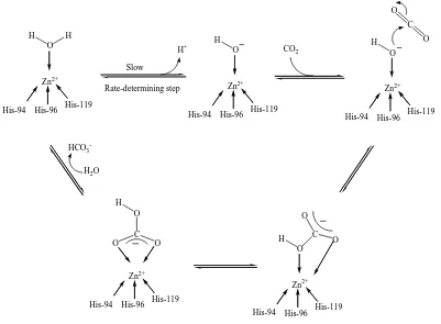
Figure 1.3 illustrates the currently accepted mechanism of hydration of CO2 by
Carbonic Anhydrase II.10 The mechanism comprises the following steps: (i) deprotonation of the zinc bound water molecule (via a His-64 shuttle) to give the active
zinc hydroxide derivative [(His)3Zn-OH]+, (ii) nucleophilic attack of the zinc-bound
hydroxide at the carbon dioxide substrate to give a hydrogen carbonate intermediate
[(His)3Zn-OCO2H]+, and (iii) displacement of the bicarbonate anion by H2O to complete
O C O O H His-119 His-96 His-94 C O O HCO3
-H2O
O H His-119 His-96 His-94 O H His-119 His-96 His-94 CO2 H+ Zn2+ Zn2+ Zn2+ O H His-119 His-96 His-94 H Zn2+ Slow Rate-determining step His-119 His-96 His-94 O O C O H Zn2+
Figure 1.3 The mechanism of hydration of CO2 by CAII
1.5 Carbonic Anhydrase Mimics
As the native enzyme contains a zinc ion in its active site, zinc complexes were
extensively studied in order to detect model compounds that would mimic the function of
enzyme. One of the first models was reported in 1975 by Wooley.20,21 Although this model was intriguing and very convincing about the essential role of zinc ions, the “fatal”
drawbacks of 1 are that zinc is tetra-coordinated, and relatively high pKa value (8.7) of
the bound water molecule (against 7.3 in Carbonic Anhydrase). Consequently, complex
N N
N
N Zn2+
OH2
1
Thus, it appeared of considerable interest to design tridental ligands which
incorporate the requisite three donor groups to mimic the three protein residues that bind
zinc at the catalytic center. The fact that tridental ligands enforce facial binding with
single relevant binding conformation provides them the ideal environment as Carbonic
Anhydrase mimics (Figure 1.4). The possibility of incorporation substituents that
directly influence the steric environment about the metal center makes them even more
attractive.
X
Z M Y
Figure 1.4 Facial binding of a tripodal ligand.
Figure 1.5 shows the common tripodal ligands 2-5 which were synthesized and studied as synthetic analogues of Carbonic Anhydrase. However, none of them possess
Carbonic Anhydrase -like properties. Without bulky subsitituents on the heteratomatic
rings, these ligands tend to form inactive 2:1 sandwich complexes even if an excess of
NH N
OH N NH HN
N
N N N
OMe
N
OH N HN HN N
N
H B
H
N N
N N
N N
2 3
4 5
Figure 1.5 Structures of common tri-podal ligands
Structural modifications of these tripodal ligands were extensively studied to inhibit
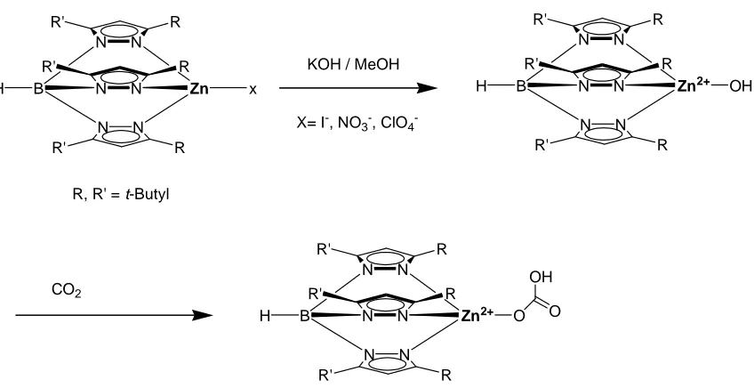
the formation of 2:1 complexes.27-31 A successful synthetic analogue of Carbonic Anhydrase was obtained by Gerard Parkin through modifying the structure of
tris(pyrazolyl)borate 5. Thus, with bulky tert-butyl substituents on the 3-position of the pyrazolyl groups, the ligand has the tendency to favor a tetrahedral coordination (Figure
1.6).32 A tetrahedral zinc hydroxide complex of tris(pyrazolyl)borate was also isolated by reaction of monomeric zinc complex with KOH in methanol. This active species which
can hydrate CO2 to bicarbonate bound to zinc. However, the zinc hydroxide species
N N
N N
N N
B Zn x
H R R' R R' R R' N N N N N N
B Zn2+ OH
H R R' R R' R R'
X= I-, NO3-, ClO4 -KOH / MeOH
CO2
N N
N N
N N
B Zn2+ O
H R R' R R' R R' O OH R, R' = t-Butyl
Figure 1.6 Reaction between the zinc hydroxide complex of 5 (R, R’ = t-Butyl) with CO2.
A benzene platform with three histidine side chains 6 has been synthesized in Vahrenkamp’s group.33 The results from potentiometric titration demonstrated the formation of tetrahedrally coordinated zinc complex [L-Zn-OH2]2+, with the pKa of the
bound water molecule 6.2.
By incorporating three imidazole groups on the lower rim of calix[6]arene, Reinaud
and co-workers generated a novel supramolecular system as Carbonic Anhydrase
mimics.34,35 Upon reaction with zinc ion, an air-stable dicationic zinc-aqua complex [Zn(X6Me3Imme3)(H2O)](ClO4)2 was obtained (Figure 1.7). The highly acidic Zn2+
center was constrained in a tetrahedral environment with a labile site oriented toward the
inside of the calix[6]arene structure. The hydrophobic pocket acted as a selective
molecular funnel for neutral molecules. 1H NMR spectroscopy studies showed the easy exchange of the bound water for amines, alcohols, amides, or nitriles.
O OCH3
N N
3
Zn OH2
N
N
N N N N Zn(ClO4)2
7
Figure 1.7 Synthesis of the novel biomimetic calix[6]arene-based zinc complexes
In an approach to mimic Carbonic Anhydrase in both tetrahedral zinc coordination
geometry and a hydrophobic cavity around metal center, Walton and co-workers reported
the synthesis and metal binding studies of a family of
1,3,5-tris(acrylideneamino)cyclohexane-based ligands 8-9.36,37 Upon addition of zinc ion, the ligand adopted a face-capping imine-N3 coordination geometry with the metal cation
N N
N
ZnSO4
R
R R
8 R =
9 R =
O
N N Zn N
OH2
R R
2+
R
Another easily synthesized family of tripodal ligands that may act as Carbonic
Anhydrase mimics are the tris(2-pyridyl)methanols e.g. 10. Previous investigations of zinc complexation reveal a strong tendency to form inactive [ZnL2]2+ sandwich-type
complex instead of a tetrahedral coordinated species (Figure 1.8).38,39
N N N
OH
N N
N
OH N
N N
OH
Zn2+
Zn2+
N N N
OH Zn2+
OH2
Zn2+
10
Figure 1.8 Formation of [ML2]2+ complex of tris(pyridyl)methanol ligand
One approach to modify and control the metal binding behavior of the
tris(2-pyridyl)methanol ligand is to generate a cavity around the metal complex. Thus, by
tailoring a hydrophobic dendimeric cleft around tris-(2-pyridyl) methanol ligands, it is
N N N
O
O O O
O O O O O O O O O O O O O O O O O O O O
O O O
O O O O O O O O O O O O O O O O O O O O O O O O O O O O O O O O O O O O O O O O O O O O O O O O O O O O Zn X 11
A recently reported zinc complex of the macrocyclic triamine ([12]ane N3) is one of
the simplest models for the Carbonic Anhydrase active site.41,42 The zinc ion is coordinated to the three nitrogen atoms of the macrocyclic ligand, and a water molecule,
as shown by X-ray crystallographic and NMR studies. The complex possesses a pKa
value of 7.3, very similar to CA.43
N N N H H H Zn2+ OH2 12
4-nitro-phenylacetate at pH 8.5 reveal a second order reaction catalyzed by zinc complex,
and the rate constant was determined to be 4.1 × 10⁻² M-1⋅s-1, compared to 4 × 10² M-1⋅s-1 of Carbonic Anhydrase II.
An alcohol-pendant cyclen 13 was synthesized later by the same group.45 Its zinc complex has the pKa value similar to complex 12. However, complex 13a is a more
efficient catalyst for the hydrolysis of esters. The enhancement of the catalytic activity is
due to the assistant from the tethered alcohol during the reaction. Detailed mechanical
studies have shown that the tethered alcoholic OH of 13a deprotonated and then directly attacked the electrophilic ester carboxyl group of 4-nitrophenyl acetate, to yield an
acyl-intermediate 13b. It was then quickly hydrolyzed, to complete the hydrolysis and regenerate the initial alkoxide complex 13a (Figure 1.9). The second rate constant was determined to be 0.14 M-1⋅s-1 at pH 9.3 and 25 °C, which was four times faster than the corresponding value of 12.
N N
N H
H Zn2+
O
N N
N H H
OH
Zn2+
O
NO2
O
N N
N H
H Zn2+
OH O O
O NO2
-CH3COOH
13 13a 13b
1.6 Cation Binding Receptors
1.6.1 Crown Ethers
The first crown-ether dibenzo-18-crown-6 15, was synthesized by Nobel Prize winner Charles Petersen in 1967 (Figure 1.10).46 The product was formed as an unexpected by-product during a preparation of bis[2-(O-hydroxyphenoxy)ether] ether 14. Small amount of catechol as impunities in the starting material led to the discovery of a new family of
compounds – the crown ethers.
OH
O
Cl Cl
OH
OH
+
OH
O O
O HO
O
O O
O O O
Cl Cl
+
NaOH / BuOH
NaOH / BuOH
14
15
Figure 1.10 Synthesis of bis[2-(O-hydrxyphenoxy)ether] ether 14 and dibenzo 18-crown-6 15
Its complexation properties with metal ions were quickly discovered (Figure 1.11).
This initial result rapidly led to the synthesis of other crown ethers with different size or
different donor atoms. Methods of evaluating their metal complexation properties were
O O O O O O O O O O O O K+ K+ 15
Figure 1.11 Binding of K+ by dibenzo-18-crown-6 15
The synthesis of crown ethers generally utilizes Williamson ether reaction.47,48 The use of alkali and alkali earth cations as template greatly facilitates their formation. The
template effect arises from complexation of the crown ether’s precursor around the metal
ion. The efficiency of metal ions is closely related to the strength of interaction between
metal ions and the product. Mandolini and co-workers have investigated the effect of
metal ions on the synthesis of benzo-18-crown-6 16 in methanol and dimethylsulfoxide. It was found that K+ was 1500 times more efficient than Na+ as a template because the potassium ion pre-organizes the linear backbone in a position favorable for the
nucleophilic attack to occur (Figure 1.12).
H O O O O O O O O O O O O Br K MeO O O O O O O Br K K 16
Figure 1.12 Templated synthesis of crown ether 16
The ability of selective cation binding is the most remarkable property of crown
display differences in their complexation abilities. Izatt and co-workers have made a
detailed study of the effect of the crown size on the binding stabilities with a variety of
univalent and bivalent cations.53 Comparison of these binding stabilities revealed that log
K values for complexes with 15-crown-5 17 were much lower that those with the ligand 18-crown-6 18 for all cations studied, except for those of small cations, such as Na+, Li+, and NH4+. The maximum stability for complexes with 18 occurs when the dimension of
the metal ion best matches the ligand cavity.
O O
O O
O O
O O
O O
O
17 18
On the other hand, the larger crown ethers dicyclohexyl-21-crown-7 19 and dicyclohexyl-24-crown-8 20 bind Cs+ more strongly than those of smaller macrocycles and are generally selective for Cs+ over all other cations.54-56
O
O O
O O O O
O
O O
O O O O
O
19 20
O O
O
O O
O
21
Solvents also have a significant effect on selective binding of cations. Agostiano
and coworkers have noted that the complexation ability of dicyclohexyl-18-crown-6 21
reversed in alcoholic solvents.47 The complexation constants of 21 in protic solvents increase regularly for all alkali cations in the order: water < methanol < ethanol <
1-propanol.
The replacement of one or more oxygen atoms in crown ether by other donors such as
nitrogen and sulfur dramatically changes the complexation properties of the ligands.47 Frensdorf has studied the effect of nitrogen substituents on the complexation stabilities of
18 (Figure 1.13). Substitution of one oxygen by nitrogen results in macrocycle 22 which has less affinity for K+. Dinitrogen substituted macrocycle 23 binds K+ even weaker. However, replacing oxygen with nitrogen atoms results in increased binding constants for
Ag+. Similar results were obtained with crown ethers containing sulfur donor atom.
O O
O
O O
O
O N H
O
O O
O
O N H
O
O O
H N
18 22 23
log K for K+: 6.1 3.9 2.0 log K for Ag+: 1.6 3.3 7.8
1.6.2 Cryptands
Cryptands are macrobicycles capable of ion encapsulation due to their cage-like
three dimensional structures. Their macrobicyclic cavities are more rigid and restricted
than crown ethers, and thus, have higher selectivity for cations. Generally, the metal ion
whose ionic radius best matches the size of the cryptand cavity will form the most stable
complex.
Because of their rigidity, cryptands are pre-organized for cation binding.
Consequently, cryptand binds cation much more strongly than corresponding crown
ethers.58-61 For example, ligand 23 and 24 have the same number of the donor atoms, while ligand 24 binds K+ 103 times stronger than ligand 23 (log K = 2.0 and 5.4 for ligand
23 and 24 respectively).58
O O N O N
O
24
Micheloni and co-workers have synthezied a variety of new small aza cages and have
studied their ligation properties.62-64 Small aza cages are highly preorganized molecules that possess three dimensional cavities of fixed sizes. These preorganized small cavities
allow selective encapsulation of metal ions of appropriate sizes. Especially strong and
N N N
N CH3
H3C HN
N N N N H3C HN
25 26
Bradshaw and co-workers have synthesized a series of new cryptands.65,66 Binding studies with a variety of cations demonstrated that these ligands had high selectivity of
K+ over Na+. The highest selectivity was observed on ligand 27 which had selectivity factor of 6.17.
O O
N O O O N O
27
More recently, Bradshaw and co-workers reported novel benzene-bridged
O
O O
O
O O
O O
O
O O
O
O
O O
O
O O
O O
O
28 29
1.6.3 Calixarenes
Calixarenes are cavity-shaped cyclic oligomers made up of phenol units. Their
easily synthized and versatile host frameworks make them very attractive in
supramolecular chemistry.69,70 Calixarenes offer many interesting possibilities in host-guest chemistry, particularly in ion complexation. The upper and lower rims of
calixarenes can be easily functionalized to be the host of a variety of guest molecules. As
hosts for cations, the phenolic oxygen atoms at the lower rim of calixarenes can complex
to cations, either in hydroxyl form, or as alkyl ether derivatives.
The selectivity of calixarenes toward cations is mainly a function of the cavity
dimensions and the nature of the binding groups. Arnaud-Neu and co-workers have
studied cation complexing properties of lower rim modified calix[4]arenes.71 Their binding studies with alkali and alkali earth metal ions demonstrated that the acid
derivative 32 was a much stronger binder than the ester 31 or original hydroxyl derivative
30. The stepwise substitution of the phenonlic hydrogen in 30 or ester function in 31 by a carboxyl acid led to an enhancement of the stability of the resulting complex, but a
R R
R
R 30 R = OH
31 R = OCH2CO2CH2CH3
32 R = OCH2CO2H
The cavity size of calixarenes has a significant effect on the selectivity of metal ions.
In general, calix[4]arenes show a high selectivity for Na+ over the other alkali metal cations. Barrett and co-workers extensively studied the effect of cavity size on the cation
selectivity.72
R R
R R
n 33 n = 1 R = OCH2CO2CH2CH3
34 n = 2 R = OCH2CO2CH2CH3
Table 1.1 Binding constants (M-1) of ligands 33, 34 with alkali cations
Ligands Li+ Na+ K+ Rb+ Cs+
33 1.0 4.4 5.3 5.6 5.5
34 2.6 5.0 2.4 3.1 2.7
Further modification of calixarenes resulted in diooxocalix[4]arene 3573 and homocalixarene 3674, both of which exhibit selectivity for larger cations over small ones. For example, ligand 35 has log K value of 2.70 and 0 for Sr2+ and Ca2+ respectively. Ligand 36 has the similar selectivity of Sr2+ over Ca2+ (log K = 1.22 and 0 respectively).
O
O O
O
O O O
O O
O O
O
OH
OH
OH HO HO
1.7 Anion Receptors
Synthetic anion receptors were developed much later then cation receptors. 75-79 The most prominent property of anions distinguishing them from any other guest species is
their negative charges. Correspondingly, anions have a relative large radius and high free
energies of salvation. Furthermore, anions occur in a range of shapes and geometries.
For example, halides are spherical, NO3- is planar, SCN- and N3- are linear.1 The intrinsic
properties of anions make it difficulty in designing multidentate receptors with
appropriately situated Lewis acidic or other acceptor sites. However, due to its important
biological role, considerable attention has been focused on supramolecular anion
complexation in recent years.
1.7.1 Positively Charged Anion Receptors
The first synthetic anion receptor was reported by C. H. Park and H. E. Simmonds in
1968.80,81 They found that the macrocycles 37 and 38, when protonated at the bridgehead nitrogen atom, were able to bind halide anions within the cavity (Figure 1.14). The
penetration of the halides anions into the macrocyclic structure was later confirmed by
X-ray crystallography. This sharply observed result of the noncovalent encapsulation of
anion opened the door of anion receptors and had a lasting effect on future development
N N
(CH2)n (CH2)n
(CH2)n
(CH2)n (CH2)n
(CH2)n Cl -HCl
N H H N
37 n = 9
38 n = 10
Figure 1.14 Complexation of Cl- by protonated forms of ligands 37 and 38
Due to the easy synthesis of azamacrocycles that were widely used as cation
complexing agents, positively charged azamacrocycles as anion receptors were quickly
developed.82-84 Host-guest binding in these molecules depends on the relative proton affinities in interconnected multiple equilibria. In water as a solvent, protonation
equilibria are readily established and the corresponding pKa values of the individual
proton can be determined by potentiometric titrations. It is no surprise, therefore, that
water is the solvent of choice to study anion binding of most of the protonated
azamacrocycles.
An aesthetically pleasing macrotricyclic ligand 39 was synthesized by Graf and Lehn.85,86 Compound 39 may be regarded as arising from four fused triaza 18 crown-6 rings. This spherical molecule is a highly versatile example for both cation and anion
receptor, depending on the pH of the medium. At neutral pH, compound 39 has basic properties that enable it to bind strongly to cations. Once the four nitrogen atoms are
protonated, it becomes an anion binder. The tetra-protonated species has a remarkable
O
N
O N O N
O
N O O
39
1.7.2 Neutral Amide Based Receptors
The hydrogen-bonding amide groups have been employed to produce a wide range
of receptors capable of binding anions. Choi and Hamilton have recently reported the
synthesis and anion binding properties of a new family of cyclic triamides.87
N N
N
O O
O
H
H
H R
R R
40 R = CO2Et
41 R = NHBoc
The triamide macrocycles 40 and 41 have rigid structures in which three amide groups are projected into the central cavity. The convergent arrangement of dipoles
enables the macrocyle to bind anions with size and shape selectivity. NMR evidence
switching to a 1:1 binding mode at higher I- concentration. The peak of amide proton had an initial up-field shift followed by a down-field shift in the 1H NMR spectra.
The binding behaviors of the macrocycles were solvent-dependent. Increasing the
polarity of solvent by increasing the percentage of DMSO-d6 in CDCl3, diminished
binding. The effect was so great with halides that at 50% DMSO-d6 / CDCl3, the M2I
complex formation could not be observed.
A three-dimensional polyamide 42 has been reported by Brown-James and co-workers.88 Strong bindings of ligand 42 with anions were observed in CDCl3 (log K >
105). However, in more polar solvent DMSO-d6, 1H NMR titrations revealed a 1:1 binding model between 42 and anions and quantitative binding results were able to obtain. The highest binding constant was observed with anion F- (log K = 5.0), followed by Cl- (log K = 3.47), CH3COO- (log K = 3.38) and H2PO4- (log K = 3.30). The crystal
structure analysis of its F- complex demonstrated that F- was centered in the cryptand cavity with hydrogen bonds to all six amide protons.
N NH
N
NH N N
HN HN
N
O O
N
H N
O O
O O
H
42
sandwich type 2:1 complexes with many anions. According to crystal structure analysis,
the guest resides in a cavity formed by the aggregation of two
perfectly-shape-complementary molecules of 43. A quantitative determination of binding stabilities of anions revealed that complex formation was cooperative. K1, the stability constant of the
1:1 complex, was in every case significantly smaller than K2, the equilibrium constant
from the 1:1 to the 2:1 complex.
NH NH HN N N N O O N N O O O O N NH NH HN N N N O O N N O O O O N NH
(CH2)4
O NH NH HN N N N O O N N O O O O N O 43 44
Cl- > NO3-. A comparison of the complexation stabilities of 44 with these of cyclopeptide
30 that forms 1:1 anion complexes showed that the presence of the second binding site increased complexation stability by a factor of 100-350.
1.7.3 Neutral Pyrrole Based Receptors
The calixpyrrole (named because of its resemblance to calixarenes) was first
reported in the 1880’s. However, its anion binding properties were not studied until
1996. Although, calix[4]pyrrole 45 possesses only a very small cavity, it can form four hydrogen bonds with anions, and was found to be able to bind small anions, such as F -and Cl-.91
H N
HN N H NH
45
The compound is attractive because it is readily obtained in one step from the
condensation of pyrrole and formaldehyde. However, its relatively small cavity prevents
it from binding large anions effectively. To explore the area of pyrrole based anion
receptors, Sessler and co-workers reported the synthesis and anion binding properties of
N
H NH
NH HN
NH HN
n
46 n = 1
47 n = 2 N
H NH
+ O MsOH
Figure 1.15 The synthesis of bipyrrole based macrocycles 46, 47.
Macrocycles 46 and 47 were synthesized in one step by the condensation of bipyrrole with acetone in the presence of a catalytic amount of methanesulfonic acid.
Binding studies revealed that macrocycle 46 bound large halide anions in a 1:1 binding model, with affinities that were substantially enhanced compared to those of
calix[4]pyrrole 45.
Expanding pyrrolic anion receptors into the third dimension, Sessler and co-workers
have reported the synthesis and binding studies of a family of calix[4]pyrroles bearing a
single side diether straps 48-50.93 The straps were expected to provide additional hydrogen bonding sites and thus allow specific modulation of the inherent anion affinity.
The results of NMR titrations revealed enchanced affinity for Cl- and Br- relative to calix[4]pyrrole. The anion binding ability of strapped calix[4]pyrrole can be effectively
N
O O
n n
N N
N
H
H H
H
48 n = 1
49 n = 2
50 n = 3
1.7.4 Urea Based Receptors
Ureas are excellent hydrogen bond donors, and, thus, widely used in the design of
synthetic receptors for anions. Gunnlangsson has employed anthracene derivatives 51-53
with aromatic or aliphatic thiourea moieties as fluorescent photo-electron transfer
chemsensor for anions.94
A titration experiment using fluorescence spectroscopy demonstrated that the
fluorescence of 51 was quenched upon addition of AcO- in a solvent such as DMSO, CHCl3 or CH3CN. The highest degree of quenching was observed in DMSO. However,
no binding was observed in the highly competitive hydrogen bonding solvent EtOH.
Titration of 51 with common anions demonstrated that compound 51 selectively bound AcO-, F-, H2PO4-. Thus, the fluorescence of 51 was quenched by AcO-, F- and H2PO4-,
HN S
N H
R 51 R =
52 R =
53 R = CH3
CF3
Another family of chemsensors based on thioureas were reported by Hong and
co-workers.95 The UV-Vis absorption of 54 in chloroform undergoes a red shift upon addition of H2PO4- anion. Clear isosbestic were observed on UV-Vis spectra, which
demonstrated a 1:1 complexation. Compound 54, with both azophenyl and nitrophenyl chemophores, has high selectivity of H2PO4- over other common anions. The degree of a
red shift was determined to be H2PO4- >> AcO- ≅ F- > Br- ≅ Cl- > I-. In the case of
compound 55 with only an azophenyl group as the chromophore, the red shift upon complexation with H2PO4-, F- and AcO- were similar, and thus no discrimination of
anions was obtained.
N N NO2
HN HN NH
NH
O2N NO2
S S
N N NO2
HN HN NH
NH
S S
Hong and co-workers have continued to synthesize anion selective receptors.96 Macrocycles 56, 57 contain three thiourea units as linkers between aromatic groups. Both ligands displayed high affinities with anions. Macrocycle 57, with extra ethyl groups, is conformationally more rigid. The binding results demonstrated that 57 had higher affinities with anions then 56, with higher selectivity of AcO- (K = 5300 M-1) over H2PO4- (K = 1600 M-1).
HN HN NH
NH
S S
H N HN
S R
R
R R R
R R
R R
56 R = H
57 R = Et
1.8 Ditopic Receptors
The concept of ditopic receptor is relatively new in supramolecular chemistry. Those
receptors simultaneously bind cation and anion, and thus often exhibit cooperative and
allosteric effects whereby the association of one influence the binding affinity of the
counter ion.97-101 Such systems have potential as new selective extraction and transportation reagents for ion pair species of environmental importance and for
1.8.1 Simultaneous Complexation of Inorganic Ion Pairs
To date, the design of ion-pair receptors have been based on hydrogen bonding,
positively charged groups or Lewis acids to coordinate the anion, and crown ether or
modified calixarenes to bind the cation. One early example of ditopic receptors was
provided by Reetz and coworkers, who linked a Lewis acidic boron center to a crown
ether.102,103
Compound 58 is capable of coordinating K+ ⋅ F- simultaneously. The crystal structure of the KF complex showed that K+ was bound by the crown ether moiety, while the F- was held by a combination of orbital overlap with the Lewis acidic boron atom and an electrostatic interaction with K+.
O O O
O O O B
O O
58
Beer and coworkers have synthesized a series of calixarenes-based ditopic
receptors.104-106 Calix[4]arene 59, which was substituted at the lower rim by two benzo-15-crown-5, had very low affinity for anions alone. However, in the presence of K+ or NH4+ cations, a sandwich complex is formed by the two crown ether units. This resulted
of the cation, preorganized 59 to bind anions. Tetrahedrally shaped H2PO4- and HSO4
-were found to be particularly strongly bound.107
O
O HO
OH
HN O O NH
O O
O O
O
O O
O O O
59
Another calixerene-based ditopic receptor was synthesized by Reinhoudt and
co-workers.108 Receptor 60 consists of a calix[4]arene with cation-binding ester groups at the lower rim and anion-binding urea groups at the upper rim. In chloroform, compound
60 adopts a pinched conformation due to intramolecular hydrogen binding between two urea groups. However, when sodium ion are added, cation binding to the ester groups of
the calixarene alters the calix conformation, thereby breaks the hydrogen bond between
O
O O
N N
O
O O O O O O
O O N O N O H H H H H H O O O N N O
O O O O O O
O O N O H H H N O H H
H Cl
-NaCl
Na+
60
Figure 1.16 Simultaneous complexation of NaCl by calix[4]arene 60
In another approach to the problem of ditopic receptor preparation, Beer and
coworkers generated a di-calix[4]arene compound 61 connected by ethyl linkers, with two urea groups on one of the upper rims.109
M+ O O O O O O HO N N HO 61 O N Hex O N Hex H H H H X
Smith and coworkers have synthesized a pre-organised macrobicyclic receptor 62
with crown ether as metal binding site and bridging amide groups for anion bindings.110 Binding studies of compound 62 revealed a weak binding with halide ions in a solvent system DMSO/CD3CN (3:1). However, in the presence of one molar of Na+ or K+,
halide affinities were significantly increased. The crystal structure of NaCl complex
showed that in the solid state compound 62 bound NaCl as a solvent separated ion pair, with Na+ bound within the crown unit, while Cl- was hydrogen-bonded to the two amide residues (Figure 1.17).
Cl
-Na+ NaCl
NH HN
N N
O O
O O
O O O
O O O
NH HN
N N
O O
O O
O O O
O O O
62
Figure 1.17 Simultaneous complexation of NaCl by ligand 62
Cl
-K+ KCl
NH HN O O
N N
O O O O
NH HN O O
N N
O O O O
63
Figure 1.18 Simultaneous complexation of KCl by ligand 63
1.8.2 Simultaneous Complexation of Organic-pairs 1.8.2.1 Carboxyl Salts and Zwitterionic Amino Acids.
Mendoza and coworkers have synthesized a carboxylate receptor 64 containing a guanidinium group (Figure 1.19).112
N N
N
O O O
O H H
O O
NO2 N
N
N
O O O
O H H
p-nitrobenzoate
64
Figure 1.19 Complexation of p-nitrobenzoate with ligand 64
Receptor 64 shows a preference for extracting sodium p-nitrobenzoate and Boc protected amino acids with aromatic side chains from water into chloroform. The
gauanidium-carboxylate interaction is further enhanced by π-π stacking. However, free
structure modification of 64 was then undertaken by incorporating a crown ether unit as a binding site for ammonium cation.113 The results of extraction experiments revealed that receptor 65 had much higher affinities of amino acids. Thus, amino acids were extracted from aqueous solution into chloroform phase containing 1 equivalent of ligand 65, with strong preference for amino acid with aromatic side chain, such as phenylalanine and
tryptophan.
N N
N
O O O
O H H
O O
O O O
N
O O NH3 N
N
N
O O O
O H H
O O O
N
O O
phenylalanine
65
Figure 1.20 Complexation of phenylalanine with ligand 65
A three–point binding mode has been proposed (Figure 1.20), with carboxylate
bound by the guanidinium unit, ammonium cation by the crown, and π-π interaction
between the phenyl side chain of the amino acid and receptor’s naphthalene unit
enchancing the strength of complex formation.
More recently, Sessler’s group has synthesized two ditopic receptors based on the
structure of sapphyrin and porphyrin.114-116 The sapphyrin-lasalocid conjugate was found to be able to selectively transport aromatic amino acids. In direct competition
times faster than L-tyrosine. On the other hand, none of the amino acids under
investigation was transported efficiently by the porphyrin-lasalocid conjugate.
N
HN
HN NH
N O
HN NH
OH C2H5
CH3
H C2H5
H O
C2H5
CH3
OH H3C
HO
O
CH3
66
1.8.2.2 Tetraalkylammonium Cations
Among the various weak noncovalent forces that provide the basis for molecular
recognition, the cation-π interaction between quaternary ammonium cations and aromatic
hosts is receiving attention in recent years.117-122 The synthetic receptors for binding quaternary ammoniums studies as far are mostly cyclic molecules, such as cyclophanes,
crytophanes, calixarenes.
The binding between quaternary ammonium cations and host molecules generally
occurs in organic solvents of low polarity. The nature of the counter anion is expected to
influence host-guest association, either by cation-anion association or active participation
One approach to the design of receptors for quaternary ammonium cations employs
cyclic peptides.123 Stefan Kubik demonstrated that cyclopeptide 67 was able to bind ammonium cations by cation-π interactions. For the n-butyltrimethylammonium iodide
complex, an associate constant of 300 M-1 has been determined in chloroform. Besides cations, cyclopeptide also binds anions via peptide N-H hydrogen bonds. Anion
complexation results in an increase of the cation affinity by a factor of 103-104. Thus, cyclopepetide binds n-butyltrimethylammonium tosylate with Ka = 3.88 × 10⁶ M-1. The
positive coorperativity of counter anions could be correlated with the preorganization of
the cyclic-peptide by the anion as well as electrostatic interactions between anionic and
cationic substrates in the final complex.
NH NH
HN
N H
NH HN
O O
O O
O
O
R1 = R2 = R3 = CH2CH2COOCH(CH3)2
67
R1 R2
R3
Roelens and coworkers have extensively studied the binding properties of macrocycle
68 with tetramethyl ammomium.124 The results from 1H NMR titrations in CDCl3
revealed a dramatic variation of binding constants for the same complex with different
counter anions. The author hypothesized that the anion effect could be correlated to the
O
O O
O
O
O O
O
68
1.9 Enantioselective Receptors
The development of enantioselective receptors continues to be a challenging
endeavor for supramolecular chemists.125-127 As a special case of molecular recognition, enantioselective recognition involves discrimination between enantiomers of a guest by a
chiral receptor or a chiral matrix.
The formation of the stable complex between host and guest molecules is a primary
requirement for enantiomeric recognition. No recognition is observed if complexes are
not formed. Interaction between host and guest species results in a proper conformation
of the diastereomeric complexes, creating an appropriate environment for enantiomeric
recognition. In principal, enantiomeric recognition stems from the steric repulsion
between the substitutes at the chiral portions of the host and guest species. A reasonably
large steric repulsion results in good enantiomeric recognition.
To date, most of the enantiomeric recognitions are based on hydrogen bonding.128-133 The most studied host systems are the macrocylic crown ethers and cryptands. Other
Amino acids and their derivatives are the most studied guest molecules, due to their
significance in biology and readily availability of pure enantiomers.
1.9.1 Enantiomeric Recognition of Amino Acid Derivatives through Ammomium Cation Binding.
The first chiral macrocyclic compounds were reported by Cram and co-workers in
1973.144 Compound 69 has no specific chiral carbon atom. However, the relative orientation of the two binapthyl groups result in a twisted, chiral conformation. Ligand
69 was found to be able to recognize D-enantiomer assessed by two phase liquid-liquid extraction experiments. The chiral recognition factors range from 3 for
C6H5CH(CO2Me)NH3+, down to 1 for C6H5CH(CO2H)NH3+.
O O
O O
O O
69
Since then, many binapthyl containing macrocycles have been synthesized and their
chiral recognition properties studied.145-150 A dramatic increase in chiral recognition provided by attachment of two methyl groups onto the binapthyl group of compound 69
has been observed. Compound 70 has the chiral recognition factor of 31 for C6H5CH(CO2Me)NH3+, compare to a chiral recognition factor of 3 with host 69.146 The
groups with one enantiomer of ammonium cations in the less stable diastereomeric
complexes.
O O
O O
O O
70
A C3-symmetrical macrocycle 71 as selector of amino acid derivatives was recently
reported by Shinkai and co-workers.143 A 1:1 complexation mode between compound 71
and primary ammonium cations was determined by 1H NMR titration experiments. The association constants for the L-configured guest were greater than S-isomers. The largest
chiral discrimination (74% ee) was observed for the picrate salt of phenylalanine ethyl
esters. The results proved their molecular design concept that a C3-symmetrical skeleton
would be very effective for chiral recognition of optically active alkyl ammonium ions.
OH
O O
O
OH HO
71
By incorporating a terpyridine unit into a crown ether macrocycle, Kwong and
titration demonstrated that compound 72 bound primary ammonium salts in a 1:2 binding model, with stronger binding with S-enantiomers than R-enantiomers (KObs = 4.2 × 10¹⁰
and 1.2 ×10¹⁰ M-1 for the S and R phenyl glycine respectively).
N
N N
O O
O
O O
O
72
Although numerous synthetic receptors have developed for recognition of α-amino
acid derivatives, little progress has been made for the enantiomeric recognition of β
-amino acid.152 To this contribution, Ahn and coworkers reported a rational approach to the recognition of β-chiral primary ammonium ions through bifurcated hydrogen
bonding. The C3-symmertric tripodal oxazoline receptor 73 was found to be able to
enantioselectively bind ammonium salts of β-chiral amines (Figure 1.21). The chiral
recognition factors of a variety of guests studied ranged from a high of 5 down to a low
of 1. A hydrogen bond donor group in the β position of the guest molecule plays a
N O N O N O Ph Ph Ph NH3
H3C
OH H 73 O N N O N
O HHNH OH
H
Figure 1.21 Enantiomeric recognition of amino acid derivatives by receptor 73
1.9.2 Enantiomeric Recognition of Amino Acid Derivatives through Carboxylate Binding.
In general, the molecular recognition of caboxylate anions has been based on the
multi-hydrogen bond interactions between host and guest.153,154 One approach to design a receptor of carboxylates is by incorporating a guanidinium moiety. Guanidinium salts
remain protonated over a wide pH range (pKa = 13.5), and the binding of carboxylate
combines an electrostatic interaction with a bidentate hydrogen binding pattern (Figure
1.22). N N N R R R O O H H R R
Figure 1.22 Binding model between guanidinium salts and carboxylate
Davis and coworker have described enantioselective carboxylate receptors 74, 75


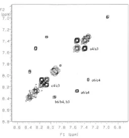
![Figure 2 in DMSO. The labeled cross peaks correspond to the exchange between free ligand (a), [ZnL].6 1H NMR EXSY spectrum of a 1:1 mixture (1 mM) of ligand 84 and zinc perchlorate 2+ (b) and [ZnL2]2+ (c)](https://thumb-us.123doks.com/thumbv2/123dok_us/8931616.1846754/75.612.194.424.74.317/figure-labeled-correspond-exchange-spectrum-mixture-ligand-perchlorate.webp)
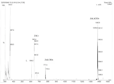
![Figure 2.10 Change of the percentages of [ZnL]2+ in aqueous solution as a function of time](https://thumb-us.123doks.com/thumbv2/123dok_us/8931616.1846754/82.612.176.437.349.560/figure-change-percentages-znl-aqueous-solution-function-time.webp)

