A multiparticulate system combining pH sensitive property and specific biodegradability has been investigated to prepare and evaluate ES-100 coated chitosan microspheres for colon targeting of ibuprofen. Chitosan microspheres were prepared by emulsion cross-linking method using different ratios of ibuprofen and chitosan (1:1, 1:1.5, 1:2 ), at emulsifier concentrations of 0.5%, 0.75%, 1% w/v and cross linking agent at concentrations of 8, 9, and 10 ml. Eudragit coating of ibuprofen chitosan microspheres was performed by coacervation phase separation technique with different core: coat ratio of 1:4 and 1:5. Chitosan microspheres and eudragit coated chitosan microspheres were evaluated for surface morphology, particle size measurement and size distribution, percentage drug entrapment and in vitro drug release in simulated gastrointestinal fluid. The size of the core microspheres ranged from 90 to 130 µm and that of coated microspheres ranged from 130 to 165 µm. The core microspheres sustained the release for 8 hrs in a pH progression medium mimicking the condition of gastro intestinal tract. The release studies of coated microspheres were performed in a similar dissolution medium as mentioned above. In acidic medium the release rate was much slower, however, the drug was released faster at pH 7.4 and the release was sustained up to 24 hrs. There was no significant change in the particulate shape and drug
o o o o
content after storage at 5 ± 2 C/Ambient, 25 ± 2 C/ 60% RH and 40 ± 2 C/ 75 % RH for a period of 6 months.
It is concluded from the present investigation that eudragit coated chitosan microspheres serve as promising controlled release carriers for colon-targeted delivery of ibuprofen.
Keywords: Ibuprofen multiparticulate system, Colon targeted drug delivery, Coacervation phase separation, Chitosan cross-linking, Eudragit–S-100 coating, pH sensitive drug delivery.
ABSTRACT
Submitted: 11/06/2012 Revised: 01/09/2012 Accepted: 15/12/20131,2,3,4 INTRODUCTION
Colon is being extensively investigated as a site for delivery of drugs. Colon is susceptible to many diseased states including constipation, irritable bowel diseases, intestinal amoebiasis, ulcerative colitis, crohn's disease, carcinomas etc.
At present these diseases are often poorly and inefficiently managed by oral drugs, which are largely absorbed before they reach the colon or by rectal administration which is less acceptable as a route of administration. Oral colon specific drug delivery systems (CDDS) have been developed by means of combination of one or more controlled release mechanisms. These targeted drug delivery systems hardly release drug in the upper part of the gastrointestinal (GI) tract,
5, 6
but rapidly release the drug in the colon . Ulcerative colitis and Crohn's disease are two distinct disorders classified under inflammatory bowel diseases. Inflammatory bowel diseases (IBD) can effectively be treated by the local delivery of drugs to the large intestine. The treatment of IBD with anti-inflammatory drugs is particularly improved by their local delivery to the bowel. Many anti-inflammatory drugs have life-threatening adverse effects on gastro-intestinal tract such
Development of Colon Targeted Multiparticulate Drug Delivery of Ibuprofen
Yashaswini V.K and Swamy N.G.N*
Department of pharmaceutics, Government College of pharmacy, Bangalore-560027. Karnataka, India.
*Address for Correspondence:
Dr. N.G.Nanjundaswamy, Department of pharmaceutics, Government college of pharmacy, Bangalore -560027, Karnataka, India
E-mail: ngnswami@yahoo.co.in
as haematemesis, peptic ulcer, severe gastric pain and vomiting. The conventional tablet dosage form of an anti-inflammatory drug provides minimal amount of the drug in the colon with undesirable side effects in the gastro intestinal tract. The treatment will be effective if the drug substances are targeted directly to the site of action in the colon. Better treatment may be possible with a lower dose of the drug. The latest approaches of drug delivery to the colon include pH dependent system, time dependent system, microbiologically controlled system, prodrug approach, polysaccharide based approach, multiparticulate system and luminal pressure
7
controlled system . Development of once a day sustained release multiparticulate formulation of an anti-inflammatory drug decreases the adverse effects, cost of treatment and enhances patient compliance. Natural polymers when used as coating agents for multiparticulate formulations, enhance the biocompatibility and biodegradability of the formulation. A large number of polysaccharides such as pectin, amylase, guar gum, chitosan, inulin, cyclodextrins, chondroitin sulphate, dextrans and locust bean gum have been investigated for their use in colon targeted drug delivery
8 9
multiparticulates of ibuprofen for better treatment of IBD with reduced adverse effects and better patient compliance.
EXPERIMENTAL
Materials
Ibuprofen was obtained as gift sample from Ce-Chem Ltd. Chitosan was obtained from chemika bio chemika reagents. Eudragit S-100 was obtained from Evonik Roehmpharma. Glutaraldehyde and span 80 – SD fine chemicals Ltd, liquid paraffin - CDH (P) Ltd, sodium hydroxide, disodium hydrogen phosphate and potassium dihydrogen phosphate-Karnataka fine chemicals, hydrochloric acid, n- hexane-Loba chemie, potassium chloride-Burgoyne Burbidges and Co. Ethyl acetate of Merck specialities Pvt Ltd were used in the study.
Method
Emulsion cross- linking method
Chitosan microspheres containing ibuprofen were prepared by Emulsion Cross-Linking Technique. Chitosan was used as the polymer and was cross linked using glutaraldehyde as
10
per the method described by Thanoo BC et al.
The chitosan solution was prepared in 5% aqueous acetic acid in which the drug was dispersed. The resultant mixture was extruded through a syringe (no. 20) into 100 ml of liquid paraffin (heavy and light, 1:1 ratio) containing 0.2%w/v of
11
dioctylsodium sulfosuccinate (DSS). The stirring was performed using a propeller at 1200 rpm. After 2min, 4ml of glutaraldehyde saturated toluene was added into the dispersion. Then, after 15min, additional 4ml of 25% aqueous glutaraldehyde was added drop by drop and stirring was continued for additional 3h. The microspheres thus obtained were filtered and washed several times with n-hexane to remove traces of oil. They were then washed with plenty of ice cold water to remove the acetic acid and glutaraldehyde. The
0
microspheres were then dried in hot air oven at 50 C overnight and stored in desiccators at room temperature.
Process variables in the preparation of microspheres:
The following variables were studied to standardize the preparation of microspheres. The microspheres were prepared using above discussed procedure. The process variables are.
1. Polymer: Drug ratio (1:1, 1.5:1, 2:1) 2. Surfactant: DSS (0.5 %, 0.75 %, 1 % w/v) 3. Volume of cross linking agent (8 ml, 9 ml, 10 ml)
Different formulations were formulated with varying polymer: drug ratio (1:1, 1.5:1 2:1), DSS (0.5% to 1%), cross-linking agent volume (8 ml, 9 ml, 10 ml) and keeping a speed of rotation of 1200 rpm for all the formulations.
Preparation of Eudragit S-100 Coated Chitosan
12,13
Microspheres
Drug loaded chitosan microspheres were used as the core material for the preparation of double coated system. Coacervation phase separation technique was adopted here. A known amount of the microspheres (50 mg) of optimized formulations F and F was dispersed in an ethyl acetate 3 5
(25ml) solution containing ES-100 (200, 250, 200, 250 mg) respectively and 0.2% w/v of span 80. This mixture was agitated for 5 min at 600 rpm. Subsequently 50 ml of n-hexane (as the non-solvent) was poured into the polymeric solution containing the core material at the rate of 1 ml/min. The medium was stirred for 60 min to complete the process of microparticle coating. Coated microspheres were then washed with an excess of n-hexane, filtered and dried overnight at room temperature.
Formulation Drug: Polymer Conc. of DSS Amount of
cross-Code ratio w/w linking agent
F1 1:1 0.5% 8ml
F2 1:1.5 0.5% 8ml
F3 1:2 0.5% 8ml
F4 1:2 0.75% 8ml
F5 1:2 1.0% 8ml
F6 1:2 0.5% 9ml
F7 1:2 0.5% 10ml
Table 1: Formulation Details of Uncoated Ibuprofen Microspheres
Formulation Ibuprofen core: Amount of Agitation speed
ES-100 Span 80
F8 1:4 0.2% 600 rpm
F9 1:5
F10 1:4
F11 1:5
Table 2: Formulation Details of ES-100 Coated Ibuprofen Microspheres
Evaluation of Uncoated and Coated Microspheres Containing Ibuprofen
13 14
Drug Encapsulation Efficiency of Uncoated and Coated Ibuprofen Microspheres;
entrapment within the coated microspheres, formulation containing drug equivalent to 50 mg of drug was allowed to equilibrate in 50 ml of pH 7.4 phosphate buffer for 24 hrs. 1ml of the above solution was diluted to 50 ml with pH 7.4 phosphate buffer. The resulting solution was analyzed using a UV spectrophotometry by recording absorbance at 223.5 nm against the reagent blank prepared from microspheres containing all materials except the drug.
Microsphere Morphology and Particle Size Distribution of Microspheres:
15
The shape and surface characteristics of chitosan microspheres was assessed by scanning electron microscopy
14
(SEM) and size distribution of microspheres was determined by optical microscopy. The particle diameters of more than 200 microspheres were measured randomly. The average
16
particle size was determined by using Edmundson's equation.
In vitro Drug Release Studies in Simulated
1
Gastrointestinal Fluids
Standard calibration curve of ibuprofen was done in simulated gastric fluid (pH1.2) and in phosphate buffer (pH7.4). Ibuprofen showed maximum absorbance at 222 nm in simulated gastric fluid and obeyed Beer's law within the concentration range of 4-28 mcg/ml. In phosphate buffer, ibuprofen showed maximum absorbance at 223.5 nm and obeyed Beer's law in the concentration range of 4-20 mcg/ml. Eudragit-coated chitosan microspheres and uncoated chitosan microspheres were evaluated for the in vitro drug release in simulated GI fluids (SGF). The drug release study of microspheres was performed by the Basket method (model TDT-08L, Electrolab) specified in USP I. Microspheres (equivalent to 50 mg of drug) were weighed accurately and filled in gelatin capsule. Capsule was taken in the basket which was immersed in 500 ml of dissolution medium (SGF).
0 0
The basket was rotated at 100 rpm at 37 C ± 0.5 C. Perfect sink conditions were maintained during the drug dissolution study period. The simulation of GI transit condition was achieved by altering the pH of dissolution medium at different time intervals. The pH of the dissolution medium was kept 1.2 for 2 hrs using 0.1 N HCl. Then KH PO (1.7 g) and 2 4
Na HPO .2H O (2.2 g) were added to the dissolution medium, 2 4 2
the pH was adjusted to 4.5 with 1.0M NaOH, and the release rate study was continued for an additional 2 hrs. After 4 hrs, the pH of the dissolution medium was adjusted to 7.4 with 0.1 N NaOH and release studies were extended up to 24 hours. 5ml samples were withdrawn from the dissolution medium at various time intervals using a pipette fitted with a treated cellophane membrane. The samples pipetted out were suitably diluted with 1.2 pH buffer and the rate of ibuprofen release was analyzed with the aid of an UV
spectrophotometer and absorbance was recorded at 222 nm. The receptor volume was maintained constant by replacing with an equivalent amount of SGF. The concentration of ibuprofen in the samples were calculated by reference to the calibration curve. All dissolution studies were performed in triplicate.
18
Stability Studies
The selected formulations were packed in their final (amber coloured glass) containers fitted with a tightly closed cap.
o o o o
They were stored at 5 C ± 2 C/Ambient, 25 C ± 2 C/ 60% RH
o o
and 40 C ± 2 C/75% RH for a period of six months as per International Conference of Harmonization (ICH) guidelines. Physical stability was analyzed by appearance and chemical stability by estimating drug content. The samples were withdrawn at the end of 15, 30, 60, 90 and 180 days and the drug content was analyzed spectrophotometrically by recording absorbance at 222 nm.
RESULTS AND DISCUSSION
Percentage Yield
It was observed that as the drug to polymer ratio in the formulation increased, the product yield decreased. The low percentage yield in some formulations may be due to microspheres lost during the washing process. A 100% yield could not be achieved principally due to adhesion of microspheres to the stirring rod of the homogenizer. The percentage yield were found to be in the range of 75.56 to 85.25% with varying formulation parameters. For uncoated formulations, percentage yield of 75.56(F1), 77.23(F2), 80.62(F3), 82.66(F4), 85.25(F5), 81.95(F6) 84.76 (F7) were obtained. For coated formulations, the percentage yield was 75.79(F8), 78.65(F9), 81.73(F10), 85.69(F11).
Percentage Drug Entrapment
The percentage drug entrapment in the formulation with varying drug to polymer ratio was found to be in the range of 75.96 to 81.66 .The incorporation efficiency decreased progressively with increase in the drug concentration, suggesting that an insufficient amount of chitosan was available to entrap the drug and increased extent of drug diffusion to the external phase due to greater flux at higher drug content.
been able to cover the entire organic droplet surface. Thus, some of the droplets would tend to aggregate till the surface area was decreased to such a point that the available amount of surfactant was able to coat the entire surface of the agglomerate and form a stable emulsion resulting in a larger microparticle size. This would also explain the smaller droplet size at higher surfactant concentration resulting in a greater surface area for rapid solvent evaporation and rapid hardening of microspheres, and further decreased drug diffusion to the external phase. As a result, the higher entrapment efficiency was obtained at higher surfactant concentrations.
The decrease in the incorporation efficiency with an increase in the volume of cross linking agent could be attributed to incomplete emulsification as a result of higher viscosity of the external oil phase, as the cross-linking agent was present in the external phase.
Particle Size
The prepared microspheres were in the range of 90 to130 µm.
The higher ratio of drug and polymer was associated with increase in microsphere size. This could be due to higher viscosity at higher drug concentration and increased drug content of the emulsion droplet at higher drug concentration. As the concentration of surfactant increased from 0.5 to 1.0% w/v, the particle size reduced. Increasing the surfactant concentration from 0.5 to 1.5% w/v exhibited a reversal in trend between particle sizes. Microspheres fabricated with 0.5% w/v span 80 showed the largest particle size while those fabricated with 1.0% w/v showed lowest particle size. The decrease in particle size could be attributed to the decreased emulsion droplet size during the formation of microparticles. When the surfactant was added in small concentrations; it may not have been able to cover the entire droplet surface. Thus some of the droplets would tend to aggregate till the surface area was decreased to such a point that the available amount of surfactant was able to coat the entire surface of the agglomerate and form a stable emulsion resulting in a larger micro particle size. This would also explain the smaller droplet size at higher surfactant concentration resulting in a greater surface area for rapid solvent evaporation and rapid hardening of microspheres and further decreased drug diffusion to the external phase, as a result, the higher entrapment efficiency was obtained at higher surfactant concentration.
In vitro Drug Release Studies
The in vitro release of ibuprofen from the prepared
microspheres exhibited a biphasic mechanism. Initially the microspheres exhibited a burst effect, which was due to presence of drug particles on the surface of the microspheres. This was followed by a slow release phase, due to drug release
occurring by matrix erosion and drug diffusion occurring from the inner core of the chitosan. The release rate of drug from all uncoated ibuprofen-loaded chitosan microspheres showed considerable drug release at pH 1.2, as chitosan is a non-enteric polymer.
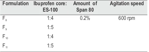
The release of drug decreased with increase in polymer: drug ratio of 1:1, 1.5:1, 2:1. The higher drug concentration appears to have enhanced drug diffusion and resulted in significantly higher release flux. Percentage cumulative drug release of formulations were found to be 99.85 (F1), 98.32(F2), 96.95(F3), 98.45 (F4), 99.22(F5), 88.21(F6), 80.22(F7). Due to the smaller particle size obtained at higher surfactant concentrations, a larger drug release rate was observed from uncoated Ibuprofen-loaded chitosan microparticles prepared using higher surfactant concentrations
The probable drug release mechanisms from the chitosan microspheres involve the following processes; i) water penetration into the microspheres, ii) chitosan swelling, gelling and dissolution of the drug and iii) diffusion of the active compound through the chitosan matrix.
Evaluation of ES-100 Coated Ibuprofen Microspheres
Percentage Drug Entrapment
The values of the entrapment efficiency were found to decrease with increase in the initial drug loading, which can be ascribed to better drug entrapment within the cores with increase in ES-100 levels. Percentage drug entrapment of formulations were found to be F8 (68.21), F9 (73.18), F10 (77.99), F11 (84.13).The drug loss could be attributed due to coating process. When the ibuprofen loaded microspheres were suspended in the polymeric solution during the process of coating with ES-100, some extraction of the drug from drug loaded ibuprofen microspheres may have occurred by partitioning in to coacervation phase.
Particle Size
There was an increase in mean diameter of the ES-100 coated microspheres compared to uncoated microspheres. The mean particle size of microspheres ranged from 93 µm to 126 µm for uncoated microspheres, 151.7 µm for 1:4, 154.95 µm for 1:5 in formulation F8 and F9, 157.5µm for 1:4, 162.8 µm for 1:5 core: coat ratio in formulation F10 and F11 . This increase in particle size of the microspheres can be attributed to an increase in viscosity with increasing polymer concentrations, which resulted in larger emulsion droplets and finally in greater microsphere size.
In Vitro Drug Release Studies
release at pH 1.2 which indicates that upon eudragit S-100 coating, the release of drug can be retarded successfully in an acidic environment.
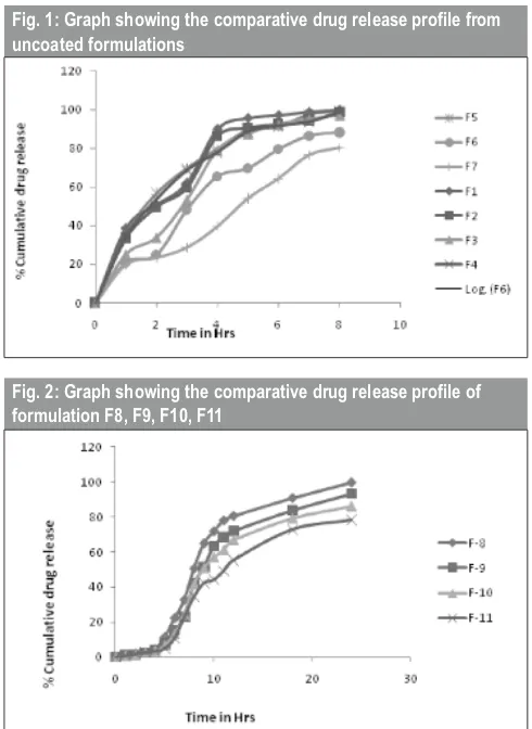
The cumulative percentage drug release from eudragit-coated ibuprofen microspheres showed the desired rate, as there was no measurable drug release observed up to 2 hrs in SGF (pH 1.2), while at pH 4.5, the ibuprofen release was quite insignificant (>2%) up to 4 hrs. Ibuprofen release from eudragit-coated chitosan microspheres in SGF followed the order 99.55 (F8)> 93.15 (F9)>85.98(F10)>78.19 (F11). In pH progression medium mimicking GIT up to 24 h, the release of ibuprofen from ES-100 chitosan microspheres decreased as the ES-100 concentration increased, suggesting that drug release could be controlled by varying the ES-100 concentration. The results might also be explained by the fact that the higher ES-100 content resulted in larger particles with proportionately less drug, so that the drug–polymer ratio was changed and thus release was reduced. The drug release profile from uncoated microspheres is shown in fig 1; the release of drug from coated formulation is shown in fig 2.
Scanning Electron Microscopy
The SEM photographs of uncoated microspheres and microspheres coated with ES-100 were examined using
Fig. 1: Graph showing the comparative drug release profile from uncoated formulations
Fig. 2: Graph showing the comparative drug release profile of formulation F8, F9, F10, F11
SEM-JEOL, JSM-840A scanning electron microscope. The photographs are displayed in fig 3, 4, 5. The drug loaded microspheres were round and smooth in appearance .
Fig. 4: SEM of Coated Microspheres
(Low magnification)
Fig. 5: SEM of Coated Microspheres
(High magnification)
Evaluation of Stability of Coated and Uncoated Microspheres
Stability studies were performed as per ICH guidelines. The results indicated that there was no evident change in the physical appearance of formulations at the end of the 6
o o
months storage period at 5 ± 2 C/ Ambient, 25 ± 2 C/ 60% RH
o
and 40 ± 2 C/ 75% RH.
Four formulations coded F3, F5, F8, F10 were chosen for stability studies. At fixed time intervals, drug content determination of these formulations showed that there were no significant changes in the values when compared to the initial formulations. Thus we may conclude that the drug does not undergo degradation on storage. These results imply good stability of the product on long-term storage.
CONCLUSION
The results of this investigation indicate that emulsion cross-linking method can successfully be employed to fabricate ibuprofen loaded chitosan multiparticles with particle size in the range of 90 to130 µm. Generally all formulations demonstrated their applicability in vitro as a promising device for pH-dependent colon delivery of ibuprofen. The entrapment efficiency and in vitro drug release kinetics were found to be greatly influenced by formulation variables such as concentration of drug, concentration of surfactant and volume of cross linking agent. These findings indicate that these variables can be suitably altered to achieve the desired controlled release profile of ibuprofen. Double microencapsulation technique employing chitosan matrix and ES-100 coating show promise for site specific and controlled delivery of ibuprofen in inflammatory infections.
ACKNOWLEDGEMENTS
The authors wish to thank 1) Ce Chem. Pharmaceuticals Pvt Ltd for sparing gift sample of Ibuprofen. 2) The Principal, Government College of Pharmacy, Bangalore, for providing the laboratory facilities to carry out research work.
REFERENCES
1. Pirjo Nykanen. Development of Multiple-Unit Oral Formulations for Colon Specific Drug Delivery Using Enteric Polymers and Organic Acids as Excipients, Division of Biopharmaceutics and Pharmacokinetics. Department of Pharmacy, University of Helsinki: Academic Dissertation 2003;1-44.
2. Sarasija S, Hota A. Colon specific drug delivery systems. Indian J. Pharm. Sci. 2000; 62(1):1-8.
3. Sinha VR, Rachnakumaria. Microbiologically triggered drug delivery to colon. Eur. J.Pharm.Sci.2003; 18:3-18.
4. Chourasia MK, Jain SK.Pharmaceutical approaches to colon targeted drug delivery systems.J Pharm Pharmaceut Sci2003; 6(1): 33-66. 5. Umadevi SK, Thiruganes R, Suresh S. Journal of Pharmaceutical
Research and Healthcare (2010);2:46-65.
6. Watts PJ and IIIum L. Colonic drug delivery. Drug Dev. Ind.Pharm.1997;23: 893-913.
7. Surender Verma, Vipin Kumar, Mishra DN, Singh SK. Colon targeted drug delivery: current and novel perspectives. INDIAN DRUGS 2012; 3(5):1274-84.
8. Ghosh S, and Barik BB. IJMMS. 2009; 1(9):375-82.
9. Sir Collen Dollery Therapeutic drug.Jan 1999,vol I, 11-14, Churchil Livingstone.
10. Thanoo BC, Sunny MC, Jayakrishnan A. Cross-linked chitosan microspheres: preparation and evaluation as a matrix for the controlled release of pharmaceuticals. J. Pharm. Pharmacol. 1992; 44: 283-6. 11. Patil SB, Murthy RSR. Preparation and in vitro evaluation of
mucoadhesive Chitosan microspheres of amlodipine besylate for nasal administration.Indian J. Pharm. Sci. 2006; 68(1): 64-7.
12. Swamy N.G.N., Abbas Z. Mucoadhesive microspheres containing amlodipine besylate for nasal administration; improvised preparation and in vitro characterization. INDIAN DRUGS 2011;48(10): 30-9. 13. Fatemeh Atyabi, Rudabeh Vahabzadeh, Rassoul Dinarvand.
Preparation of Ethyl cellulose Coated Gelatin Microspheres as a Multiparticulate Colonic Delivery System for 5-Aminosalicilic Acid. Iran. J. Pharm. Res. 2004; 2: 81-6.
14. Shivakumar HN, Sarasija Suresh, Desai BG. Design and evaluation of pH sensitive multiparticulate system for chronotherapeutic delivery of diltiazepam hydrochloride. Indian J.Pharm. Sci. 2006; 68(6): 781-7. 15. Chaurasia M, Chourasia MK, Jain NK, Jain A, Soni Vet al.
Cross-LinkedGuar Gum Microspheres: A viable approach for improved delivery of Anticancer Drugs for the treatment of Colorectal Cancer. AAPS PharmSciTech 2006;7(3): E1-E9.
16. Dandagi PM, Mastiholimath VS, Gadad AP, IIiger SR. Mucoadhesive Microspheres of Propranolol Hydrochloride for Nasal Delivery. Indian J. Pharm. Sci. 2007; 69(3): 402-7.
17. Amol Paharia, et al, Eudragit-coated Pectin Microspheres of 5-Fluorouracil for Colon Targeting. AAPS PharmSciTech 2007; 8(1): E1-E7.
18. N.G.N. Swamy, Z. Abbas. Preparation and in vitro characterization of mucoadhesive polyvinyl alcohol microspheres containing amlodipine besylate for nasal administration. Ind J Pharm Edu Res 2012; 46(1): 52-8.