http://www.sciencepublishinggroup.com/j/ijmi doi: 10.11648/j.ijmi.20170504.11
ISSN: 2330-8303 (Print); ISSN: 2330-832X (Online)
Differentiating Benign from Malignant Cervical Lymph
Nodes with Sonoelastography
Abdelmoniem Mahmoud Mohie-Eldine
1, Nabila El-Gazzar
2, Emad Mohamed Mashaly
1,
Reda Abd-Elsameea Alarabawy
1, Sherief Abd-ElSalam
21
Radiological Department, Tanta University, Tanta, Egypt 2Tropical Medicine Departement, Tanta University, Tanta, Egypt
Email address:
sherif_tropical@yahoo.com (S. Abd-ElSalam)
To cite this article:
Abdelmoniem Mahmoud Mohie-Eldine, Nabila El-Gazzar, Emad Mohamed Mashaly, Reda Abd-Elsameea Alarabawy, Sherief Abd-ElSalam. Differentiating Benign from Malignant Cervical Lymph Nodes with Sonoelastography. International Journal of Medical Imaging.
Vol. 5, No. 4, 2017, pp. 42-46. doi: 10.11648/j.ijmi.20170504.11
Received: May 3, 2017; Accepted: June 3, 2017; Published: October 18, 2017
Abstract:
In the clinical practice, there is many overlap in diagnostic criteria between benign and malignant lymph nodes by grayscale and Doppler sonography. This study was carried out on 30 patients who suffered from enlarged cervical lymph nodes examined with B-mode US, Doppler US and Sonoelastography in the Sonoelastography unit of the Radiology department of Tanta university hospitals to prospectively estimate the accuracy of sonoelastography in the differentiation between benign and malignant cervical lymph nodes. Sonoelastography had the highest sensitivity (92.1%), specificity (95.5%) and accuracy (93.3%) in the differentiation between benign and malignant cervical lymph nodes, so Sonoelastography is a promising technique that is easy and rapid non-invasive to perform and help identifying cervical lesions that are likely to be malignant to avoid hazards that found in tissue biopsy especially in patients with bleeding disorders.Keywords:
Lymphadenopathy, Elastography, Sonoelastography1. Introduction
Of the 450 lymph nodes inside the human body, the head and neck contain 60 - 70. Those nodes display reactive enlargement in response to infection (e.g., infection of upper aero-digestive tract). They also go through expansion while they are secondarily involved in head and neck cancer. Differentiation between reactive and metastatic lymphadenopathy is important [1].
Diverse imaging modalities are used to evaluate the possible etiology of neck nodes enlargement which include CT, MRI and ultrasonography. Among them, sonography is the least expensive and time consuming exam and ultrasound guided fine needle aspiration (US-FNA) can be done simultaneously to achieve cloth for cytological assessment.
A few characteristics have been documented to assist expect malignant cervical lymphadenopathy. However, no single criterion had satisfactory sensitivity and specificity for predicting this disease entity [2].
One of the differentiating criteria among benign and malignant lymph nodes is hardness (elasticity) [1]
Elasticity (hardness) varies in different types of tissue (fat, collagen, and so forth) and within the same tissue in different pathologic states (inflammatory, malignant) [1].
Amongst different imaging modalities, ultrasound has the very best sensitivity in the evaluation of malignant cervical nodes, whereas PET/CThas the highest specificity in the diagnosis [3].
B-mode ultrasound has sensitivity and specificity reaching 85.7% and 90%, respectively, [4]. While, US elastography has sensitivity and specificity achieving 83% and 100%, respectively. Mixed b-mode sonography and US elastography has sensitivity and specificity attaining 92% and 94%, respectively [1].
diagnosis [3].
2. Methods
The study was conducted on 30 patients who suffered from enlarged cervical lymph nodes (13 male and 17 female), they were referred from Tanta university hospital clinics within 10 months period from May 2015 to February 2016, after obtaining their informed consent and within the approved protocol of Tanta Faculty of medicine ethical committee.
All patient names were hidden and replaced by code numbers to maintain privacy of the patients. Real time B-mode ultrasonography, Doppler US and Elastographic examination of the cervical lymph nodes was done for the study patients in the Sonoelastography unit of the Radiology department of Tanta university hospitals.
The study included patients with enlarged cervical lymph nodes, diagnosed by clinical examination. However, any patient with known cause of cervical lymphadenopathy was excluded from the study.
All patients were subjected to detailed history taking, full clinical examination and radiological imaging including Real-time B-mode ultrasonography, Doppler US and Elastographic examination of the cervical lymph nodes was done using Toshiba Aplio 500 Machine in the Sonoelastography unit of the Radiology department of Tanta university hospitals and they underwent laboratory investigations e.g. Total leukocyte count with differential leukocyte count, platelet count, prothrombin time and activity. Confirmatory Lymph node biopsy of the enlarged cervical lymph nodes was finally made.
In the case of suspected cases of metastatic cervical lymph nodes, they underwent abdomino-pelvic, neck and breast ultrasound, chest X-ray, mammography and contrast enhanced computed tomography of thehead, neck, chest, abdomen and pelvis.
3. Results
This study was prospectively carried on 30 patients (13 male and 17 female). The mean age for both benign and malignant cervical lymph nodepatients was (56.24 years ± 12.45 SD); the mean age washigher for malignant LNs (59.37 years ± 9.68 SD) than for benign LNs (42.78 years ± 10.36 SD), (P < 0.003). Their ages ranged from 28 to 72 years.
Detailed description of the number and percentage for each pathological cervical lymph nodes entity within benign and
malignant categories are illustrated in Table 1.
Table 1. The number and percentage of each pathological result of the enlarged cervical lymph nodes.
Final pathological diagnosis of the
enlarged cervical lymph nodes Number & Percentage
Chronic non-specific lymphadenitis (Reactive
lymph nodes) 11 (36.67%)
Benign 11 (36.67%)
Lymphoma 10 (33.33%)
Metastasis from Thyroid carcinoma 7 (23.33%)
Metastasis from Laryngeal carcinoma 2 (6.67%)
Malignant 19 (63.33%)
Total 30 (100%)
For assessment of the elastographic Strain ratio (Strain index), it wasfound that; the mean strain ratio for both malignant and benign LNs was 4.14 ± 3.19 with a range of (0.89 – 7.33). We found that the mean strain ratio for malignant LNs (4.61 ± 2.52; range: 1.46 ± 7.33) was significantly greater than that for benign LNs (mean, 1.19 ± 0.24; range: (0.89-1.48).
In this study, the assessment of the lymph node strain ratio was according to Lyshchik, et al., (2007) [9]. So, it was considered LNs with strain ratio less than 1.5 as benign and LNs with strain ratio more than or equal 1.5 as malignant.
After revising the pathological results; it was found that 12/30 (40%) of LNs were diagnosed as benign by elastography strain ratio; out of which 11/12 (91.7%) were confirmed to bebenign (true negative) by pathology and 1/12 (8.3%) were proved to be malignant by pathology (false negative). On the other hand, 18/30 (60%) of LNs were diagnosed as malignant by strain ratio and all of them (100%) were confirmed to be malignant by pathology (truepositive). These results revealed that the calculated sensitivity of elastography strain ratio was 94.7% and specificity was 100%, PPV and NPV were 100% and 91.7%, respectivly and the total accuracy was 96.7%, Table 2.
Table 2. Analysis of false positive and false negative diagnosis by strain ratio.
Number PathologicalDiagnosis
1 False Negative
1 Lymphoma
0 False positive
0 -
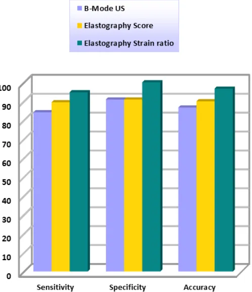
The diagnostic performance of the cervical lymph nodes B-Mode sonography, elastographic pattern (score) and elastography strain ratio are shown in Table 3 and Figure 1.
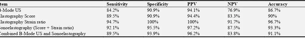
Table 3. Comparison between the diagnostic performance of the lymph node B-Mode sonography, elastographyscore and strain ratio, total Sonoelastography and Combined B-Mode US and Sonoelastography.
Item Sensitivity Specificity PPV NPV Accuracy
B-Mode US 84.2% 90.9% 94.1% 76.9% 86.7%
Elastography Score 89.5% 90.9% 94.4% 83.3% 90%
Elastography Strain ratio 94.7% 100% 100% 91.7% 96.7%
Sonoelastography (Score + Strain ratio) 92.1% 95.5% 97.2% 87.5% 93.3%
Figure 1. Comparison between the diagnostic performance of the Lymph node B-Mode sonography, Elastography Score and Strain ratio.
Elastography strain ratio shows the highest sensitivity, specificity and accuracy. Elastography score and strain ratio show higher sensitivity, specificity and accuracy compared to B-Mode sonography.
4. Discussion
Sonoelastography visualizes the hardness of the lesion by reflecting the biological characteristics. The palpation results vary with the experience of the examiners. If the stiffness of the tissues can be visualized through some instrument and quantitatively measured, the elasticity information of the tissues will be reflected more objectively [4].
Two different methods could be obtained for sonoelastography evaluation; qualitative elastography by the scoring method and quantitative elastography by strain ratio measurement. By means of scoring system, operator should score the target lesion according to the proportion of blue areas in the lesions. It is semi-objective and it depends on different factors such as the operator's experience and scoring system (5 or 4 point) [5].
Strain ratio (SR) measurement has been considered to be more accurate than scoring method because it could estimate the difference between stiffness of the lesions and the surrounding tissue [6].
One of the advantages of the SR method is being quantitative, in cases that the scores are the same visually, the SR could be different [6, 7].
In this study we found that benign cervical lymphadenopathy gives strain ratio that ranges from 0.89 to
1.48, whereas malignant cervical lymph nodes give strain ratio that ranges from 1.46 to 7.33. The mean strain ratio of malignant cervical LNs is significantly higher than the mean strain ratio of malignant cervical LNs.
There was a significant difference in the mean strain ratio between the benign and malignant cervical lymph nodes; 1.19 ± 0.24 versus 4.61 ± 2.52, respectively (P < 0.01). Also, we found that all the benign cervical lymph nodes were less than 1.5 times stiffer than the surrounding muscles; however, 94.7% of the malignant cervical lymph nodes were more than 1.5 times stiffer than the surrounding muscles, (P < 0.01). When the diagnostic accuracy of a strain ratio greater than 1.5 was calculated, results showed that this criterion had 94.7% sensitivity, 100% specificity, 100% positive predictive value, 91.7% negative predictive value and the highest overall accuracy (96.7%) of all the diagnostic criteria examined.
diagnostic accuracy of a strain index greater than 1.5 was calculated, results showed that this criterion had 85% sensitivity, 98% specificity, 96% positive predictive value, 90% negative predictive value and the highest overall accuracy (92%) of all the diagnostic criteria examined.
Our results show positive correlation between conventional B-Mode sonography, elastography score and strain ratio; in agreement with Hefeda and Badawy. (2014) &choi et al., (2015) and Lyshchik, et al., (2007) [8, 9, 11, 12].
5. Conclusion
Finally, the study concluded that Sonoelastography can improve the performance of conventional sonography in the diagnosis of enlarged cervical lymph nodes by increasing the overall sensitivity, specificity and accuracy of cervical lymph node ultrasonography.
Sonoelastography is a rich and rapidly developing technique that promises to improve diagnosis of many different lymph node diseases. As with most new technologies, diagnostic criteria and quality control mechanisms are still being established. Interested Radiologists are encouraged to begin training themselves on the properuse of this emerging methodology as most new ultrasound machines will have this option.
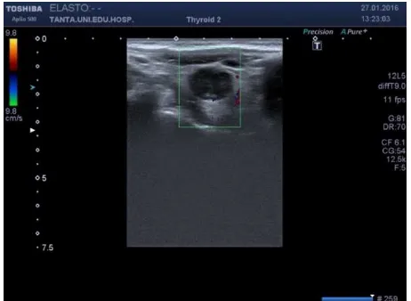
Case [1]
Figure 2. Case 1: B-Mode ultrasound of enlarged cervical lymph nodemeasuring 18 mm x 6.1 mm, withregular border, preserved hilum, and hypoechoic cortex.
Figure 3. Case 1: color Doppler ultrasound shows hilar vascularity of the enlarged cervical lymph node.
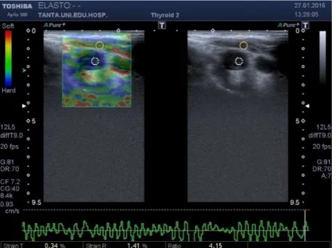
Figure 4. Case 1: color elastogram of enlarged cervical lymph node gives pattern 2 elastography score and strain ratio is 1.25.
Case 1 figures 2, 3, 4; show enlarged cervical lymph node measuring 4.2 mm x 15 mm, regular border, preserved hilum, hypoechoic cortex, with hilar vascularity, gives pattern 1 elastography and strain ratio is 0.89, Final histopathological diagnosis was Reactive lymphoid hyperplasia.
Case 2
Figure 5. Case 2: B-Mode ultrasound of enlarged cervical lymph node measuring 16.2 mm x 13.7 mm, rounded shape, hypoechoic with absent hilum.
Figure 7. Case 2: color elastogram of enlarged cervical lymph node gives pattern 3 elastography score and strain ratio is 4.15.
Case 2 figures, 5, 6, 7; show enlarged cervical lymph node measuring 16.2 mm x 13.7 mm, regular border, abscent hilum, hypoechoic, apparently avascular, gives pattern 3 elastographyand strain ratio is 4.15, Final Histopathological diagnosis was Non-Hodjkin's Lymphoma.
References
[1] Alam F, Naito K, Horiguchi J, et al., (2008): “Accuracy of sonographicelastography in the differential diagnosis of enlarged cervical lymph nodes: comparison with conventional B-mode sonography”. Am J Roentgenol; 191: 604–10.
[2] Liao L, Wang T, Young Y, et al., (2010): “Real-time and computerized sonographic scoring system for predicting malignant cervical lymphadenopathy”. Head Neck journal; 32: 594–598.
[3] Ahuja A, Ying M, Ho S, et al., (2008): “Ultrasound of malignant cervical lymph nodes”. Cancer Imaging; 8 (1): 48– 56.
[4] Sureshkannan P, Vijayprabhu A, and John R. (2011): “Role of ultrasound in detection of metastatic neck nodes in patients with oral cancer”. Indian J Dent Res; 22 (3): 419–423.
[5] Zhang Y, Lv Q, Yin Y, et al., (2009): “The value of ultrasound elastography in differential diagnosis of superficial lymph nodes”. Front Med China; 3 (3): 368–374.
[6] Mahsa G, Mehdi M, Kamran A, et al., (2014): “Sonoelastography for differentiating benign and malignant cervical lymph nodes: A Systematic Review and Meta-Analysis”. Int J Preventive Med.; 5 (12): 1521–1528.
[7] Teng DK, Wang H, Lin YQ, et al., (2012): “Value of ultrasound elastography in assessment of enlarged cervical lymph nodes”. Asian Pac J Cancer Prev.; 13: 2081–2085.
[8] Hefeda M, and Badawy M. (2014): “Can ultrasound elastography distinguish metastatic from reactive lymph nodes in patients with primary head and neck cancers?”. The Egyptian Journal of Radiology and Nuclear Medicine; 45 (3): 715–722.
[9] Lyshchik A, Higashi T, Asato R, et al., (2007): “Cervical lymph node metastases: diagnosis at sonoelastography: initial experience”. Radiology; 243 (1): 258 –267.
[10] Cheng K, Choi Y, Shim W, et al. (2016): Virtual touch tissue imaging quantification shear wave elastography: prospective assessment of cervical lymph nodes. Ultrasound in medicine & biology, 42 (2), 378-386.
[11] Choi J, Lee J and Baek J. (2015): Ultrasound elastography for evaluation of cervical lymph nodes. Ultrasonography, 34 (3), 157-164.