1: Department of molecular Medicine. School of Advanced Technologies in Medicine, Tehran University of Medical Sciences, Tehran, Iran. 2: Molecular and Medicine Research Center, Arak University of Medical Sciences, Arak, Iran.
3:Department of Parasitology, Arak University of Medical Sciences, Arak, Iran. Department of Immunology, Faculty of Medicine, Tehran University of Medical Sciences, Tehran, Iran.
4:Department of Immunology, Faculty of Medicine, Tehran University of Medical Sciences, Tehran, Iran.
5:Department of Immunology, Faculty of Medicine, Cellular and Molecular Research Center, Kurdistan University of Medical Sciences, Sanandaj, Iran.
6: Department of Molecular Medicine, School of Advanced Technologies in Medicine, Tehran University of Medical Sciences, Tehran, Iran.
A Simplified Protocol for the Purification of
Schwann Cells and Exosome Isolation
from C57BL/6 Mice
Mana Shojapour
1, Ghasem Mosayebi
2, Reza Hajihossein
3,
Farshid Noorbakhsh
4,
Aram Mokarizadeh
5, Mohammad Hossein Ghahremani*
6Abstract
Background: The purification of Schwann cells has proven to be a difficult process, with most methods requiring
the use of special equipment. However, obtaining a sufficient number and high purity of Schwann cells is an integral aspect in their use for clinical application. Therefore, the aim of this study was to establish a simple and effective protocol for the isolation and purification of Schwann cells from the sciatic nerve of C57BL/6 mice. Furthermore, we aimed to provide a protocol for the isolation of exosomes from these cells.
Methods: To purify Schwann cells, we used a combination of in situ nerve pre-degeneration and fetal bovine
serum. To determine the most effective method of cell purification, we treated the culture with varying concentrations of fetal bovine serum and examined which concentration provided the highest Schwann cell purity. Exosomes were then isolated from Schwann cells through a process of repeated centrifugation and filtration steps.
Results: We were able to increase the purified population of Schwann cells from C57BL/6 mice by reducing
the concentration of FBS. The purity of Schwann cells at FBS concentrations of 10%, 5%, and 2% were 93.42%, 91.25%, and 97.83%, respectively.
Conclusions:When using a concentration of 2% FBS, we obtained the highest purification yield of Schwann
cells. Our protocol does not require special equipment or materials. We have created a protocol that is simple, fast, and safe while providing a high yield of purified Schwann cells. The exosome isolation method described in this paper is an appropriate approach with a high quality and yield.
Keywords: C57bl/6 mice, Exosome, Pre-degeneration, Purification, Schwann cells
Introduction
Schwann cells are the main type of glial cells within the peripheral nervous system (PNS). These cells play vital roles in development, homeostasis, the conduction of nerve impulses along axons, areinvolved in antigen presentation, as well as the
maintenance of healthy axons and axonal regeneration following PNS injury (1). Many studies have demonstrated the potential clinical application for Schwann cells in a variety of diseases and medical conditions. Schwann cells
have shown promise in the treatment of demyelinating disorders and spinal cord injuries through autologous cell transplantation. They have also been shown to be a useful tool in the construction of artificial nerve tube conduits, and in peripheral nerve tissue engineering. Furthermore, Schwann cells have been shown to be a valuable resource for the treatment of central nervous system damage (2-6). Through improving and simplifying the way in which pure and high cell density Schwann cell cultures are obtained, we can equip scientists with the proper tools to better understand the biological properties and molecular mechanisms of Schwann cells. As we improve our understanding of Schwann cell biology, we can improve the success of these cells in clinical applications. However, the success of these biological and clinical studies begins with the proper isolation, purification, and expansion of Schwann cells. Results of previous studies have shown that Schwann cell purification is difficult due to frequent contamination by fibroblasts. As fibroblasts have a faster rate of proliferation than Schwann cells, they quickly become the predominant cell type in culture. Furthermore, most of the reported methods for Schwann cell purification are complicated, expensive, and time-consuming (7, 8).
In general, the application of exosomes as cell-free therapeutics has been shown to be more advantageous when compared to cell therapy (9-11). In recent years, interest in exosomes has grown due to their potential diagnostic and therapeutic relevance. Exosomes are small membrane vesicles between 50–1,000 nm in diameter and are secreted by most cells to the extracellular environment. These small vesicles are assumed to play important roles in intercellular communication (12, 13). Lopez-verrilli and colleagues have shown that Schwann cell-derived exosomes are capable of enhancing axonal regeneration in vitro and enhancing regeneration after sciatic nerve injury in vivo (14, 15).
The aim of the present study was to successfully purify and expand Schwann cells from C57BL/6 mice, as well as to isolate the exosomes from these Schwann cells.
Materials and methods
Animals
C57BL/6 mice (6–8 weeks old) were obtained from the Pasteur Institute of Iran (Karaj, Iran). All animal treatments and experiments were conducted according to the ethics guidelines for research on animals approved by the Institutional Animal Ethics Committee of the Tehran Medical Science School, which follows the United States National Institute for Health guidelines for the care and use of experimental animals.
Preparation of poly-L-lysine coated plates
For primary cell cultures, poly-L-lysine coated plates were prepared according to the manufacturer's instructions (Sigma, USA). Under sterile conditions, 500 ul of the poly-L-lysine solution (Sigma, USA) was added to completely cover the culture surface. The plates were then incubated for 18 hours at room temperature. The coating solution was removed in its entirety, the plates were rinsed two times with phosphate-buffered saline (PBS 0.15 M; pH 7.4) and the surfaces were set aside to allow for drying.
In situ nerve degeneration
For pre-degeneration, C57BL/6 mice (6–8 weeks old) were anesthetized with a combination of ketamine (80 mg/kg; Sigma, USA) and xylazine (10 mg/kg; Sigma, USA) and injected intraperitoneally. The hair from the posterior thigh was shaved, the exposed skin was disinfected with 70% ethanol. The right sciatic nerves were cut,
thenthe skin with the incision was sewn shut with absorbable sutures. After surgery, animals were monitored until full recovery, then placed into a clean home cage. Mice were left for seven days to promote the formation of Wallerian degeneration with the maximum number of Schwann cells.
Primary culture
Seven days following the cutting of the sciatic nerve, animals were euthanatized and the body was sterilized with 70% ethanol. The following process requires a sterile environment, equipment and cell culture tools. The disconnected distal nerve segment was resected and nerves were washed
three times with ice-cold PBS supplemented with 2% penicillin/streptomycin. The nerve was then transferred into a cell culture plate containing, serum-free Dulbecco's modified Eagle's medium (DMEM) (Gibco, Scotland, UK). The surrounding membranes and muscular tissue were carefully removed under a stereomicroscope, then the epineurium was stripped off with fine forceps. The nerves were washed with ice-cold PBS and cut into pieces 1 mm in length with a scalpel. Finally, the nerve pieces were collected in a 15-ml conical tube containing 2 ml of DMEM and centrifuged at 200 G for 10 min. Enzymatic digestion was performed with 0.05% collagenase-A (Roche, Germany) solution for 2 hours at 37 °C. Any remaining cells were mechanically dispersed. Enzymatic activity was halted by adding 300 ul of fetal bovine serum (FBS) and centrifuged at 5 min at 1500g rpm. The cells were counted using a hemocytometer. Later, 1.2–2 ×10 4 cells were seeded into poly-L-lysine- coated culture 35-mm dishes, and incubated at 37 °C with 5% CO2 for 2 days. The cells were allowed to reach high densities.
Passage and cell culture
Cultures were divided into three groups once they reached 70%–90% confluency. The culture medium was supplemented with varying concentrations of FBS. Groups were divided into group numbers 1, 2 and 3, each receiving a concentration of either 10%, 5%, and 2% FBS, respectively. The culture medium was replaced every 48 hours until the cells became confluent. The cells were then counted at a magnification of 40X in 6 random fields. Cells that displayed bipolar or tripolar morphology and contained oval nuclei were recognized as Schwann cells, whereas flat or polygonal cells with a large cytoplasm were recognized as fibroblasts.
On the seventh day of the experiment, the cells were trypsinized and the total number of cells was counted using a hemocytometer. The purity of Schwann cell cultures was calculated by determining the ratios of the number of Schwann cells to the total cell counts. To confirm and characterize Schwann cells in culture, cells were identified using immunohistochemical staining with anti-S100 protein. Cultures were fixed for 30 min in 4%
paraformaldehyde, washed three times with PBS, incubated, and washed again with PBS. Cells were then incubated overnight with anti-S100 antibody (sigma, USA), then washed and incubated with a secondary antibody (sigma, USA) for 1 hour at 37 °C. After a final wash in PBS, cells were visualized under a fluorescence microscope (Olympus).
Exosome purification
Once cell cultures reached 85–90% confluency, cells were washed three times using PBS to remove any remaining FBS. This was then followed by a 24-48 hours’ incubation with DMEM without FBS. The cell suspension was transferred to conical tubes. After the incubation period, cells were placed in a swinging bucket centrifuge at 300 ×g for 10 min and 2,000 × g for 30 min at 4 °C to pellet the cells and debris. Supernatant was filtered using a 0.22-μm filter to remove particles larger than 200 nm. The filtrate was concentrated using a Centricon Plus-70 centrifugal filter device (Millipore, USA). Following a final centrifugation step at 70000 x g for 60 minutes at 4 °C, the supernatant was discarded and the pellet of exosomes was resuspended in 100-200 ml of filtered PBS and stored at -80 °C for further analysis. The size and shape of purified exosomes were evaluated using transmission electron microscopy (TEM).
Transmission electron microscopy (TEM)
A small drop of the exosomes was deposited on the carbon coated grid and fixed in 1% glutaraldehyde. The grids were also stained with freshly prepared 1% (v/v) uranyl acetate (Merck, Germany). Transmission electron microscopy (LEO906, Germany) was performed at 80 kV for imaging.
R
esults
The primary cell culture of C57BL/6 isolated Schwann cells
On the first day of primary cell culture, Schwann cells proliferated more quickly than fibroblasts. In terms of morphology, fibroblasts are characterized
by a flat shape, with a large cytoplasm-to-nucleus ratio, and poor refraction rates. Schwann cells have a bipolar or tripolar spindle shape, smaller size, and a stronger refraction rate. Based on this morphological identification of Schwann and fibroblast cells, we determined there to be few fibroblasts present among the Schwann cells (Fig. 1).
Fig. 1. Primary culture (24 h) after digestion, showing a large number of cells attached to the bottom plate (4×).
Schwann cell purification
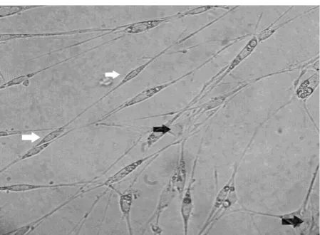
After 48 hours of primary cell culture, Schwann cell purification was performed by changing the concentration of FBS. In the first group, the numbers of fibroblasts increased after 72 and proliferated more quickly than Schwann cells. In groups 2 and 3, the concentration of FBS was reduced, and on days 5 and 6, the number of fibroblasts significantly changed (Fig. 2). The purity of Schwann cells at FBS concentrations of group 1 (10%), group 2 (5%), and group 3 (2%) were 93.42%, 91.25%, and 97.83%, respectively.
Fig. 2. Two types of cells are present: Fibroblasts (black arrow) with flat polymorphic forms, larger nuclei, and larger cytoplasm. Schwann cells (white arrow) had bipolar or tripolar spindle shapes and smaller sizes (20×).
Exosome identification by electron microscopy
We evaluated purified exosomes for size and shape using transmission electron microscopy. The range of the diameter of the exosomes was from 40–150 nm. The exosomes were also round in shape (Fig. 3).
Fig. 3. Transmission electron microscopy image of extracellular vesicles isolated from supernatants of Schwann cell culture at 80 kV (×60,000).
Discussion
Many studies have investigated the ways in which Schwann cells can be effectively isolated and purified, however, each have been met with limitations. Schwann cell isolation and purification has been achieved through differential detachment with complex collagenases and dispase (16) as well as through differential adhesion methods (17). These methods are time-consuming and have the possibility of contamination and loss of cell numbers. Methods involving treatment with antimitotic agents have been shown to have a negative effect on the functionality of Schwann cells (18, 19). The use of antibody-dependent complement-mediated cytolysis is too expensive to obtain large amounts of purified cells (20). The immunoselection method is restricted in its utility because it requires special equipment (21, 22). It should also be noted that most studies have been conducted in rats.
In this study, we aimed to obtain large numbers of Schwann cells from C57BL/6 mice in a short period of time, using simple techniques, without the need for special equipment. Furthermore, we aimed to increase the yield of Schwann cells using in situ nerve
degeneration, as well as attain a greater purity of Schwann cells through testing different concentrations of FBS.
In a study on mice that used Forskolin as a mitogenic agent, the addition of pharmacological agents influenced gene expression. Altering the cells in this way reduces the use of these cells for clinical application (23). In studies on neonatal mice using differential detachment with collagenase during the enzymatic treatment, increase the possible risk of loss of cells and contamination (8).
According to previous research, in situ nerve pre-degeneration is an appropriate and standardized method to increase the cell count and purity of Schwann cells (25). During the Wallerian degeneration, Schwann cells become activated, dedifferentiate, proliferate and promote axonal regeneration (26, 27). Research has shown that the best time for maximum pre-degeneration is about 7 days. Results have indicated that the yield of Schwann cells will be several times higher, and that the purity of Schwann cells can be increased (21). With this in mind, our study applied the pre-degeneration method as well as examined the effect of FBS concentration on increasing the purity of the Schwann cells in culture.
Past studies have shown that the proliferation rates of Schwann cells in a 35-mm dish were higher than the proliferation of cells in 25- flasks (28). Therefore, we used 35-mm dishes for our Schwann cell cultures.
Past studies have also shown that proliferation of cultured Schwann cells was dependent on the nature of the substrate in which they were cultured (28). We used the poly-L-lysine solution as the plate coating.
Research has highlighted the initial cell density to be an important factor in increasing cell purity (28, 29). Cultures with lower initial seeding
density have been shown to result in a decrease of fibroblast interference. Therefore, in this study, our culture groups used a cell density of 1.2–2 ×10 4 cells on poly-L-Lysine coated dishes.
Here we found that Schwann cells proliferate more quickly during the first 48 hours after plating and become the predominant cell type in primary culture. Following this time period, fibroblasts became the dominant cell population within culture. Therefore, after 48 hours, by decreasing the FBS concentration, fibroblast proliferation was decreased.
It should also be noted that the Schwann cell proliferation rate significantly decreased after 3 weeks. This is because the cells enter into a quiescent state and have a possibility of spontaneous transformation in culture (30). Therefore, we did not use consecutive passages of cells. Following the first passage, cells were frozen and stored in liquid nitrogen.
Exosomes are bilayer membranous nano-vesicles with endogenous origin. Exosomes participate in several biological activities. The application of exosomes has opened a new avenue for diagnostic and therapeutic development. Here, the isolation of exosomes from purified Schwann cells is based on repeated centrifugation and filtration steps.
In conclusion, our results have shown to be consistent with other rat studies in regard to obtaining a high purity of Schwann cells in 2% FBS (29, 31). Our protocol requires no special equipment or materials. It is a safe, rapid, and simple method of obtaining high purity and yield.
Acknowledgements
This study was supported by the Tehran University of Medical Sciences, Tehran, Iran. Grant No.25961.
References
1. Bhatheja K, Field J. Schwann cells: origins and role in axonal maintenance and regeneration. The international journal of biochemistry & cell biology. 2006;38(12):1995-9.
2. Guenard V, Kleitman N, Morrissey TK, Bunge RP, Aebischer P. Syngeneic Schwann cells derived
from adult nerves seeded in semipermeable guidance channels enhance peripheral nerve regeneration. The Journal of neuroscience: the official journal of the Society for Neuroscience. 1992 Sep;12(9):3310-20.
3. Rodriguez FJ, Verdu E, Ceballos D, Navarro X. Nerve guides seeded with autologous schwann cells improve nerve regeneration. Experimental neurology. 2000 Feb;161(2):571-84.
4. Vacanti CA, Bonassar LJ, Vacanti MP, Shufflebarger J. Replacement of an avulsed phalanx with tissue-engineered bone. The New England journal of medicine. 2001 May 17;344(20):1511-4. 5. Oudega M, Xu XM. Schwann cell transplantation for repair of the adult spinal cord. J Neurotrauma. 2006 Mar-Apr;23(3-4):453-67. 6. Agudo M, Woodhoo A, Webber D, Mirsky R, Jessen KR, McMahon SB. Schwann cell precursors transplanted into the injured spinal cord multiply, integrate and are permissive for axon growth. Glia. 2008 Sep;56(12):1263-70.
7. Wei Y, Zhou J, Zheng Z, Wang A, Ao Q, Gong Y, et al. An improved method for isolating Schwann cells from postnatal rat sciatic nerves. Cell and tissue research. 2009 Sep;337(3):361-9.
8. Jin YQ, Liu W, Hong TH, Cao Y. Efficient Schwann cell purification by differential cell detachment using multiplex collagenase treatment. Journal of neuroscience methods. 2008 May 15;170(1):140-8.
9. Lai RC, Arslan F, Lee MM, Sze NS, Choo A, Chen TS, et al. Exosome secreted by MSC reduces myocardial ischemia/reperfusion injury. Stem Cell Res. 2010 May;4(3):214-22.
10. Farsad K. Exosomes: novel organelles implicated in immunomodulation and apoptosis. Yale J Biol Med. 2002 Mar-Apr;75(2):95-101. 11. Bruno S, Grange C, Deregibus MC, Calogero RA, Saviozzi S, Collino F, et al. Mesenchymal stem cell-derived microvesicles protect against acute tubular injury. J Am Soc Nephrol. 2009 May;20(5):1053-67.
12. Camussi G, Deregibus M-C, Bruno S, Grange C, Fonsato V, Tetta C. Exosome/microvesicle-mediated epigenetic reprogramming of cells. Am J Cancer Res. 2011; 1(1): 98–110.
13.Simons M, Raposo G. Exosomes--vesicular carriers for intercellular communication. Curr Opin Cell Biol. 2009 Aug;21(4):575-81.
14.Lopez-Verrilli MA, Court FA. Transfer of Vesicles from Schwann Cells to Axons: a Novel Mechanism of Communication in the Peripheral Nervous System. Front Physiol. 2012 Jun 13;3:205. 15. Lopez-Verrilli MA, Picou F, Court FA. Schwann cell-derived exosomes enhance axonal regeneration in the peripheral nervous system. Glia. 2013;61(11):1795-806.
16. Wu W, Jin YQ, Kretlow JD, Xu L, Duan HC, Qi ZL. Purification of Schwann cells from adult rats by differential detachment. Biotechnology letters. 2009 Nov;31(11):1703-8.
17. Kreider BQ, Messing A, Doan H, Kim SU, Lisak RP, Pleasure DE. Enrichment of Schwann cell cultures from neonatal rat sciatic nerve by differential adhesion. Brain research. 1981 Mar 2;207(2):433-44.
18.Morrissey TK, Kleitman N, Bunge RP. Isolation and functional characterization of Schwann cells derived from adult peripheral nerve. The Journal of neuroscience: the official journal of the Society for Neuroscience. 1991 Aug;11(8):2433-42.
19. Casella GT, Bunge RP, Wood PM. Improved method for harvesting human Schwann cells from mature peripheral nerve and expansion in vitro. Glia. 1996 Aug;17(4):327-38.
20. Brockes JP, Fields KL, Raff MC. Studies on cultured rat Schwann cells. I. Establishment of purified populations from cultures of peripheral nerve. Brain research. 1979 Apr 6;165(1):105-18. 21. Kraus A, Tager J, Kohler K, Manoli T, Haerle M, Werdin F, et al. Efficacy of various durations of in vitro predegeneration on the cell count and purity of rat Schwann-cell cultures. Journal of neurotrauma. 2010 Jan;27(1):197-203.
22. Manent J, Oguievetskaia K, Bayer J, Ratner N, Giovannini M. Magnetic cell sorting for enriching Schwann cells from adult mouse peripheral nerves. Journal of neuroscience methods. 2003 Mar 15;123(2):167-73.
23. Zhu J, Qin J, Shen Z, Kretlow JD, Wang X, Liu Z, et al. Dispase rapidly and effectively purifies Schwann cells from newborn mice and adult rats. Neural regeneration research. 2012 Feb 5;7(4):256-60.
24. Fu SY, Gordon T. The cellular and molecular basis of peripheral nerve regeneration. Molecular neurobiology. 1997 Feb-Apr;14(1-2):67-116. 25. Keilhoff G, Fansa H, Schneider W, Wolf G. In vivo predegeneration of peripheral nerves: an effective technique to obtain activated Schwann cells for nerve conduits. Journal of neuroscience methods. 1999 Jul 1;89(1):17-24.
26.Bampton ET, Ma CH, Tolkovsky AM, Taylor JS. Osteonectin is a Schwann cell-secreted factor that promotes retinal ganglion cell survival and process outgrowth. The European journal of neuroscience. 2005 May;21(10):2611-23.
27. Hoke A, Cheng C, Zochodne DW. Expression of glial cell line-derived neurotrophic factor family of growth factors in peripheral nerve injury in rats. Neuroreport. 2000 Jun 5;11(8):1651-4.
28.Honkanen H, Lahti O, Nissinen M, Myllyla RM, Kangas S, Paivalainen S, et al. Isolation, purification and expansion of myelination-competent, neonatal mouse Schwann cells. The European journal of neuroscience. 2007 Aug;26(4):953-64.
29. Hedayatpour A, Sobhani A, Bayati V, Abdolvahhabi MA, Shokrgozar MA, Barbarestani M. A method for isolation and cultivation of adult Schwann cells for nerve conduit. Archives of Iranian medicine. 2007 Oct;10(4):474-80.
30. Haynes LW, Rushton JA, Perrins MF, Dyer JK, Jones R, Howell R. Diploid and hyperdiploid rat Schwann cell strains displaying negative autoregulation of growth in vitro and myelin sheath-formation in vivo. Journal of neuroscience methods. 1994 Jun;52(2):119-27.
31. Komiyama T, Nakao Y, Toyama Y, Asou H, Vacanti CA, Vacanti MP. A novel technique to isolate adult Schwann cells for an artificial nerve conduit. Journal of neuroscience methods. 2003 Jan 30;122(2):195-200.