R E S E A R C H A R T I C L E
Open Access
Theme trends and knowledge structure on
choroidal neovascularization: a quantitative
and co-word analysis
Fangkun Zhao
1,2,3, Bei Shi
4, Ruixin Liu
1,5, Wenkai Zhou
1,2,3, Dong Shi
1,2,3and Jinsong Zhang
1,2,3*Abstract
Background:The distribution pattern and knowledge structure of choroidal neovascularization (CNV) was surveyed based on literatures in PubMed.
Methods:Published scientific papers about CNV were retrieved from Jan 1st, 2012 to May 31st, 2017. Extracted MeSH terms were analyzed quantitatively by using Bibliographic Item Co-Occurrence Matrix Builder (BICOMB) and high-frequency MeSH terms were identified. Hierarchical cluster analysis was conducted by SPSS 19.0 according to the MeSH term-source article matrix. High-frequency MeSH terms co-occurrence matrix was constructed to support strategic diagram and social network analysis (SNA).
Results:According to the searching strategy, all together 2366 papers were included, and the number of annual papers changed slightly from Jan 1st, 2012 to May 31st, 2017. Among all the extracted MeSH terms, 44 high-frequency MeSH terms were identified and hotspots were clustered into 6 categories. In the strategic diagram, clinical drug therapy, pathology and diagnosis related researches of CNV were well developed. In contrast, the metabolism, etiology, complications, prevention and control of CNV in animal models, and genetics related researches of CNV were relatively immature, which offers potential research space for future study. As for the SNA result, the position status of each component was described by the centrality values.
Conclusions:The studies on CNV are relatively divergent and the 6 research categories concluded from this study could reflect the publication trends on CNV to some extent. By providing a quantitative bibliometric research across a 5-year span, it could help to depict an overall command of the latest topics and provide some hints for researchers when launching new projects.
Keywords:Choroidal neovascularization, Bibliometric analysis, Co-word analysis, Social network analysis
Background
Choroidal neovascularization (CNV) is defined as a process of blood vessel growth abnormality from the choroid layer into the retina layer [1] and this symptom could result in a sudden deterioration of central vision, metamorphopsia, or even worse, hemorrhage of new blood vessels. CNV formation is the pathological termin-ation in a set of chorioretinal diseases, such as age-re-lated macular degeneration (AMD), pathological myopia, polypoidal choroidal vasculopathy (PCV) [2]. The
pathogenic mechanism of CNV development is yet not well understood. However, studies have shown that vas-cular endothelium growth factor (VEGF) plays an essen-tial role in the development of CNV [3]. Anti-VEGF agents are proved to be useful for improving the clinical outcome of wet AMD. However, CNV membranes can-not subside completely after anti-VEGF intravitreal injections, and visual acuity can be enhanced in only 30–40% of the patients after treatment [4].
Bibliometry is used to make quantitative analysis and decipher the hot topics of literatures. Hence, bibliometry is helpful for scientists to monitor the growth and pat-terns of a specific scientific field. Methods such as co-citation analysis and co-word analysis can be used to * Correspondence:[email protected]
1Department of Ophthalmology, The Fourth Affiliated Hospital of China
Medical University, Shenyang, China
2Eye hospital of China Medical University, Shenyang, China
Full list of author information is available at the end of the article
reveal the hot topics of researches [5]. Co-word analysis is a content analysis method based on the principle that a selected literature can be represented by a set of professional words [6]. In this analysis, the relationship of two interested professional words is defined by the frequency of their co-existence in the same article. This relationship is then used to deduce the research focus and framework by categorizing the words into different areas using statistical analyses including cluster analysis, factor analysis and multidimensional scaling analysis. In a field of interest, cluster analysis has been adapted com-prehensively to acquire the research themes [7].
Hierarchical cluster analysis is a widely used classifica-tion technique in many scientific areas. With this hier-archical cluster analysis, using a grouping algorithm and similarity measurement, a dendrogram can be generated and clusters can be categorized [8]. The density and cen-trality of each cluster can be calculated according to the result of the hierarchical cluster analysis. In addition, a strategic diagram was applied to interpret the tendency of these clusters.
Social network analysis (SNA) is a method to study the relationship among a set of factors and to analyze the connections with regard to network theory that in-cludes nodes (representing extracted MeSH terms in this study) and ties (representing the relationship of these MeSH terms in this study) [9]. In intricate networks, recognizing the impactful nodes is of great theoretical and practical importance. Centrality measurement is an important measuring method used for analyzing net-works, and degree, betweenness and closeness centrality are the three most widely accepted indexes which are established to compare the centrality of nodes in net-works [10]. A node’s degree centrality is the number of direct links it has with other nodes in the network, which can reflect how important that node is to the net-work to some extent. Betweenness measures the influ-ence of a given node in a network. It is calculated as how frequently a node lies on the geodesic paths of other nodes in the network. Closeness centrality is de-fined as the inverse sum of shortest distances from a node to all other nodes, which means the higher close-ness centrality is, the closer the node to the others [11].
Aiming to provide an intuitional knowledge structure in the bibliometric perspective for future researchers, we tried, for the first time, to have a quantitative analysis of the research characteristics and popular topics in a wider field of “CNV”. In this study, cluster analysis based on MeSH terms co-occurrence and strategic diagram were used to provide a picture of the research status and emerging issues of CNV in retinal disease. We also ap-plied SNA analysis to provide a visible knowledge struc-ture of the relationship between CNV and its etiology, diagnosis, treatment, etc.
Methods
Data collection
Data were retrieved and downloaded from PubMed, a biomedical literature database developed by the US National Center for Biotechnology information. Articles from PubMed are indexed with MeSH (Medical Subjects Headings) terms, a set of normalized words that can reflect the content of articles. Based on those MeSH words, the co-word clustering analysis can be performed [7]. In this study, relevant articles were retrieved by searching PubMed without the restriction of language. Retrieval strategy employed was“choroidal neovasculari-zation”[MeSH]. The publication scope was limited from Jan 1st, 2012- May 31st, 2017 and a total of 2366 articles were retrieved. The primary search was conducted by two investigators independently screened these publica-tions based on titles, abstracts and the full text in some cases. The concordance rate between these two investiga-tors was 0.90, indicating a strong agreement [12]. Any dis-crepancies were discussed until a consensus was reached.
Each publication downloaded from PubMed contained the following items: title, author, institution, country, publication year and MeSH terms. These data were saved as XML format.
Data extraction and matrix setup
Bibliographic Item Co-Occurrence Matrix Builder (BICOMB) [7], can accurately extract and count the bibliographic information from worldwide databases to generate a co-occurrence matrix, and provide basic data for subsequent statistical analysis. This soft was employed to determine the distribution of the publica-tion year, journals and the frequency ranking of major MeSH terms/MeSH subheadings of the included publi-cations. In addition, the frequency of MeSH terms was recorded and sorted. In this study, MeSH terms, with an occurrence greater than or equal to 20 times, were de-fined as high-frequency MeSH terms. Thereafter, 44 high-frequency MeSH terms were extracted from the in-cluded publications to represent the research hot spots of CNV. According to the co-occurrence situation of these high-frequency MeSH terms in the same article, a MeSH term-source article matrix was built with MeSH terms as the row name and source articles as the column names. The hierarchical cluster analysis was set up based on this MeSH term-source article matrix. Meanwhile, a 44*44 high-frequency MeSH terms co-occurrence matrix was constructed to support further co-word analysis of strategic diagram and centrality description with SNA.
was applied to construct clustering relationship dendro-gram. These high-frequency MeSH terms were com-bined according to the similarity degrees. With the help of semantic relationships among the MeSH terms and the content of the representative papers in each cluster, the basic framework of research hot spots of CNV was drawn and analyzed.
Strategic diagram analysis
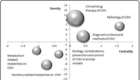
A strategic diagram is a two-dimensional space built by plotting themes according to their centrality and density along two axes [13]. The X-axis represents centrality or the external cohesion index, namely the central position of the theme within the overall network. The Y-axis rep-resents density or the internal cohesion index, namely the conceptual development of the theme [14]. Four quadrants were generated with the X- and Y-axis. The above mentioned six categories were then allocated into these four quadrants according to the results of the clus-tering analysis. Furthermore, excel was used to generate a strategic diagram.
Social network analysis
The high-frequency MeSH terms co-occurrence matrix was imported into the Ucinet 6.0 (Analytic Technologies Co., Lexington, KY, USA) software, after which the SNA method was used to analyze the themes and knowledge structure of CNV. To visualize the network structure, the MeSH term networks were displayed in two dimen-sional maps by the software NetDraw2.084. The nodes of the network are the major MeSH terms/MeSH sub-headings, and the links represent the co-occurrence fre-quency of these terms. To understand the structure of the network on CNV, we evaluated the location of these MeSH terms in the network by measuring the degree, betweenness and closeness centralities of each node.
Results
Distribution characteristics of relevant literatures
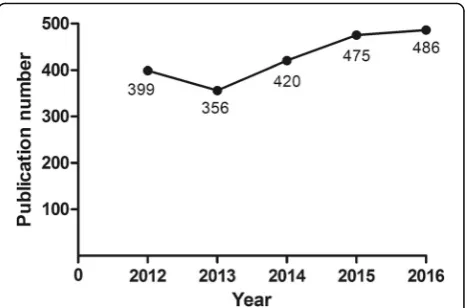
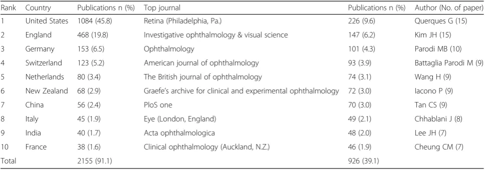
Based on the search strategy, a total of 2366 publications (Jan. 1st, 2012-May. 31st, 2017) were included in this study. In the past 5 years, researchers were paying in-creasing attention on CNV. As shown in Fig. 1, the an-nual publication of articles has gradually increased from 399 in 2012, to 486 in 2016 in the fields of CNV. Altogether, 358 journals have been involved in this field. Table 1 displays the top ten productive journals, which are considered as the core journals in this research area. Among the top ten journals, the top three journals are Retina, Investigative Ophthalmology & Visual Science (IOVS) and Ophthalmology, and these three journals consist more than 20% of the total searched literatures in this field. The US is by far the greatest contributor of ophthalmic researches and institutions from US and
England have conducted more than 60% of the re-searches in this specific area.
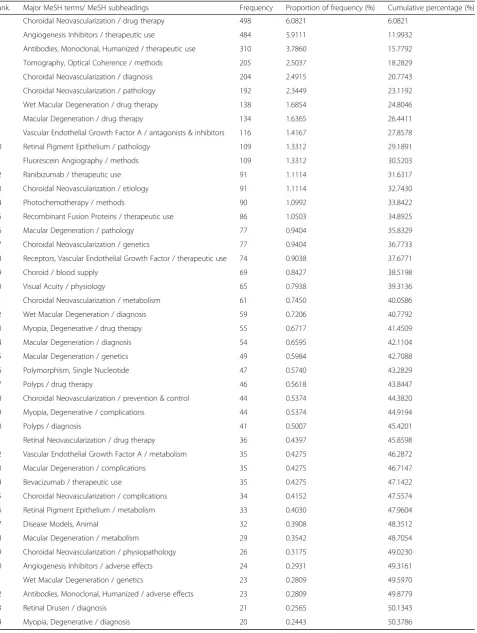
Research hot spots concluded by MeSH term clusters For the included publications, there were 1983 MeSH terms with a cumulative frequency of 8188 times. As shown in Table 2, the cumulative frequency percentage of the 44 high-frequency MeSH terms accounts for 50. 38% (4125/8188) of the total MeSH terms. These MeSH terms could represent the research hot spots on CNV in this past 5 years.
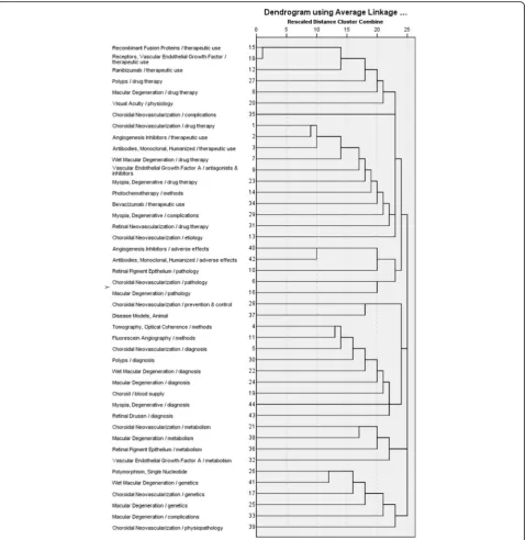
Based on the hierarchical clustering analysis, the MeSH terms were analyzed and classified into 6 categor-ies (Fig. 2 & Table 3). These categories include: (1) Clinical drug therapy of age-related macular degener-ation (AMD), PCV and degenerative myopia, the drugs of which include vascular endothelial growth factor A (VEGFA) inhibitors, and monoclonal antibodies and re-combinant fusion proteins; (2) Study on etiology, com-plications, prevention and control of CNV in animal models; (3) Pathology of CNV and adverse effect of angiogenesis inhibitor; (4) Diagnostic methods, including optical coherence tomography (OCT) and fundus fluor-escein angiography (FFA), and diagnostic criteria of CNV, polyps, drusen and degenerative myopia; (5) Metabolism related researches on CNV, retinal pigment epithelium (RPE) and macular degeneration; (6) Genetics related researches on macular degeneration and degen-erative myopia. These six categories could represent the major research topics in these 5 years.
Strategy diagram for CNV
Motor-themes are those with both strong centrality and high density as shown in Quadrant I (upper-right). Specialized themes are those in Quadrant II (upper-left) and are defined as those with inadequate external inter-actions but high density. Quandrant III (lower-left)
contains themes with weak density and inadequate cen-trality, and these themes are usually considered to be ei-ther appearing or vanishing. The last quadrant, Quandrant IV (lower-right), contains themes with strong centrality but lacking of internal maturation. [13]. In strategic diagrams, themes are represented by spheres of different areas, which are organized in different quad-rants according to their internal and external cohesion (density and centrality, respectively). As shown in Fig.3, the area of the spheres is proportional to the number of high-frequency MeSH terms. Cluster No. 1, 3 and 4 lo-cate in Quadrant I, representing that researches on clin-ical drug therapy, diagnostic criteria and methods, as well as the pathology of CNV are in the core status with high density and centrality. Cluster No. 2, 5 and 6 locate in Quadrant III, indicating that researches on etiology, complications, prevention and control of CNV in animal models, as well as metabolism and genetics related stud-ies on CNV are not mature, namely on the edge of the research field.
Social network analysis of CNV
An SNA is presented in Tables4and 5, and degree, be-tweenness and closeness centrality are applied to analyze the SNA network structure.
In the network of “CNV”, 14 MeSH terms are shown to have a degree centrality more than the mean value of 225.091, and the top ten high-frequency MeSH terms are also included. Among these ten high-frequency MeSH terms, “Angiogenesis Inhibitors/therapeutic use” displays the highest degree centrality of 1374.
The top two betweenness centrality values listed in Table 4 are 41.27 and 38.64, representing “Choroidal Neovascularization/pathology”and“Angiogenesis Inhibi-tors/therapeutic use”, respectively. These two MeSH terms have the strongest mediating role in the network. As shown in Table 5, the mean betweenness centralization is 9.795. MeSH terms “Macular
degeneration/pathology”, “choroid/blood supply”, and “Vascular Endothelial Growth Factor A/metabolism” show relative higher betweenness centrality value of 29. 47, 18.72 and 14.203, respectively. Whereas, the degree centralities of these MeSH terms are 124, 140 and 54, respectively, which are far lower than their mean degree centrality value of 225.091.
As shown in Table4, MeSH terms“Choroidal Neovas-cularization/pathology” and “Angiogenesis Inhibitors/ therapeutic use”both present the top two closeness cen-trality value of 41.5.
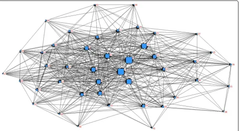
To understand easier, SNA was drawn based on the betweenness centrality. As seen in Fig. 4, the size of nodes indicates the MeSH terms betweenness centrality and the thickness of lines represents the co-occurrence frequency.
Discussion
MeSH terms can reveal the most accurate content of these literatures and a large collection of MeSH terms can reflect the research status of the discipline and trends. Vision Disorders, Glaucoma, Diabetic Retinop-athy, Macular Degeneration, and Cataract are the most frequent MeSH terms related to eye diseases [15]. Through the statistical analysis with BICOMB software, the distribution characteristics of the literatures on “choroidal neovascularization”[MeSH] in recent 5 years maintains a fluctuating increase with a slight decline in 2013. Moreover, our analysis revealed that the US and England are the biggest contributors in the research of CNV, which is consistent with the bibliometric results of other fields [16–18]. This could be explained by the rea-son that English is the first language in these two countries.
In order to systematically examine the fundamental knowledge structure of CNV, this study integrated co-word analysis and SNA based on Bibliometry. According to the co-word analysis, closely related MeSH terms can Table 1Temporal distribution of publications on CNV in PubMed (2012-May 2017)
Rank Country Publications n (%) Top journal Publications n (%) Author (No. of paper)
1 United States 1084 (45.8) Retina (Philadelphia, Pa.) 226 (9.6) Querques G (15)
2 England 468 (19.8) Investigative ophthalmology & visual science 147 (6.2) Kim JH (15)
3 Germany 153 (6.5) Ophthalmology 101 (4.3) Parodi MB (10)
4 Switzerland 123 (5.2) American journal of ophthalmology 93 (3.9) Battaglia Parodi M (9)
5 Netherlands 80 (3.4) The British journal of ophthalmology 74 (3.1) Wang H (9)
6 New Zealand 68 (2.9) Graefe’s archive for clinical and experimental ophthalmology 72 (3.0) Iacono P (9)
7 China 56 (2.4) PloS one 70 (3.0) Tan CS (9)
8 Italy 45 (1.9) Eye (London, England) 49 (2.1) Chhablani J (8)
9 India 40 (1.7) Acta ophthalmologica 48 (2.0) Lee JH (7)
10 France 38 (1.6) Clinical ophthalmology (Auckland, N.Z.) 46 (1.9) Cheung CM (7)
Table 2High-frequency MeSH terms from the included papers on CNV (n= 1983)
Rank. Major MeSH terms/ MeSH subheadings Frequency Proportion of frequency (%) Cumulative percentage (%)
1 Choroidal Neovascularization / drug therapy 498 6.0821 6.0821
2 Angiogenesis Inhibitors / therapeutic use 484 5.9111 11.9932
3 Antibodies, Monoclonal, Humanized / therapeutic use 310 3.7860 15.7792
4 Tomography, Optical Coherence / methods 205 2.5037 18.2829
5 Choroidal Neovascularization / diagnosis 204 2.4915 20.7743
6 Choroidal Neovascularization / pathology 192 2.3449 23.1192
7 Wet Macular Degeneration / drug therapy 138 1.6854 24.8046
8 Macular Degeneration / drug therapy 134 1.6365 26.4411
9 Vascular Endothelial Growth Factor A / antagonists & inhibitors 116 1.4167 27.8578
10 Retinal Pigment Epithelium / pathology 109 1.3312 29.1891
11 Fluorescein Angiography / methods 109 1.3312 30.5203
12 Ranibizumab / therapeutic use 91 1.1114 31.6317
13 Choroidal Neovascularization / etiology 91 1.1114 32.7430
14 Photochemotherapy / methods 90 1.0992 33.8422
15 Recombinant Fusion Proteins / therapeutic use 86 1.0503 34.8925
16 Macular Degeneration / pathology 77 0.9404 35.8329
17 Choroidal Neovascularization / genetics 77 0.9404 36.7733
18 Receptors, Vascular Endothelial Growth Factor / therapeutic use 74 0.9038 37.6771
19 Choroid / blood supply 69 0.8427 38.5198
20 Visual Acuity / physiology 65 0.7938 39.3136
21 Choroidal Neovascularization / metabolism 61 0.7450 40.0586
22 Wet Macular Degeneration / diagnosis 59 0.7206 40.7792
23 Myopia, Degenerative / drug therapy 55 0.6717 41.4509
24 Macular Degeneration / diagnosis 54 0.6595 42.1104
25 Macular Degeneration / genetics 49 0.5984 42.7088
26 Polymorphism, Single Nucleotide 47 0.5740 43.2829
27 Polyps / drug therapy 46 0.5618 43.8447
28 Choroidal Neovascularization / prevention & control 44 0.5374 44.3820
29 Myopia, Degenerative / complications 44 0.5374 44.9194
30 Polyps / diagnosis 41 0.5007 45.4201
31 Retinal Neovascularization / drug therapy 36 0.4397 45.8598
32 Vascular Endothelial Growth Factor A / metabolism 35 0.4275 46.2872
33 Macular Degeneration / complications 35 0.4275 46.7147
34 Bevacizumab / therapeutic use 35 0.4275 47.1422
35 Choroidal Neovascularization / complications 34 0.4152 47.5574
36 Retinal Pigment Epithelium / metabolism 33 0.4030 47.9604
37 Disease Models, Animal 32 0.3908 48.3512
38 Macular Degeneration / metabolism 29 0.3542 48.7054
39 Choroidal Neovascularization / physiopathology 26 0.3175 49.0230
40 Angiogenesis Inhibitors / adverse effects 24 0.2931 49.3161
41 Wet Macular Degeneration / genetics 23 0.2809 49.5970
42 Antibodies, Monoclonal, Humanized / adverse effects 23 0.2809 49.8779
43 Retinal Drusen / diagnosis 21 0.2565 50.1343
Fig. 2Dendrogram of 44 high-frequency MeSH terms
Table 3Cluster analysis of MeSH terms
Cluster Number of MeSH termsa Cluster analysis
1 15,18, 12,27,8,20,1,2,3,7,9,23,14,34,29,31 Clinical drug therapy of AMD, PCV and degenerative myopia
2 35,13,28,37 Study on etiology, complications, prevention and control of CNV in animal models
3 40,42,10,6,16 Pathology of CNV and adverse effect of angiogenesis inhibitor
4 4,11,5,30,22,24,19,44,43 Diagnostic criteria and methods of CNV, polyps, drusen and degenerative myopia.
5 21,38,36,32 Metabolism related researches on CNV
6 26,41,17,25,33,39 Genetics related researches on macular degeneration and degenerative myopia a
be gathered and form clusters. Cluster 1 relates to the drug therapy of wet macular degeneration, degenerative myopia and PCV. Drug therapies mainly focus on mono-clonal antibody and recombinant fusion proteins. In the angiogenesis process, VEGF-A stimulates the growth of the abnormal blood vessels. Medications that can block this protein include ranibizumab, bevacizumab and aflibercept. Ranibizumab is a humanized, monoclonal, VEGF-specific antibody fragment that can prevent it from binding to its receptor, thus inhibiting angiogen-esis. Bevacizumab is a VEGF-specific full-length human-ized monoclonal antibody. The effectiveness and safety of these two medications for the treatment in neovascu-lar AMD have been analyzed to be simineovascu-lar [19]. Aflibercept is a novel fusion protein which binds to VEGF-A, VEGF-B and placental growth factor (PIGF). Since it possesses stronger binding affinity for VEGF than the previous mentioned two medications, it allows longer intervals between treatments [20]. Cluster 3 re-lates to the pathology of CNV, adverse effects of angio-genesis inhibitors and monoclonal antibodies. Ocular neovascularization includes retinal neovascularization and subretinal or choroidal neovascularization. DR and retinal vein occlusions are the most prevalent ischemic retinopathies relate to retinal neovascularization. Subret-inal or choroidal neovascularization occurs in diseases of the outer retina and Bruch’s membrane, the most prevalent of which is AMD [21]. CNV is a common pathological process in a heterogeneous variety of chor-ioretinal disease. Any pathologic changes that involve RPE and damages to Bruch’s membrane can be compli-cated by CNV [22]. Experimental evidences showed that pathogenesis of CNV involve the angiogenesis of vascu-lar component and inflammation of extravascuvascu-lar com-ponent [23]. Today, intravitreal VEGF inhibitors are the mainstay of treatment worldwide. It also covers emer-ging therapies including radiation, latest generation anti-VEGF agents and combination therapies [24]. The most frequently reported ocular adverse events after intravit-real injection are inflammation and increased intraocular
pressure [19]. Cluster 4 relates to the diagnostic methods and criteria of CNV in PCV, wet macular degeneration and degenerative myopia. With the help of diagnostic imaging methods such as OCT and FFA, it is feasible to detect subtle exudation in some individuals who have experienced a recent change in visual acuity [19]. By means of SD OCT (spectral domain optical coherence tomography), outer retinal tubulations (ORTs) mainly present circular or ovoid shape and hyperreflective ma-terial can also be observed in the border. ORTs are the common symbols in eyes with CNV and geographic at-rophy [25]. The emergence of these structures usually indicates the next anti-VEGF drugs application should be considered. However, their non-detection means re-peated intravitreal injection of anti-VEGF drugs is tem-porarily unneeded [26]. These three clusters (Cluster 1, 3, 4) located in Quadrant I, which demonstrates these research hotspots are centralized and well developed.
Cluster 2 relates to etiology, prevention and complica-tions of CNV in animal models. In the eye, many diseases involve angiogenesis, including CNV in AMD and retinal neovascularization in DR and retinopathy of prematurity (ROP) [27]. Numerous studies in mouse models have helped to elucidate the molecular patho-genesis underlying retinal, subretinal and choroidal neo-vascularization. Currently, three animal models are commonly recognized to study ocular neovascularization in diseases of AMD, proliferative diabetic retinopathy (PDR) and ROP [2]. The most commonly used mouse model for analyzing AMD caused by CNV formation is the laser-induced CNV model, even though without the existence of macula in mice. In this model, due to the le-sions caused by laser photocoagulation, new blood ves-sels are formed from choroid to subretinal space, presenting major characteristics of wet AMD. PDR is a most typical form of retinal neovascularization causing vision loss in patients. Factors including persistent high blood pressure, hyperglycemia and hypoxia cause initial damages to retinal capillaries. Subsequently, abnormal neovascularization is established both along the retina, as well as inside the vitreous. The most typical animal model for PDR can be represented by streptozotocin (STZ) intraperitoneal administration in mice. Oxygen-induced retinopathy (OIR) is the typical animal model for studying the ROP. This model is based on the dis-ease mechanisms of the ROP and is used to investigate and analyze the abnormal neovascularization caused by ischemia [2]. Cluster 5 relates to metabolism on CNV. Oxidative stress typically causes endothelial cell dysfunc-tion, pericyte apoptosis and angiogenesis, which further result in retinopathy [28]. Angiogenesis is a complex process whereby interactions between stimulatory and inhibitory factors result in new blood vessel formation. Antiangiogenic therapies function either by blocking
Table 4Individual centrality of CNV research from 2012-May 2017
No. Major MeSH terms / MeSH subheadings Degree Betweenness Closeness
1 Choroidal Neovascularization / drug therapy 1259 22.29 39.50
2 Angiogenesis Inhibitors / therapeutic use 1374 38.64 41.50
3 Antibodies, Monoclonal, Humanized / therapeutic use 818 28.90 40.00
4 Tomography, Optical Coherence / methods 462 18.61 38.50
5 Choroidal Neovascularization / diagnosis 437 12.57 37.00
6 Choroidal Neovascularization / pathology 331 41.27 41.50
7 Wet Macular Degeneration / drug therapy 404 13.67 36.50
8 Macular Degeneration / drug therapy 336 18.31 36.00
9 Vascular Endothelial Growth Factor A / antagonists & inhibitors 350 23.07 39.00
10 Retinal Pigment Epithelium / pathology 242 24.35 38.50
11 Fluorescein Angiography / methods 272 9.26 34.50
12 Ranibizumab / therapeutic use 307 8.82 36.00
13 Choroidal Neovascularization / etiology 97 11.03 36.00
14 Photochemotherapy / methods 219 4.74 34.50
15 Recombinant Fusion Proteins / therapeutic use 322 7.36 36.00
16 Macular Degeneration / pathology 124 29.47 38.00
17 Choroidal Neovascularization / genetics 96 8.05 32.50
18 Receptors, Vascular Endothelial Growth Factor / therapeutic use 292 5.88 35.50
19 Choroid / blood supply 140 18.72 38.00
20 Visual Acuity / physiology 186 6.62 35.50
21 Choroidal Neovascularization / metabolism 57 1.73 27.00
22 Wet Macular Degeneration / diagnosis 135 5.98 33.00
23 Myopia, Degenerative / drug therapy 188 5.38 33.50
24 Macular Degeneration / diagnosis 113 5.46 33.00
25 Macular Degeneration / genetics 65 2.26 30.00
26 Polymorphism, Single Nucleotide 87 3.84 30.50
27 Polyps / drug therapy 173 0.16 29.00
28 Choroidal Neovascularization / prevention & control 40 5.44 30.50
29 Myopia, Degenerative / complications 116 3.11 32.00
30 Polyps / diagnosis 110 0.59 26.83
31 Retinal Neovascularization / drug therapy 90 1.59 30.00
32 Vascular Endothelial Growth Factor A / metabolism 54 14.20 33.50
33 Macular Degeneration / complications 63 4.65 32.50
34 Bevacizumab / therapeutic use 123 2.83 32.50
35 Choroidal Neovascularization / complications 48 3.97 33.00
36 Retinal Pigment Epithelium / metabolism 39 4.63 29.00
37 Disease Models, Animal 44 0.92 28.50
38 Macular Degeneration / metabolism 26 0.79 24.67
39 Choroidal Neovascularization / physiopathology 35 6.35 30.50
40 Angiogenesis Inhibitors / adverse effects 48 0.34 27.50
41 Wet Macular Degeneration / genetics 38 0.12 25.33
42 Antibodies, Monoclonal, Humanized / adverse effects 62 2.52 30.00
43 Retinal Drusen / diagnosis 38 1.04 27.50
stimulatory factors or by promoting inhibitory factors, thus, disrupting the formation of new vessels. Cluster 6 relates to genetics researches on macular degeneration. Gene therapy is a mean of treating diseases and disor-ders caused by gene abnormal expression through the insertion of specific genes in vivo [29]. Recently, the clustered regularly interspaced short palindromic repeats (CRISPR)/CRISPR-associated protein 9 (Cas9) system has been developed as a novel genome-editing tool in numerous medical aspects including ocular diseases [30]. Currently, limitations of gene therapy include inef-ficient and unsustainable target gene expression inside the cells, and concerns of using viral vectors for target gene delivery. However, it is believed to be an evolving technique with comprehensive applications soon in the future. These three clusters located in Quadrant III indi-cate that these existing research hotspots are peripheral and undeveloped, and further study on these themes are recommended in the future.
The SNA result shows that the top ten high-frequency MeSH terms also possess relatively high degree
centrality. According to the measurement of the degree centrality, we conclude that MeSH terms such as “Angiogenesis Inhibitors/therapeutic use” have the most number of direct connections with other components and lead the development in the field of retinopathy. As for the betweenness centrality in this study, “Choroidal Neovascularization/pathology”and“Angiogenesis Inhibi-tors/therapeutic use” are in the hub position of the whole network, which represent these dominant compo-nents have the greatest potential on controlling the co-occurrence of other components. However, hotspots on genetics and the metabolism of CNV are along the edge of the network. This phenomenon indicates that pathology and treatment of CNV put researches on ret-inopathy in motion, whereas the researches about genet-ics and metabolism of CNV are the emerging field. The MeSH terms “Macular degeneration/pathology”, “ chor-oid/blood supply”, and “Vascular Endothelial Growth Factor A/metabolism” show relative high betweenness centralities, whereas the degree centralities are lower than the mean value. This demonstrates that although these components do not show distinct direct relations with other nodes, they occupy the intermediary position and are of great importance in maintaining the stability of the whole network.
Conclusions
In conclusion, hierarchical clustering analysis and stra-tegic diagram can be used to demonstrate the thematic Table 5Descriptive statistics for centrality measure about CNV
Centralization Mean ± SD Min Max Network
centralization
Degree 225.091 ± 284.624 26 1374 8.791%
Betweenness 9.795 ± 10.292 0.118 41.269 3.57%
Closeness 33.235 ± 4.393 24.667 41.500 39.80%
structure of a specific field and estimate the maturing status of each cluster, respectively. However, these two methods fail to decipher the central MeSH term and re-veal the relationship of each component. SNA makes up the deficiency of the aforementioned methods and de-picts the relationships among high-frequency MeSH terms in a system. The size of nodes and the thickness of lines represent the position of the MeSH term in the whole network. The characteristics of these methods are summarized in Supplementary Data 1.
Our study integrated the strategic diagram and SNA based on the co-word analysis of MeSH terms on CNV. Researches on the drug therapy and pathology changes are in the core status, whereas studies on metabolism and genetics are the emerging topics. Although our study could provide some hints for researches in choos-ing the research topics, the results of our analyses are af-fected by some methodological limitations that should be considered. Firstly, the majority of journals and litera-tures included in PubMed are in English thus the non-inclusion of all national journals may also influence the result to some extent. Secondly, many papers could be published in the subspecialty while having little impact on the field. Conversely, some elite journals are ac-knowledged to carry articles of generally high quality and therefore be relatively selective in the articles they publish. Thus, the papers may contribute different weight in the knowledge structure. Thirdly, the co-word analysis is based on high-frequency MeSH terms. Thus, the amount of high-frequency MeSH terms might have some influence on the clustering analysis result, and the new emerging topics with low attention may not have been included. Therefore, analyses combining multiple databases and new emerging topics should be conducted in the future studies.
Abbreviations
BICOMB:Bibliographic item co-occurrence matrix builder; CNV: Choroidal neovascularization; MeSH: Medical subject headings; SNA: Social network analysis
Acknowledgements
The authors would like to thank all reviewers for their valuable comments.
Funding
This study was supported by National Science Foundation for Young Scientists of China (Grant No.81600777). Basic Research Fund for Young Teachers of Higher Education in Liaoning province (Grant No. LQNK201717).
Availability of data and materials
The datasets analysed during the current study are available in the PubMed database.
Authors’contributions
JZ conceived the study. FZ and BS designed the study, carried out the data analyses and finished the manuscript. RL, WZ, DS participated in the interpretation of data and figures. All authors read and approved the final manuscript.
Ethics approval and consent to participate
Since this study did not meet criteria for Human or Animal Subjects Research, no formal ethics approval and consent were required in the present study.
Consent for publication
Not applicable.
Competing interests
The authors declare that they have no competing interests.
Publisher’s Note
Springer Nature remains neutral with regard to jurisdictional claims in published maps and institutional affiliations.
Author details 1
Department of Ophthalmology, The Fourth Affiliated Hospital of China Medical University, Shenyang, China.2Eye hospital of China Medical University, Shenyang, China.3Key Lens Research Laboratory of Liaoning Province, Shenyang, China.4Department of Physiology, China Medical University, Shenyang, China.5Department of Ophthalmology, Second People’s Hospital of Fuxin City, Fuxin, China.
Received: 14 August 2017 Accepted: 23 March 2018
References
1. Zhou Q, Anderson C, Zhang H, Li X, Inglis F, Jayagopal A, Wang S. Repression of choroidal neovascularization through actin cytoskeleton pathways by microRNA-24. Mol Ther. 2014;22(2):378–89.
2. Sulaiman RS, Basavarajappa HD, Corson TW. Natural product inhibitors of ocular angiogenesis. Exp Eye Res. 2014;129:161–71.
3. Andreoli CM, Miller JW. Anti-vascular endothelial growth factor therapy for ocular neovascular disease. Curr Opin Ophthalmol. 2007;18(6):502–8. 4. Zampros I, Praidou A, Brazitikos P, Ekonomidis P, Androudi S. Antivascular
endothelial growth factor agents for neovascular age-related macular degeneration. J Ophthalmol. 2012;2012:319728.
5. Yao Q, Chen K, Yao L, Lyu PH, Yang TA, Luo F, Chen SQ, He LY, Liu ZY. Scientometric trends and knowledge maps of global health systems research. Health Res Policy Syst. 2014;12:26.
6. Hong Y, Yao Q, Yang Y, Feng JJ, Wu SD, Ji WX, Yao L, Liu ZY. Knowledge structure and theme trends analysis on general practitioner research: a co-word perspective. BMC Fam Pract. 2016;17:10.
7. Li F, Li M, Guan P, Ma S, Cui L. Mapping publication trends and identifying hot spots of research on internet health information seeking behavior: a quantitative and co-word biclustering analysis. J Med Internet Res. 2015;17(3):e81. 8. Leal W, Llanos EJ, Restrepo G, Suarez CF, Patarroyo ME. How frequently do
clusters occur in hierarchical clustering analysis? A graph theoretical approach to studying ties in proximity. J Cheminform. 2016;8:4.
9. Zhang C, Yu Q, Fan Q, Duan Z. Research collaboration in health management research communities. BMC Med Inform Decis Mak. 2013;13:52.
10. Piraveenan M, Prokopenko M, Hossain L. Percolation centrality: quantifying graph-theoretic impact of nodes during percolation in networks. PLoS One. 2013;8(1):e53095.
11. Gao C, Lan X, Zhang X, Deng Y. A bio-inspired methodology of identifying influential nodes in complex networks. PLoS One. 2013;8(6):e66732. 12. Landis JR, Koch GG. The measurement of observer agreement for
categorical data. Biometrics. 1977;33(1):159–74.
13. Viedma-Del-Jesus MI, Perakakis P, Munoz MA, Lopez-Herrera AG, Vila J. Sketching the first 45 years of the journalPsychophysiology(1964-2008): a co-word-based analysis. Psychophysiology. 2011;48(8):1029–36. 14. Li HY, Cui L, Cui M. Hot topics in Chinese herbal drugs research
documented in PubMed/MEDLINE by authors inside China and outside of China in the past 10 years: based on co-word cluster analysis. J Altern Complement Med. 2009;15(7):779–85.
15. Boudry C, Denion E, Mortemousque B, Mouriaux F. Trends and topics in eye disease research in PubMed from 2010 to 2014. PeerJ. 2016;4:e1557. 16. Glynn RW, Scutaru C, Kerin MJ, Sweeney KJ. Breast cancer research output,
1945-2008: a bibliometric and density-equalizing analysis. Breast Cancer Res. 2010;12(6):R108.
17. Zhang XC, Huang DS, Li F. Cancer nursing research output and topics in the first decade of the 21st century: results of a bibliometric and co-word cluster analysis. Asian Pac J Cancer Prev. 2011;12(8):2055–8.
19. Solomon SD, Lindsley K, Vedula SS, Krzystolik MG, Hawkins BS. Anti-vascular endothelial growth factor for neovascular age-related macular
degeneration. Cochrane Database Syst Rev. 2014;8:CD005139. 20. Holash J, Davis S, Papadopoulos N, Croll SD, Ho L, Russell M, Boland P,
Leidich R, Hylton D, Burova E, et al. VEGF-trap: a VEGF blocker with potent antitumor effects. Proc Natl Acad Sci U S A. 2002;99(17):11393–8. 21. Campochiaro PA. Molecular pathogenesis of retinal and choroidal vascular
diseases. Prog Retin Eye Res. 2015;49:67–81.
22. Campa C, Costagliola C, Incorvaia C, Sheridan C, Semeraro F, De Nadai K, Sebastiani A, Parmeggiani F. Inflammatory mediators and angiogenic factors in choroidal neovascularization: pathogenetic interactions and therapeutic implications. Mediat Inflamm. 2010;2010.https://doi.org/10.1155/2010/ 546826.
23. Spaide RF. Rationale for combination therapy in age-related macular degeneration. Retina. 2009;29(6 Suppl):S5–7.
24. Villegas VM, Aranguren LA, Kovach JL, Schwartz SG, Flynn HW Jr. Current advances in the treatment of neovascular age-related macular degeneration. Expert Opin Drug Deliv. 2017;14(2):273–82.
25. Preti RC, Govetto A, Filho RGA, Cabral Zacharias L, Gianotti Pimentel S, Takahashi WY, Monteiro MLR, Hubschman JP, Sarraf D. OPTICAL COHERENCE TOMOGRAPHY ANALYSIS OF OUTER RETINAL TUBULATIONS: sequential evolution and pathophysiological insights. Retina. 2017.https:// doi.org/10.1097/IAE.0000000000001810.
26. Matuskova V. Retinal tubulation. Ceska a Slovenska Oftalmologie. 2015; 71(2):83–6.
27. Zhang SX, Ma JX. Ocular neovascularization: implication of endogenous angiogenic inhibitors and potential therapy. Prog Retin Eye Res. 2007;26(1):1–37. 28. Li C, Miao X, Li F, Wang S, Liu Q, Wang Y, Sun J. Oxidative stress-related
mechanisms and antioxidant therapy in diabetic retinopathy. Oxidative Med Cell Longev. 2017;2017:9702820.
29. Dalkara D, Goureau O, Marazova K, Sahel JA. Let there be light: gene and cell therapy for blindness. Hum Gene Ther. 2016;27(2):134–47. 30. Hung SS, McCaughey T, Swann O, Pebay A, Hewitt AW. Genome
engineering in ophthalmology: application of CRISPR/Cas to the treatment of eye disease. Prog Retin Eye Res. 2016;53:1–20.
• We accept pre-submission inquiries
• Our selector tool helps you to find the most relevant journal
• We provide round the clock customer support
• Convenient online submission
• Thorough peer review
• Inclusion in PubMed and all major indexing services • Maximum visibility for your research
Submit your manuscript at www.biomedcentral.com/submit