Abstract
Objective: LIGHT (TNFSF 14) belongs to the tumor necrosis factor superfamily and is expressed by activat-ed T cells as well as various types of antigen present-ing cells. LIGHT binds to its cellular receptors TR2 and LTßR and has a co-stimulatory role in T cell acti-vation. Here, we compared the relative expression of LIGHT in different immune cells and the biological activity of immune cell-derived LIGHT on endothelial cells.
Methods and Results: Surface expression of LIGHT and mRNA production by PBMC and isolated T cells (CD4+ or CD8+) significantly increased after
stimula-tion with PMA (Phorbolester-12- Myristat-13-Acetat) + ionomycin. No LIGHT expression on PMA stimu-lated monocytes or monocytic-like THP-1 cells could be detected; differentiation of monocytes and THP-1 cells into macrophages, however, resulted in up-regula-tion of LIGHT. Supernatants of stimulated T cells contained higher concentrations of soluble LIGHT than macrophage supernatants normalized to cell numbers; release of soluble LIGHT was found to be dependent on metalloproteinase activity. Size determi-nation of released soluble LIGHT by size exclusion chromatography revealed a molecular mass of ~60 kDa, suggesting a trimeric form. Released soluble LIGHT induced expression of proinflammatory anti-gens ICAM-1, tissue factor and IL-8 in human en-dothelial cells and caused apoptosis of IFN-γ pretreat-ed endothelial cells. Soluble LIGHT was detectpretreat-ed at low levels in sera of healthy controls and was signifi-cantly enhanced in sera of patients with chronic he-patitis C and rheumatoid arthritis (24.93 ± 9.41 vs. 129.53 ± 49.14 and 172.13 ± 77.64; p< 0.0005).
Conclusion: These findings suggest that among im-mune cells activated T lymphocytes are the main source of soluble LIGHT with released amounts of soluble LIGHT markedly higher compared to platelets. Immune cell-derived membrane-bound and soluble trimeric LIGHT is biologically active, inducing proinflammatory changes in endothelial cells. En-hanced plasma levels of soluble LIGHT in patients with chronic infections suggest a role of LIGHT in systemic inflammatory activation.
Key words:LIGHT, endothelial cells, inflammation
I
NTRODUCTIONThe tumor necrosis factor related cytokines provide es-sential communication pathways that help orchestrate inflammatory and immune responses. They play an in-tegral role in regulation of innate and adaptive immu-nity [1, 2]. LIGHT belongs to the tumor necrosis su-perfamily and acts as a co-stimulatory molecule for T cells, including the enhancement of T cell proliferation and secretion of IFN-γ. LIGHT exists in a membrane-bound and soluble form. It is a ligand for TR2, LTßR and TR6, all of which are TNF receptor family mem-bers. Studies in animal models suggest that LIGHT signaling pathways may be crucial for the development of various autoimmune disorders, at least in part be-cause of their effects on T cells and T-cell homing into inflamed tissues [3, 4]. In an experimental mouse mod-el it was shown that soluble LIGHT is involved in the pathogenesis of hepatitis via LIGHT-LTßR interaction [5]. Several studies suggest that LIGHT is involved in atherogenesis via induction of proatherogenetic cy-tokines and decreasing plaque stability by inducing metalloproteinase activity in macrophages [6].
Recently, Otterdal et al. [7] reported that LIGHT was associated with platelets und released upon activa-tion. Thrombus material obtained at the site of plaque rupture in patients with STEMI (ST segment elevation myocardial infarction) contained platelet-derived LIGHT, suggesting platelets as the origin of LIGHT. In line with these findings we previously showed that the adhesion of platelets to endothelial cells is mediat-ed by platelet-LIGHT [8]. Furthermore, patients with STEMI showed enhanced plasma levels of soluble LIGHT compared to healthy controls [5, 10]. Recently, it was shown that concentrations of platelet-derived cytokines are markedly influenced by preclinical condi-tions and may be released only ex vivo [9]; raising the question if soluble LIGHT in patient sera really origi-nates from platelets or possibly from other cell types, e. g. lymphocytes or macrophages. Similarly, the origin of circulating soluble LIGHT in other human autoim-mune or inflammatory disease states (rheumatoid arthritis, infection) has not yet been studied – leaving the relative contribution of different cell types to cir-culating soluble LIGHT unresolved.
In the present study we analyzed different immune cells for expression and the release of membrane-bound and soluble LIGHT to quantify the different
Eur J Med Res (2009) 14: 147-156 © I. Holzapfel Publishers 2009
P
ROINFLAMMATORY AND
P
ROTHROMBOTIC
E
FFECTS ON
H
UMAN
V
ASCULAR
E
NDOTHELIAL
C
ELLS OF
I
MMUNE
-
CELL
-
DERIVED
LIGHT
S. Celik1*, V. Shankar1*, A. Richter1, H.-J. Hippe1, M. Akhavanpoor1, F. Bea1, C. Erbel1, S. Urban2,
N. Blank3, N. Wambsganss1, H. A. Katus1, T. J. Dengler1 1Department of Cardiology, University of Heidelberg, INF 410, Heidelberg,
2Department of Molecular Virology, Otto Meyerhof Zentrum, University of Heidelberg, INF350, Heidelberg,
3Department of Rheumatology, University of Heidelberg, INF 410, Heidelberg, Germany
sources of LIGHT. Our findings show that T lym-phocytes show high expression levels of membrane-bound and release large amounts of soluble LIGHT while monocytes and THP-1 cells only begin to express LIGHT upon differentiation into macro-phages. Release of soluble LIGHT is shown to be matrix metalloproteinase-dependent, soluble LIGHT appears to be shed as a trimeric form which is biologi-cally active.
Finally, enhanced serum levels of soluble LIGHT were detected in patients with chronic inflammatory disorders.
M
ETHODS CELLCULTUREHuman umbilical vein endothelial cells were isolated from umbilical cords by enzymatic digestion as previ-ously described [10] and serially cultured in medium.
PBMC were obtained from healthy adult volunteers by density gradient centrifugation of heparinized ve-nous blood. Mononuclear cells were collected from the interphase after Ficoll separation (Amersham Pharmacia Biotech, Freiburg, Germany). For the isola-tion of the single subpopulaisola-tions of PBMCs magnetic Micro-Beads (Miltenyi Biotec, Bergisch Gladbach, Germany) were used according to the manufacturer’s instructions. A total of 2.5 x 10⁶T cells per ml were
stimulated with 100 ng/ml PMA (Sigma, Germany) and 1 µg/ml ionomycin (Sigma) for different time of points. Human monocytic cell line THP-1 was cul-tured in RPMI medium.
THP-1 cells were differentiated by overnight incu-bation with PMA (100 ng/ml).
PMA-differentiated THP-1 macrophages were stim-ulated in 12-well plates (2.5 x 10⁶ cells/ml, Nunclon)
with TNF-α(10 ng/ml; Sigma, Germany) for 2, 4 and 16 hours.
Monocytes were isolated from PBMC using mag-netic beads and cultured in RPMI medium in the pres-ence of cytokine MCSF at a concentration 100 nM for 6 days to generate macrophages. Differentiation of monocytes into macrophages was confirmed by the surface expression of CD80 and CD86. Stimulation of macrophages was performed in 12-well plates (5 x 10⁶
cells/ml) with TNF-α (10 ng/ml) for 2, 4 and 16 hours.
For the generation of the supernatants 2.5 x 10⁶
cells in 1 ml medium of each cell were activated re-spectively for 24 and 48 hours.
LIGHTCDNA, CONSTRUCTION OF LIGHT-CONTAININGVECTOR ANDCELLTRANSFECTION Total RNA isolated from T cells was used to generate cDNA by RT-PCR (MBI-Fermentas). Total RNA was prepared from T cells using QiaAmp RNA blood mini kit. LIGHT cDNA was amplified by PCR using pfu DNA polymerase (Stratagene). The size of LIGHT cDNA was 731 bp. LIGHT cDNA was then cloned in to plasmid pcDNA 3.1D/V5-His-TOPO (Invitrogen, Germany) to generate pTOPOLIGHT. The integrity of pTOPOLIGHT was confirmed by restriction di-gestion analysis and sequencing.
Transfection of 293 cells
293 cells were transfected with plasmid pTopoLIGHT using Superfectin reagent (Qiagen) as recommended by the manufacturer.
GELEXCLUSIONCHROMATOGRAPHY
Isolation of soluble LIGHT from culture supernatant of transiently LIGHT-expressing 293 cells or PMA + ionomycin stimulated PBMC and determination of apparent molecular weights of proteins and protein complexes were achieved by size exclusion chromatog-raphy on a calibrated Superdex 200 column (1.6 _ 60 cm; Pharmacia), connected to a FPLC system (Phar-macia). Sample volumes were 0.5 ml for analytical and up to 2 ml for preparative purposes. Eluted proteins were collected in fractions of 2.2 ml, concentrations determined by ELISA subjected to SDS-PAGE, and analyzed by silver staining or immunoblotting. (3 frac-tions of isolated soluble LIGHT from PBMC super-natant were pooled and used for endothelial cell stim-ulation assays).
FILTRATION OFLIGHT CONTAININGSUPERNATANTS LIGHT containing medium (500 µl) was collected from stimulated PBMC, pipetted into the sample reser-voir of Microcon 30 kDa, 50 kDa and 100 kDa cut-off centrifugal filter device (Millipore Corp., Bedford, Massachusetts, USA) and centrifuged according to the manufacturer’s instructions. For the calculation of per-cent filtrate and retentate recovery a corresponding formula provided by the manufacturer was used.
WESTERNBLOTANALYSIS
Western blotting was performed according to standard procedures. For western blotting of the supernatants of activated PBMCs, Microcon centrifugal filters YM50 were used to concentrate soluble LIGHT and then suspended in SDS sample buffer for SDS-PAGE (12-15%). The separated proteins were transferred electrophoretically to nitrocellulose membranes, and immunodetection was carried out using specific anti-bodies against LIGHT. Peroxidase-conjugated donkey anti-rabbit were used, and detection was carried out with an enhanced chemiluminescence (ECL) reagent (Pierce). Chemiluninescence was quantified using a FluorS-MultiImager (BioRad, Munich, Germany).
FLOWCYTOMETRY
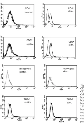
Expression of LIGHT on THP-1 cells, CD4+, CD8+
and CD14+was measured by indirect
immunofluores-cence flow cytometric analysis.
Cells were processed following standard procedures as described before [11]. A total of 5 x 10⁵cells were
stimulated with PMA (Phorbolester-12- Myristat-13-Acetat; 100 ng/ml) + ionomycin (1µg/ml) or TNF-α
REALTIME-PCR ANALYSIS OF MRNA EXPRESSION The mRNA expression of ICAM-1, IL-8, ß-Actin and LIGHT was evaluated by Real Time PCR with Light-Cycler (Roche, Germany) according to the manufac-turer`s instructions.
A long (NM_003807) and short (NM_172014) form of LIGHT RNA has been reported earlier. To distinguish both types of RNA, PCR primers which spans the region missing in the shorter form was de-signed. By using RT-PCR, the presence of longer or a shorter RNA form was detected by amplicon having product sizes of 194 bps and 84 bps respectively.
DETERMINATION OFTISSUEFACTORACTIVITY IN HUVECBYONE-STAGECLOTTINGASSAY One-stage clotting assays were performed as previously described [12]. Human endothelial cells (106/well) were
incubated with isolated soluble LIGHT for 6 hours. TF activity was calculated by comparing the measured clotting time with a standard curve made with known amounts of TF. The measured amount of TF was ex-pressed as picograms of TF/106cells ± S.D.
ELISA ASSAYS
Plasma levels of LIGHT in patients with hepatitis C and rheumatoid arthritis and LIGHT expression in the supernatants of stimulated cells was performed by ELISA according to the manufacturer’s instructions ((R&D Systems, Wiesbaden, Germany)
APOPTOSISASSAY
Endothelial cells (10 x 105 cells / well) were cultured
with endothelial cell medium and preincubated with IFN-γ (250 U/ml) for 24 hours and then stimulated with cycloheximide 2.5 µg/ml (ICN Biomedicals) in combination with TNF-α (10 ng/ml) or recombinant LIGHT (100 ng/ml).
After stimulation for 24 hours the cells were har-vested and washed in PBS following fixation in cold 70% ethanol. After fixation for at least 30 min at 4 °C, cells were washed twice with PBS and stained with 50 µg/ml RNase and 50 µg/ml propidium iodide (Sigma) and analyzed by FACS.
PATIENTS
22 patients, 15 females, age range 25 – 69 years, with rheumatoid arthritis defined by American College of Rheumatology criteria [13], were identified in rheuma-tology clinic with approval from the local research ethics committee.
10 patients (6 female; age range 18 – 50 years) with chronic hepatitis C infection (defined by positive serum antibodies to HCV by means of a second or third generation HCV enzyme linked immunosorbent assay and detectable serum HCV RNA) were recruited to the study. Plasma samples from all individuals were collected and stored at -80 °C until analysis.
16 healthy volunteers (10 female; age range 20 - 48 years) with no previous diagnosis of RA or other
chronic inflammatory diseases served as normal con-trols.
Participating subjects gave written informed con-sent. The investigation conforms to the principles out-lined in the Declaration of Helsinki.
DATAPRESENTATION ANDSTATISTICS
Comparisons between group means were performed using Mann-Whitney U-test, Student t-test or ANO-VA analysis as appropriate. Data are presented as mean + SD. P < 0.05 was considered as statistically significant.
R
ESULTSEXPRESSION OFLIGHT ONDIFFERENTACTIVATED IMMUNECELLS
Stimulation of purified CD4+ and CD8+ T cells with
PMA (100 ng/ml) + ionomycin (1µg/ml) induced a marked increase in surface expression of LIGHT. No LIGHT expression could be detected on monocytes and undifferentiated THP-1 cells after stimulation with TNF-α(10 ng/ml). All cells were stimulated for 24 hours and analyzed by flow cytometry (Fig.1).
MRNA EXPRESSION OFLIGHTINACTIVATED IMMUNECELLS
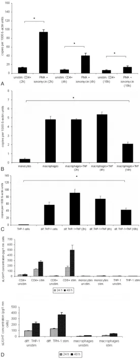
mRNA LIGHT expression in resting and stimulated T cells (PMA 100 ng/ml + ionomycin 1µg/ml ), mono-cytes, macrophages and differentiated THP-1 cells (TNF-α10 ng/ml) following specific time patterns (2, 4 and 16 hours) was evaluated by RT-PCR. LIGHT levels increased in response to stimulation with a max-imum at 2 – 4 hours after activation in T cells, macrophages and differentiated THP-1 cells (Fig. 2A – 2 C). No mRNA expression for LIGHT was seen in undifferentiated monocytes. In all mRNA / PCR ex-periments only a single band of LIGHT cDNA was detected, even using specifically designed primers to detect a potential differentially spliced mRNA form (see methods).
RELEASE OFSOLUBLELIGHTBYACTIVATEDIMMUNE CELLS
Secretion of soluble LIGHT into the supernatants of PBMC or stimulated CD4+ and CD8+ (PMA 100
ng/ml + ionomycin 1 µg/ml) monocytes, macro-phages, THP-1 cells and differentiated THP-1 (TNF-α
10 ng/ml) cells was analyzed by ELISA. Significant concentrations of soluble LIGHT could be detected in the supernatants of PBMC or CD4+and CD8+cells
and differentiated THP-1 cells, which increased markedly upon cell activation, but not in monocytes and undifferentiated 1 cells. Differentiated THP-1 and T cell supernatants contained similar amounts of soluble LIGHT. Supernatant concentrations of LIGHT were higher in the CD8+subset than in CD4+
In all cell types the specificity of ELISA results was supported by identical findings after ultrafiltration (30000 rpm, 12 h).
SOLUBLELIGHTISGENERATED THROUGH PROTEOLYTICCLEAVAGE OF MLIGHTBYMATRIX
METALLOPROTEINASEINHIBITORS
The involvement of metalloproteinases in the regula-tion of soluble LIGHT was investigated by testing the effect of a broad-spectrum synthetic metallopro-teinase inhibitor (GM6001, a hydroxamate inhibitor of MMPs). GM6001 (30µM) inhibited significantly the release of soluble LIGHT into the supernatant of pe-ripheral blood mononuclear cells and isolated CD4+T
cell subset (Fig. 3A and B) when added 30 minutes pri-or and during the stimulation with PMA 100 ng/ml + ionomycin 1 µg/ml.
MONOMERIC SIZE OFSOLUBLELIGHT Western blot analysis (reducing conditions) of lympho-cyte derived soluble LIGHT revealed a molecular mass of approximately ~23 - 25 kDa, similar to the molecu-lar mass of recombinant soluble LIGHT (25 kDa), which represents only the extracellular part (Fig. 4).
SIZEDETERMINATION OFSOLUBLELIGHT For initial estimation of the molecular mass of soluble LIGHT filters with a molecular weight cut- off values of > 100 kDa, 50 kDa and 30 kDa were used. Reten-tate fraction of soluble LIGHT in microcon filters with 30 kDa and 50 kDa molecular weight cut off was about 90%. Spinning of soluble LIGHT through a fil-ter with a 100 kDa MWCO resulted in a ~ 30% reten-tate fraction, indicating a molecular mass of soluble LIGHT between 50 – 100 kDa.
Size determination of lymphocyte-derived soluble LIGHT by size exclusion chromatography on a cali-brated Superdex 200 gel filtration column resulted in a molecular mass of approximately 60 kDa, indicating a trimeric form of soluble LIGHT. The apparent mole-cular mass of soluble LIGHT was calculated from standard curves as a mean value of three independent measurements (data not shown).
INFLAMMATORYEFFECTS OFLYMPHOCYTEDERIVED LIGHTONHUVEC
To investigate the biological activity of cell-derived sol-uble LIGHT, endothelial cells were incubated with LIGHT-containing supernatants derived from activated
Fig. 2A – 2C. mRNA expression of LIGHT in immune cells. T cells were stimulated with PMA and ionomycin for 2, 4 or 16 hours. PMA differenti-ated THP-1 cells, monocyte-derived macrophages were further stimulated with TNF-αfor 2, 4 or 16 hours. Expression of LIGHT was assessed at differ-ent time points by RT-PCR. Maximum of LIGHT expression in T cells was observed after 2 hours after stimulation (Fig. 2A). *P < 0.05 vs unstimulated cells. In differentiated THP-1 cells and monocyte-derived macrophages maximum LIGHT expression peaked after 4 hours at the mRNA-level (Fig. 2B and 2C). *P < 0.05 vs monocytes and THP-1 cells. These data correspond to one representative experiment from three performed.
A
B
C
lymphocytes (PMA 100 ng/ml + ionomycin 1µg/ml). After stimulation of HUVECs for 3 hours a marked up-regulation of ICAM-1 and IL-8 was shown by RT-PCR (Fig. 5A). Similar results were obtained with solu-ble LIGHT isolated by size exclusion chromatography (Fig. 5B). Pre-incubation of LIGHT-containing super-natants with anti-LIGHT mAb resulted in a partial
re-duction of these cytokines; with isolated soluble LIGHT antibody preincubation resulted in almost complete inhibition of up-regulation of ICAM-1 and IL-8. Membrane-bound LIGHT of stimulated PBMC was shown to induce up-regulation of ICAM-1 and IL-8 when co-incubating whole-cell PBMC with endothe-lial cells (Fig. 5C). Pre-incubation of PBMC with
anti-Fig. 4.Monomeric size of soluble LIGHT.To determine the monomoric size of soluble LIGHT, supernatants of PMA (100 ng/ml + ionomycin 1 µg/ml) activated PBMCs were collected after 48 hours and concentrated with Microcon centrifugal fil-ters YM50. Soluble LIGHT production in supernatant was then assessed by ELISA. Molecular mass of monomeric LIGHT was detected by Western blotting. 50 ng of recombinant sLIGHT were loaded in one lane as a control (Fig. 4).
왗
Fig. 5A – 5C.Induction of ICAM-1 and IL-8 by lympho-cyte-derived membrane-bound and soluble LIGHT in hu-man endothelial cells.Human endothelial cells were co-incu-bated in supernatant containing soluble LIGHT, isolated solu-ble LIGHT and stimulated (PMA 100ng/ml + ionomycin 1 µg/ml) and unstimulated whole lymphocytes for 3 hours. Thereafter, HUVECS were harvested and analyzed for expres-sion of the adheexpres-sion molecule ICAM-1 and IL-8 by real-time PCR. After stimulation ICAM-1 and IL-8 were significantly in-creased in all experiments (Fig. 5A – 5C). To confirm that the stimulation of the endothelial cells was due to LIGHT, specific anti-LIGHT-antibodies were used. After pre-incubation of whole lymphocytes and LIGHT containing medium with anti-LIGHT antibody, cytokine induction was partially abolished. After pre-treatment of isolated LIGHT containing medium with anti LIGHT, the effects were even completely abolished (Fig. 5B). These data correspond to one representative experi-ment of three performed. *P < 0.05 and **P < 0.005 vs stimu-lated cells without blocking antibody.
Fig. 3A – 3B. Matrix metalloproteinase (GM6001) mediates shedding of soluble LIGHT.30 minutes prior to activation of PBL and CD4+ subsets with PMA metalloproteinase inhibitor GM6001 (30 µM) was added to the medium. After 24 hours and 48 hours the conditioned medium was analyzed for the presence of soluble LIGHT by ELISA. MMP inhibitor markedly reduced the re-lease of soluble LIGHT in lymphocytes (Fig. 3A and 3B). Values represent the mean ± SD of soluble LIGHT analyzed in triplicate. ***P < 0.0005 vs without GM6001 treated cells.
A B
5A
LIGHT resulted in partial inhibition of ICAM-1 and IL-8 up-regulation, suggesting a substantial contribu-tion of cell-bound LIGHT to this up-regulacontribu-tion. Simi-lar results were obtained when using PMA-stimulated CD4+-T cells (data not shown). Furthermore,
induc-tion of tissue factor activity by isolated soluble LIGHT in endothelial cells could be shown by one stage-clot-ting assay (Fig. 5D), which again was inhibitable by an-tibody pre-incubation.
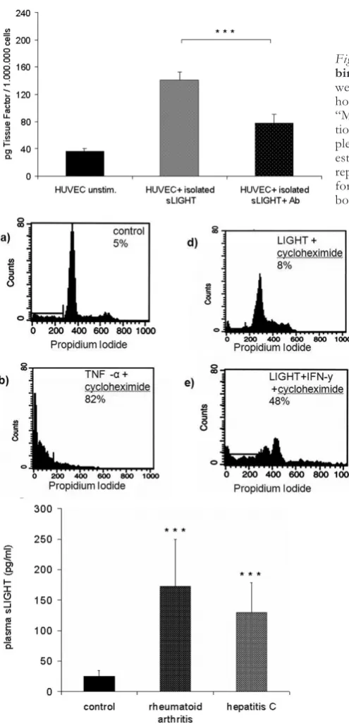
LIGHT-MEDIATEDAPOPTOSIS INHUVEC There have been many studies addressing the role of LIGHT in apoptosis, especially with tumour cell lines expressing both receptors TR2 and LTßR. LIGHT alone did not induce apoptosis in endothelial cells.
Since previous studies reported that LIGHT, in com-bination with IFN-γinduced cell death through LTßR in several cancer cell lines, endothelial cells were prein-cubated with IFN-γ (25 U/ml) for 24 hours prior to apoptosis induction. Such IFN-γpretreated endothe-lial cells were fully susceptible to LIGHT-mediated apoptosis (Fig. 6). IFN-γ alone had no effect on en-dothelial cells (not shown).
INCREASEDPLASMALEVELS OFLIGHTINHEPATITIS CANDRHEUMATOIDARTHRITISPATIENTS Involvement of LIGHT in human atherosclerosis, synovialitis of rheumatoid arthritis or pathogenesis of experimental rodent liver inflammation has been re-ported. To elucidate the potential systemic relevance
Fig. 5D. Induction of tissue factor (TF) activity by recom-binant LIGHT in human endothelial cells.Endothelial cells were stimulated with isolated soluble LIGHT, harvested after 6 hours and assayed for procoagulant activity as described under “Materials and Methods”. LIGHT induced a marked up-regula-tion of tissue factor in endothelial cells. TF activity of each sam-ple was determined based on comparison with a standard curve established with known amounts of recombinant TF. The data represent the mean of three independent experiments per-formed (Fig. 5D). ***P < 0.0005 vs stimulated cells without bocking antibody.
Fig. 6.Induction of apoptosis in endothelial cells by re-combinant LIGHT.LIGHT induced apoptosis endothe-lial cells after preincubation with IFN-γ. Endothelial cells were incubated with medium, soluble LIGHT, TNF-α, cy-cloheximide alone or in combination with TNF-α or LIGHT for 24 hours. Thereafter harvested and analyzed for propidium iodide exclusion by FACS analysis. IFN-γplus cycloheximide alone did not induce apoptosis (control). LIGHT combined with cycloheximide induced apoptosis in endothelial cells after pre-treatment with IFN-γ (Fig. 6). IFN-γalone had no effect on endothelial cells (not shown). The data represent the mean ± SD of three independent ex-periments performed.
of soluble LIGHT in inflammatory diseases like he-patitis C or rheumatoid arthritis, plasma levels of LIGHT of these patients was examined by ELISA (Fig. 7). While healthy controls showed low, but dis-tinct serum concentrations of soluble LIGHT (n = 16), patients with hepatitis C (n = 10) and rheumatoid arthritis (n= 22) showed several-fold increased plasma levels of soluble LIGHT. (Hepatitis C: 129.53 ± 49.14 pg/ml, rheumatoid arthritis: 172.13 ± 77.64 pg/ml; both p < 0.0005).
D
ISCUSSIONThe tumor necrosis factor superfamily member LIGHT plays an important role in regulating the im-mune response and LIGHT may play a prominent role in predominantly T cell mediated pathologies. Several studies have demonstrated the expression of LIGHT in different cell types and its potential role in inflam-matory diseases [15, 16]. Recently, we have shown that platelet-associated LIGHT is involved in the adhesion of platelets to endothelial cells under static and high-shear dynamic flow conditions [11]. Furthermore our result showed enhanced plasma levels of LIGHT in patients with acute myocardial infarction. These find-ings were in agreement with the results of Otterdal et al.10 and strengthen the notion of the potential role of platelet-derived LIGHT in coronary heart disease. Al-though the expression of LIGHT has been shown in different cell types, including platelets, the relative contribution of various cellular origins to soluble LIGHT as well as the relevance and biological activity of soluble LIGHT secreted by different cells has not been investigated. In this study we compared the quantitative expression of LIGHT in different im-mune cells and the biological activity of soluble LIGHT upon endothelial cells as an exemplary read-out system.
Here, we could show that among the immune cells, CD4+/CD8+ T cells are the main source of soluble
LIGHT. Circulating monocytes do not express LIGHT, but differentiation into macrophages resulted in expression of LIGHT, even more so in differentiat-ed THP-1 cells. Secretion of soluble LIGHT was ma-trix metalloproteinase-dependent and led to inflamma-tory changes in endothelial cells measured as up-regu-lation of adhesion molecules (ICAM-1), pro-coagulant factors (TF) and release of chemokines (IL-8). Size determination of soluble LIGHT revealed a molecular mass of approximately 60 kDa, suggesting a trimeric form similar to other TNFSF members. Furthermore, patients with chronic inflammatory diseases such as hepatitis C and rheumatoid arthritis showed enhanced concentrations of soluble LIGHT compared to healthy controls.
Basal cellular expression of LIGHT on mRNA and surface protein levels was significantly increased in pe-ripheral blood mononuclear cells or isolated CD4+/
CD8+T cells after stimulation. Stimulated monocytes
or monocyte-like THP-1 cells lacked expression of LIGHT, but differentiation of THP-1 cells into macro phages resulted in a marked upregulation of LIGHT. Monocyte-derived macrophages showed only a weak expression of LIGHT.
Soluble LIGHT could be detected in large amounts in the supernatants of CD4+/ CD8+T cells as well as
differentiated THP-1 cells, but not in the supernatants of THP-1 cells and monocytes. Ultrafiltration experi-ments excluded membrane fragexperi-ments of ruptured / dead cells as the source of soluble LIGHT. In recently published data involvement of platelet-derived LIGHT in acute coronary syndromes was suggested [10, 11], but based on recently published data by Ivandic et al. [12] that release of platelet-mediated cy-tokines are mostly influenced by preanalytical condi-tions through ex vivo activation, the actual relevance and contribution of platelet- derived LIGHT in ather-osclerosis might have been misinterpreted or overesti-mated. Clinical relevance of other platelet-derived in-flammatory cytokines was shown for soluble CD40 ligand [17]; increased concentrations of sCD40L were reported mostly in disorders associated with platelet activation such as acute and stable coronary artery dis-ease [18, 19]. However, Ivandic et al.12 recently re-ported that increased sCD40 L concentrations by platelets were dependent on sample preparation; no disease-related in vivo activation of platelets was ob-served.
LIGHT has been claimed that it exists in several distinct molecular forms, which are directed to distinct cellular compartments, including the extracullar space, the membrane, and the cytosol [20], however, PCR data in our experiments consistently showed a single band for LIGHT (not shown). Furthermore, our data indicate that similar to soluble CD40 ligand [21] shed-ding of soluble LIGHT is mediated by metallopro-teinase from the cell surface. Metalloprometallopro-teinase in-hibitor reduced significantly the release of soluble LIGHT and no separate mRNA or cDNA form of soluble LIGHT was detectable.
Size determination of immune cell-derived soluble LIGHT by gel exclusion chromatography and filter cartridge centrifugation revealed a molecular mass of approximately 60 kDa and provide first evidence for a trimeric form since western blot analysis shows a monomeric size of 23 – 25 kDa. It can be speculated that similar to other TNFSF members, LIGHT is as-sembled to and functions as a homotrimer. These re-sults implicate that release of LIGHT, at least partly, is due to the action of metal-dependent enzymes.
tumor formation in mice [3, 22], induction of apopto-sis in endothelial cell has not been addressed yet. Our findings indicate that soluble LIGHT is also able to in-duce apoptosis in IFN-γ-pretreated endothelial cells. IFN-γhas been reported to be involved in inflamma-tory responses, e. g. its expression in atherosclerotic plaques [23]. Since, we found that LIGHT-mediated apoptosis occurs only in IFN-γ-pretreated endothelial cells, it is likely that both, LIGHT and IFN-γ, work in concert to enhance inflammatory reactions.
Recently Dahl et al. [24] could show myocardial ex-pression of LIGHT in ischemic and non-ischemic regions of the left ventricle in patients with end-stage heart failure undergoing cardiac transplantation. In this patient collective HVEM (herpesvirus entry medi-ator) was up-regulated in circulating monocytes, ac-companied by increased expression of LIGHT in cir-culating T cells. Furthermore, LIGHT-mediated IL-6 expression in PBMC and endothelial cells could be shown in patients with chronic heart failure, under-scoring a role for LIGHT in the progression of heart failure. In our study we show increased plasma levels of soluble LIGHT in patients with chronic hepatitis C and rheumatoid arthritis as an example for inflamma-tory diseases where activated T cells play a key role. As our data show that monocytes do not express LIGHT and resident macrophages show only a slight expres-sion while circulating T cells have the highest LIGHT expression, it might be speculated that activated T cells are the main source of circulating LIGHT in in-flammatory or infectious conditions. In terms of platelet-derived LIGHT, the quantitative contribution by platelets is difficult to asses because of ex vivo acti-vation.
These findings implicate that soluble LIGHT origi-nates not only from platelets, but largely from immune cell activation during systemic inflammation or infec-tion.
In summary, rather than representing “disposal mechanism” shedding/release of soluble LIGHT rep-resents a relevant biological activation process leading to pro-inflammatory changes e. g. in endothelial cells, where the interaction with T cell activation and platelet function/adhesion may accelerate athero -thrombotic pathways. Soluble LIGHT however, does not appear to be exclusively produced by platelets and macrophages as suggested previously, but elevated LIGHT serum concentrations seen in diverse inflam-matory or infectious pathologies, most likely are de-rived from activated immune cells. The diagnostic/ prognostic efficacy of soluble LIGHT determination in various clinical states will have to be determined in more detail in future studies. Nonetheless, diagnostic determinations or therapeutic inhibition of the LIGHT pathway may become useful research develop-ments in various clinical pathologies.
R
EFERENCES1. Aggarwal BB. Signalling pathways of the TNF superfami-ly: a double-edged sword.Nat Rev Immunol. 2003 Sep; 3(9):745-56
2. Tamada K, Shimozaki K, Chapoval AI, Zhai Y, Su J, Chen SF, Hsieh SL, Nagata S, Ni J, Chen L. LIGHT, a TNF-like molecule, costimulates T cell proliferation and
is required for dendritic cell-mediated allogeneic T cell re-sponse J Immunol. 2000 Apr 15;164(8):4105-10
3. Scholz H, Sandberg W, Damas JK, Smith C, Andreassen AK, Gullestad L, Froland SS, Yndestad A, Aukrust P, Halvorsen B. Enhanced plasma levels of LIGHT in un-stable angina: possible pathogenic role in foam cell for-mation and thrombosis. Circulation. 2005;112:2121-2129. 4. Schneider K, Potter KG, Ware CF. Lymphotoxin and
LIGHT signaling pathways and target genes.Immunol Rev. 2004 Dec;202:49-66. Review
5. Anand S, Wang P, Yoshimura K, Choi IH, Hilliard A, Chen YH, Wang CR, Schulick R, Flies AS, Flies DB, Zhu G, Xu Y, Pardoll DM, Chen L, Tamada K. Essential role of TNF family molecule LIGHT as a cytokine in the pathogenesis of hepatitis. J Clin Invest. 2006 Apr;116(4): 1045-51. Epub 2006 Mar 23
6. Lee WH, Kim SH, Lee Y, Lee BB, Kwon B, Song H, Kwon BS, Park JE. Tumor necrosis factor receptor su-perfamily 14 is involved in atherogenesis by inducing proinflammatory cytokines and matrix metalloproteinas-es. Arterioscler Thromb Vasc Biol. 2001 Dec;21(12): 2004-10
7. Otterdal K, Smith C, Oie E, Pedersen TM, Yndestad A, Stang E, Endresen K, Solum NO, Aukrust P, Damas JK. Platelet-derived LIGHT induces inflammatory responses in endothelial cells and monocytes. Blood. 2006.
8. Celik S, Langer H, Stellos K, May AE, Shankar V, Kurz K, Katus HA, Gawaz MP, Dengler TJ. Platelet-associated LIGHT (TNFSF14) mediates adhesion of platelets to hu-man vascular endothelium. Thromb Haemost. 2007 Oct; 98(4):798-805
9. Ivandic BT, Spanuth E, Haase D, Lestin HG, Katus HA. Increased plasma concentrations of soluble CD40 ligand in acute coronary syndrome depend on in vitro platelet activationClin Chem. 2007 Jul;53(7):1231-4. Epub 2007 May 10
10. Pober JS, Bevilacqua MP, Mendrick DL, Lapierre LA, Fiers W, Gimbrone MA, Jr. Two distinct monokines, in-terleukin 1 and tumor necrosis factor, each independently induce biosynthesis and transient expression of the same antigen on the surface of cultured human vascular en-dothelial cells. J Immunol. 1986; 136:1680-1687.
11. Kluger MS, Johnson DR, Pober JS. Mechanism of sus-tained E-selectin expression in cultured human dermal microvascular endothelial cells. J Immunol. 1997;158:887-896.
12. Nawroth PP, Stern DM, Kisiel W, Bach R.Cellular re-quirements for tissue factor generation by bovine aortic endothelial cells in cultureThromb Res. 1985 Dec 1;40(5): 677-91
13. Arnett FC, Edworth SM, Bloch DA, McShane DJ, Fries JF, Cooper NS, Healey LA, Kaplan SR, Liang MH, Luthra HS (1988) The American Rheumatism Association 1987 revised criteria for the classification of rheumatoid arthritis. Arthritis Rheumatism 31:315–324
14. Mauri DN, Ebner R, Montgomery RI, Kochel KD, Che-ung TC, Yu GL, Ruben S, Murphy M, Eisenberg RJ, Co-hen GH, Spear PG, Ware CF. LIGHT, a new member of the TNF superfamily, and lymphotoxin alpha are ligands for herpesvirus entry mediator. Immunity. 1998;8:21-30. 15. Anand S, Wang P, Yoshimura K, Choi IH, Hilliard A,
Chen YH, Wang CR, Schulick R, Flies AS, Flies DB, Zhu G, Xu Y, Pardoll DM, Chen L, Tamada K. Essential role of TNF family molecule LIGHT as a cytokine in the pathogenesis of hepatitis. J Clin Invest. 2006 Apr;116(4): 1045-51. Epub 2006 Mar 23
17. Aukrust P, Muller F, Ueland T, Berget T, Aaser E, Brunsvig A, Solum NO, Forfang K, Froland SS, Gullestad L. Enhanced levels of soluble and membrane-bound CD40 ligand in patients with unstable angina. Pos-sible reflection of T lymphocyte and platelet involvement in the pathogenesis of acute coronary syndromes. Circula-tion. 1999;100:614-620)
18. Garlichs CD, John S, Schmeisser A, Eskafi S, Stumpf C, Karl M, Goppelt-Struebe M, Schmieder R, Daniel WG. Upregulation of CD40 and CD40 ligand (CD154) in pa-tients with moderate hypercholesterolemia. Circulation. 2001 Nov 13;104(20):2395-400
19. Marx N, Imhof A, Froehlich J, Siam L, Ittner J, Wierse G, Schmidt A, Maerz W, Hombach V, Koenig W. Effect of rosiglitazone treatment on soluble CD40L in patients with type 2 diabetes and coronary artery disease. Circula-tion. 2003 Apr 22;107(15):1954-7. Epub 2003 Apr 14. 20. Granger SW, Butrovich KD, Houshmand P, Edwards
WR, Ware CF.Genomic characterization of LIGHT re-veals linkage to an immune response locus on chromo-some 19p13.3 and distinct isoforms generated by alter-nate splicing or proteolysis J Immunol. 2001 Nov 1;167(9):5122-8
21. Contin C, Pitard V, Itai T, Nagata S, Moreau JF, Déchanet-Merville J. Membrane-anchored CD40 is processed by the tumor necrosis factor-alpha-converting enzyme. Implications for CD40 signaling. J Biol Chem. 2003 Aug 29;278(35):32801-9. Epub 2003 Jun 16
22. Zhai Y, Guo R, Hsu TL, Yu GL, Ni J, Kwon BS, Jiang GW, Lu J, Tan J, Ugustus M, Carter K, Rojas L, Zhu F,
Lincoln C, Endress G, Xing L, Wang S, Oh KO, Gentz R, Ruben S, Lippman ME, Hsieh SL, Yang D. LIGHT, a novel ligand for lymphotoxin beta receptor and TR2/HVEM induces apoptosis and suppresses in vivo tumor formation via gene transfer. J Clin Invest. 1998 Sep 15;102(6):1142-51
23. Tellides G, Tereb DA, Kirkiles-Smith NC, Kim RW, Wil-son JH, Schechner JS, Lorber MI, Pober JS.Interferon-gamma elicits arteriosclerosis in the absence of leuko-cytes.Nature. 2000 Jan 13;403(6766):207-11.
24. Dahl CP, Gullestad L, Fevang B, Holm AM, Landrø L, Vinge LE, Fiane AE, Sandberg WJ, Otterdal K, Frøland SS, Damås JK, Halvorsen B, Aukrust P, Oie E, Yndestad A. Increased expression of LIGHT/TNFSF14 and its re-ceptors in experimental and clinical heart failure. Eur J Heart Fail. 2008 Apr;10(4):352-9. Epub 2008 Mar 18 Received: March 8, 2009 / Accepted: March 12, 2009
Address for correspondence: Thomas Dengler, M.D. Department of Cardiology University of Heidelberg Im Neuenheimer Feld 410 69120 Heidelberg Germany
Phone: +49-(0)6221-56-38681 Fax: +49-(0)6221-56-5515,