R E S E A R C H
Open Access
Gliomatosis peritonei: a series of eight
cases and review of the literature
Dan Wang
1, Cong-wei Jia
2, Rui-e Feng
2, Hong-hui Shi
1*and Juan Sun
3Abstract
Background:Gliomatosis peritonei (GP) is a rare condition characterized by mature glial tissue implants widespread in the peritoneum. The GP is often associated with ovarian teratoma. However, little is known about the characteristics and prognosis of GP. The purpose of this study was to describe the features, treatment, and prognosis of GP. Additionally, we review previously reported cases of GP, summarizing the presently known data.
Methods:From January 2000 to January 2016, cases of ovarian teratoma and GP treated at Peking Union Medical
College Hospital were reviewed. We assessed the pathology, treatments, and outcomes along with prognostic information. Additionally, the literature regarding this clinical condition was also reviewed.
Results:Eight patients met the inclusion criteria. Patients had a median age of 20 (range, 15–25) years. GP was diagnosed as the primary tumor in 6 patients and at a secondary surgery in two patients. The primary ovarian tumor consisted of immature teratoma (n= 7) and mature teratoma (n= 1). Grades of immature ovarian teratoma were 2, grade 1; 3, grade 2; and 2, grade 3. Tumors mean had a size of 20.4 (range, 11–30) cm. The median follow-up time was 60.5 (range, 3–144) months. All cases had conservative surgery and seven of them had macroscopic residual disease postoperatively. During the study period, the eight patients remained alive and asymptomatic. Three patients in the study experienced spontaneous pregnancy. After reviewing the existing literature, a total of 14 patients with nodal gliomatosis were present and 10 of them were alive. According to the literature review, five articles reported more than five cases. Of a total of 67 patients, 60 of them remained alive.
Conclusion:The prognosis of immature ovarian teratoma with GP is favorable. Complete resection of GP is often
difficult. Residual peritoneal disease in GP can be asymptomatic and quiescent over a long period. A more conservative surgical approach may be carried out in patients with massive peritoneal spread after the presence of metastatic immature elements is excluded. Owing to the risk of recurrence and malignant transformation of GP, a long-term follow-up is necessary for patients with residual peritoneal disease.
Keywords:Ovarian teratoma, Gliomatosis peritonei, Prognosis
Background
Gliomatosis peritonei (GP) is a rare disease character-ized by many peritoneal and omental implants com-posed of mature glial tissue. It is mainly associated with ovarian teratomas, especially in immature ovarian tera-toma. Thus far, only about 100 cases have been reported [1]. According to the WHO grading system for imma-ture ovarian teratoma, GP is considered as grade 0. In
fact, GP has long been associated with good prognosis. Because of its rarity, the prognostic effect of GP and its clinical characteristics remain to be clarified. In the present study, we report eight cases of GP diagnosed and treated at our hospital. We also reviewed the rele-vant literature to increase the understanding of this disease.
Methods
From January 2000 to January 2016, cases of ovarian teratoma with GP diagnosed at Peking Union Medical College Hospital were reviewed. Medical and patho-logical records were retrospectively reviewed to evaluate * Correspondence:honghuishi@sina.com
1
Department of Obstetrics and Gynecology, Peking Union Medical College Hospital, Chinese Academy of Medical Science and Peking Union Medical College, Shuaifuyuan NO.1, Dongchen District, Beijing 100730, People’s Republic of China
Full list of author information is available at the end of the article
the clinical features of all patients. Patients were in-cluded in the study if they met the following criteria: histological review of the first tumor by an expert path-ologist; diagnosis of ovarian teratoma (mature or imma-ture), excluding other malignant germ cell tumors; For patients who received adjuvant chemotherapy after sur-gery, immature teratoma (IMT) associated with GP at the secondary surgery were excluded; patients whose follow-up information was intact. The relevant literature was reviewed simultaneously.
Ovarian teratomas were graded according to the pub-lished guidelines. Peritoneal nodal specimens were carefully examined to exclude the co-existence of metastatic imma-ture elements. The precise surgical details were recorded. Oncologic and pregnancy outcome were analyzed.
All patients had undergone conservative surgery (con-servation of uterus and at least a portion of an ovary). Additional surgical procedures, such as peritonectomies, appendectomy, omentectomy, and lymphadenectomy were performed depending on the stage and macro-scopic situation during the operation. Some patients underwent a second surgery.
Adjuvant chemotherapy was given because of tumor rup-ture, stage IC, or advanced stage or high grade of teratoma (grade 2 or grade 3) according to the NCCN guideline. Follow-up was based on clinical examination and radiologic imaging. Patient information was updated until April 2016 with a median follow-up of 60.5 (range: 3–144) months.
Results
During the study period, eight patients that presented at our hospital met the inclusion criteria (shown in Fig. 1).
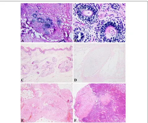
The patients’clinical and pathological features are pre-sented in Table 1. Patients had a median age of 20 (range: 15–25) years. The mean size of tumors was 20.4 (range: 11–30) cm. In the present study, seven primary ovarian tumors were immature teratomas (n= 7; grade 1, 2; grade 2, 3; and grade 3, 2) (Fig. 2 a, b), and one was a mature teratoma (n= 1) (Fig. 2c, d). The level of blood CA125 before therapy was available in seven patients, with a median CA125 level of 340.3 U/ml (range: 98.9– 673.5 U/ml).
Six patients had undergone comprehensive staging surgery, which led to the diagnosis of GP (Fig. 2e). The other two patients underwent unilateral cystectomy as the first surgery and GP was detected in the secondary surgery (cases 4 and 5). Seven patients had residual dis-ease postoperatively.
In case 4, the patient presented with abdominal disten-sion, abdominal pain, nausea, and vomiting. The B ultra-sound showed a 14-cm cystic-solid pelvic mass. She underwent a unilateral cystectomy and was diagnosed with immature ovarian teratoma (grade 2). Subsequently, she received three cycles of bleomycin, etoposide, and cisplatin (PEB). Four months after the completion of chemotherapy, the computed tomography (CT) showed masses in Douglas’pouch and on the liver surface with enlarged pelvic lymph nodes. All tumor markers were negative before the second surgery. Second surgery showed numerous miliary nodules on the surface of the intestine and masses of about 3 to 5 cm in Douglas’ pouch. The implants were so dispersed that complete re-section of the tumors was impossible. Frozen re-section of the mass in Douglas pouch showed mature glial
elements without immature tissues. Thus, growing tera-toma syndrome (GTS) was diagnosed. After the diagno-sis of GP, she did not receive further therapy. She was remained alive with persisting but asymptomatic disease 32 months after the diagnosis of GP (36 months after the diagnosis of immature ovarian teratoma).
In case 5, the patient underwent laparoscopic cystec-tomy because of ovarian cyst at another hospital. The pathologic evaluation indicated the presence of imma-ture teratoma (grade 2). However, she was not given chemotherapy at that time. Six months after the first surgery, she suffered from recurrence of the ovarian cyst. A second laparoscopic surgery was performed, revealing both ovarian cysts, as well as peritoneal and pelvic wall implants. Laparoscopic cystectomy was performed. Sur-gical pathology showed mature teratoma, associated with GP. Ten months later, a CT showed multiple masses in the abdominopelvic cavity, near the liver and spleen. The surgical procedure consisted of a subopti-mal cytoreduction. After the second surgery, the pathologic evaluation of resected tissue showed ma-ture teratoma and mama-ture peritoneal gliomatosis. She remained alive and asymptomatic 53 months after the diagnosis of GP (61 months after the diagnosis of ma-ture ovarian teratoma).
Four cases (case 2, 3, 6, and 8) of nodal gliomatosis were detected in form of glial tissue in the pelvic lymph nodes (Fig. 2f ). A summary of the cases of nodal glioma-tosis is presented in Table 2. A total of 14 patients with nodal gliomatosis were described and 10 remained alive at the time of the last follow-up. Most of the published papers on GP were case reports. Only 5 articles in the literature reported more than five cases (Table 3). For a
total of 67 patients, 60 of them were still alive at the time of the last follow-up.
Five patients were given chemotherapy (PEB or bleo-mycin, vincristine, and cisplatin [PVB]). If the dose of bleomycin reached the lifetime dose, the chemotherapy regimen was changed to PE or PV by omitting bleo-mycin. Immunohistochemical staining for glial fibrillary acidic protein (GFAP) was available in three cases. All three cases were positive for GFAP (Fig. 3a). Two of them were positive for S-100 protein (Fig. 3b). Three pa-tients (cases 5, 6, and 8) in our group experienced a spontaneous pregnancy.
Discussion
The metastatic implantation of mature glial tissue on the surface of the peritoneum, omentum, and abdominal lymph nodes is defined as GP. In a clinical setting, these widespread peritoneal, grayish tan-colored, tiny nodules encountered intraoperatively may be misdiagnosed as ovarian carcinomas or peritoneal tuberculosis. Ideal op-timal resection may not be achieved in such cases with widespread implantation. However, GP has a favorable prognosis. In the report by Yoon et al., the overall sur-vival did not differ between immature ovarian teratomas with GP and immature ovarian teratomas without GP, though patients with GP presented more frequent recur-rence and shorter recurrecur-rence-free survival [2]. All pa-tients (15 cases with GP) except one are currently alive when the articles were published.
GP is often diagnosed on HE-stained tissue sections, and its differentiation with low-grade epithelial ovarian tumors was difficult. A positive staining reaction of glial tissue for the neural marker, GFAP, is helpful. GFAP Table 1Clinical and pathological features of 8 cases with GP
case Age, y CA125 U/ml Tumor Size (cm)
Surgical Procedure (1st surgery) Primary tumor
Metastatic Tissue of the 1st surgery
Adjuvant therapy
GFAP Residual GP
Outcome
1 23 420.1 18 USO, Peritonectomies, Appendectomy, omenctomy
IMT, G1 GP PEB*3 NA YES Alive
16 months
2 25 98.9 22 USO, Peritonectomies, Omenctomy, lymphadenectomy
MT GP, Nodal,
gliomatosis
NO + YES Alive
3 months
3 16 340.4 20 USO, Peritonectomies, Omenctomy, lymphadenectomy
IMT, G2 GP, Nodal, gliomatosis
PEB*6 + YES Alive
68 months
4 18 171.7 14 cystectomy IMT, G2 NA PEB*3 NA YES Alive
38 months
5 23 NA 11 cystectomy IMT, G2 NA NO NA YES Alive
61 months
6 22 381.1 25 USO, Peritonectomies, Omenctomy, lymphadenectomy
IMT, G1 GP, Nodal, gliomatosis
NO + YES Alive
60 months
7 15 673.5 23 USO, Peritonectomies, Omenctomy, lymphadenectomy
LOV: IMT, G3, ROV: G1
GP PEB*2
PVB*2 PV*2
NA NO Alive
97 months
8 17 238.7 30 USO, Peritonectomies, Omenctomy, Lymphadenectomy, Appendectomy
IMT, G3 GP, Nodal, gliomatosis
PEB*4 NA YES Alive
immunostain confirmed the glial nature of the tissue. A strong expression often suggests tumor cells are mature and well differentiated [3].
The etiology of GP is largely unknown. According to previous reports, there are two theories about the devel-opment of GP. One relates to capsular defects of the pri-mary teratoma or dissemination via angiolymphatic channels. In some cases, GP within the omentum imme-diately adjacent to a capsular defect supports the mech-anism of spread in its inception [4]. In 11 of the 12 cases in Robboy’s report, the capsule either had a tear or was adherent to the omentum or adnexal structure [5]. In support of lymphatic dissemination, mature glial tissue has been presented in para-aortic and pelvic lymph nodes with or without the presence of GP (Table 2). In some reports, GP was only found in the second surgery
(as in case 4 in the present study). Kim et al. reviewed about 100 cases in the literature and found 9 cases of nodal gliomatosis in the pelvic or para-aortic lymph nodes with or without the presence of GP [6]. However, in their literature review, there were three cases of nodal gliomatosis presented as grades 1 to 3. As the nodal im-plantation contained immature elements, these should not be considered as nodal gliomatosis but as metastasis of the immature teratoma.
to the ovarian teratoma and arise from normal cells such as pluripotent Müllerian stem cells. It is possible that peri-toneal stem cells can differentiate into glial cells under the stimulation of some factors secreted by teratomas [8]. The occurrence of GP after ventriculoperitoneal shunt opera-tions when glial tissue is transported from cerebrospinal fluid into peritoneal cavity via shunt further support this theory [9]. However, the detailed mechanisms by which subperitoneal cells develop into glial follicles remain to be determined.
The stage and grade of the primary teratoma and the grade of its metastatic tumor are related to the prog-nosis of teratoma. Robboy and Scully reviewed the
literature and presented a large series of cases [5]. They found that the prognoses in metastasized ovarian teratomas, or in ovarian teratomas leading to periton-eal implants, are favorable when the implants are com-posed of fully mature glial tissue. Müller reviewed all cases of GP published between 1906 and 2002. Eleven cases showed adverse outcomes [1]. They found that the recurrence of disease was associated with lack of extensive histological sampling at the first surgery. Thus, all specimens need to be adequately sampled and multiple biopsies should be taken to exclude im-mature glial tissue or teratoma elements. Once the presence of immature teratoma is confirmed in the Table 2Summary of nodal gliomatosis cases reported in the literatures
Authors Age LN sites Primary tumor Treatment Outcome
Benirschke [16], 1960 18 ys Retroperitoneal,ilac,cervical axillary Mature teratoma chemoradiotherapy Dead 8 months
Nagashima [13], 1974 22ys Inguinal, mesenteric, mediastinal, cervical IMT S+ Ch Dead 8 months
Shafie [14] 1984 12 ys Omental MT S+ Ch NR 5 ys
Perrone [15] 1986 10 mo Para-arotic IMT G1 Surgery NR 9 months
Khan [17] 2005 23 ys Lymph node IMT G1 S+ Ch NA
Fang [18] 2015 20 ys Para-arotic IMT G3 S+ Ch Alive 36 months
Kim [6] 2013 34 ys Hypogastric IMT G1 S NR 9 months
Li Liang [8] 2015 18 ys Lymph node IMT G1 NA ANED 19 months
42 ys Lymph node MGCT NA AWD 23 months
10 ys Lymph node MGCT NA ANED 11 months
Present study 25 ys iliac MT Surgery Alive 3 months
16 ys Iliac IMT G2 S+ Ch Alive 68 months
22 ys iliac IMT G1 S+ Ch Alive 60 months
17 ys iliac IMT G3 S+ Ch Alive 144 months
ANEDalive with no evidence of disease,AWDalive with disease,Chchemotherapy,IMTimmature teratoma,LNlymph node,MTmature teratoma,MGCTmixed germ cell tumor,NAnot available,NRnot recurrence,Ssurgery,ysyears
Table 3Cases of ovarian teratoma associated with GP in studies that reported more than five cases
Authors Cases Median
Age ys
Ovarian neoplasm Diagnosis Recurrence Treatment Follow up
Norris [10], (1976) 7 17 IMT: G1: 5, G2-G3: 2 1st surgery: 7 NA S: 4, S + Ch: 1, S + Rx:2 5 alive, 1 dead, 1 NA
Harms [11], (1989) 13 11.5 IMT: G1:8, G2-G3: 5 1st surgery: 11, 2nd surgery: 2
NO S: 6, S + Ch: 7 13 alive
Yoon [2], (2012) 16 13 IMT: G1: 4, G2-G3: 11, MT:1
1st surgery: 15, 2nd surgery: 1
37.5 %, (6/16) S: 3, S + Ch: 13 15 alive, 1 dead
Bentivegna, [12], (2015)a 9 36 IMT: G1: 5, G2-G3: 4 1st surgery: 1,
2nd surgery: 8
22.2 %, (2/9) S: 5, S + Ch: 4 9 alive
Liang [8], (2015) 14 NA IMT: G1: 5, G2-G3: 9 1st surgery: 10, 2nd surgery: 4
NA NA 10 alive, NA: 4
Present 8 20 IMT: G1: 2, G2-G3: 5,
MT: 1
1st surgery: 6, 2nd surgery: 2
NO S: 3, S + Ch: 5 8 alive
Total 67 NA IMT: G1: 29, G2-G3: 35,
MT: 2
1st surgery: 50, 2nd surgery: 17
17.4 %, (8/46)b S: 21, S + Ch: 30, S + Rx:2, NA:14
60 alive, 2 dead, NA: 5
Chchemotherapy,IMTimmature teratoma,MTmature teratoma,NAnot available,Ssurgery,Rxradiotherapy
a
One case in the article (case 8) was consisted of mixed ovarian germ tumor (yolk sac and dysgerminoma and mature teratoma). Thus, the table shows 9 cases
b
metastatic tissue, the treatment scheme and prognosis may change.
Most of the papers on GP are cases. At present there are only 5 articles which report more than five cases (Table 3) [2, 8, 10–12]. Most cases of GP are associated with ovarian teratoma, especially with immature ovarian teratoma. In Yoon’s report, there were a total of 16 ovar-ian teratomas associated with GP [2]. Among them, 15 cases were of immature ovarian teratomas of various grades (4 cases were grade 1; 11 cases were grade 2 or 3). Liang reported the largest series of GP cases so far [8]. Sixteen of 23 cases were associated with immature ovarian teratomas. However, immature ovarian teratoma with GP showed better prognosis than would be ex-pected based on the grading of the primary immature teratoma. In Norris’s report, the survival of patients with grades 1, 2, and 3 were 82 %, 63 %, and 30 %, respect-ively [10]. In Yoon’s report, all but one case of immature teratoma with GP remained alive when their report was published, although GP showed more frequent recur-rences [2]. In Robboy’s report, 12 patients were alive and well [5]. More over, the survival of 8 patients in our series may also indicate that the presence of mature glial implants does not affect adversely the prognosis of ovar-ian teratoma.
Presence of glial tissue in lymph nodes is rare. Thus far, 14 cases have been reported in nine articles [6, 8, 13–18] (Table 2). Based on previous reports, adjuvant chemotherapy is unnecessary for patients with retroperi-toneal lymph node metastasis of mature glial tissue. The prognosis of patients with nodal gliomatosis is favorable. In our series, patients with nodal gliomatosis remained alive and well during the follow-up period.
Sometimes immature ovarian teratoma can be associ-ated with miliary spread of immature implants. After surgery and chemotherapy, the immature tissue may transfer into mature tissue. Chemotherapeutic retrocon-version has also been called growing teratoma syndrome (GTS) [19]. However, there are some differences be-tween GP and GTS. First, GP is composed of pure ma-ture implants in the peritoneum without other mama-ture nonglial tissues which can be seen in GTS. Second, GP
could be encountered in the first surgery without prior chemotherapy. At last, the management of these two terms is quite different. An optimal cytoreduction is rec-ommended for GTS to avoid complications such as bowel obstruction and perforation [20]. Because the le-sions in GP are extensive, complete resection of GP is usually difficult. Luckily residual peritoneal disease in GP can be asymptomatic and quiescent over a long period [12]. Thus, residual implants can be ignored and the therapy is mainly depended on the stage and grade of the primary ovarian teratoma. Seven cases in our series presented macroscopic residual disease at postoperatively, and all of these patients remained alive and asymptomatic during the median follow-up of 60.5 months.
Bentivegna reported 10 cases of GP, while six of them were managed by conservative surgery [12]. All of their patients were asymptomatic at the time of the last con-sultation, and half had incomplete resected macroscopic GP. These authors found residual peritoneal disease could be totally quiescent over a long period without any impact on patient outcomes. As ovarian teratoma mostly occurs in young women who wish to preserve fertility, it is important to reduce the surgical scope and reduce surgical trauma without compromising cure rate. As residual implants of GP can be ignored, a more con-servative surgical approach may be carried out in pa-tients with massive peritoneal spread after exclusion of the presence of metastatic immature elements.
Conclusion
Gliomatosis peritonei is metastatic implantation of ma-ture glial tissue on surfaces of peritoneum. It is often associated with ovarian teratoma of any grade. The prog-nosis of GP is favorable. Because GP is always present with massive peritoneal implantation, optimal resection is difficult. Although residual peritoneal disease can be totally quiescent over a long period, long-term follow-up is needed for patients with residual disease. A more con-servative surgical approach may be carried out in pa-tients with massive peritoneal spread.
Abbreviations
GFAP: glial fibrillary acidic protein; GP: Gliomatosis peritonei; GTS: growing teratoma syndrome; IMT: immature ovarian teratoma; MGCT: mixed germ cell tumor; PEB: bleomycin, etoposide, and cisplatin; PVB: bleomycin, vincristine, cisplatin; USO: unilateral salpingo-oophorectomy.
Acknowledgements
We show greatful thanks to Yang Chen for collection of the original articles about GP published many years ago. We thank Fan Yu (M.D. Candidate, Peking Union Medical College) for drawing figures.
Funding No
Availability of data and material
The dataset supporting the conclusions of this article is included within the article and its additional files.
Authors’contributions
DW have participated in the design of the study, analyzed the data, prepared the manuscript and revised it critically. HS have designed the study, responsed for the concept, analyzed the data and revised it critically for important intellectual content. CJ, RF have made paraffin sections and immunohistochemical examinations, the pathological pictures collection and their interpretation. JS have participated for the acquisition and analysis of data. All authors read and approved the final manuscript.
Competing interests
The authors declare that they have no competing interests.
Consent for publication Not applicable.
Ethics approval and consent to participate
Obstetrics and Gynecology Peking Union Medical College Hospital Human Research Ethics Committee Approval was obtained for the use of all samples.
Author details
1Department of Obstetrics and Gynecology, Peking Union Medical College
Hospital, Chinese Academy of Medical Science and Peking Union Medical College, Shuaifuyuan NO.1, Dongchen District, Beijing 100730, People’s Republic of China.2Department of Pathology, Peking Union Medical College Hospital, Chinese Academy of Medical Science and Peking Union Medical College, Shuaifuyuan NO.1, Dongchen District, Beijing 100730, People’s Republic of China.3Department of Obstetrics and Gynecology, Maternal and
Child Health Care Hospital of Zaozhuang, Wenhua Road, Shizhong District, Zaozhuang 277100, People’s Republic of China.
Received: 10 June 2016 Accepted: 20 July 2016
References
1. Muller AM, Sondgen D, Strunz R, Muller KM. Gliomatosis peritonei: a report of two cases and review of the literature. Eur J Obstet Gynecol Reprod Biol. 2002;100:213–22.
2. Yoon NR, Lee JW, Kim BG, et al. Gliomatosis peritonei is associated with frequent recurrence, but does not affect overall survival in patients with ovarian immature teratoma. Virchows Arch. 2012;461:299–304. 3. Gu S, Wu YM, Hong L, et al. Glial fibrillary acidic protein expression is an
indicator of teratoma maturation in children. World J Pediatr. 2011;7:262–5. 4. Nielsen SN, Scheithauer BW, Gaffey TA. Gliomatosis peritonei. Cancer.
1985;56:2499–503.
5. Robboy SJ, Scully RE. Ovarian teratoma with glial implants on the peritoneum. An analysis of 12 cases. Hum Pathol. 1970;1:643–53. 6. Kim NR, Lim S, Jeong J, Cho HY. Peritoneal and nodal gliomatosis with
endometriosis, accompanied with ovarian immature teratoma: a case study and literature review. Korean J Pathol. 2013;47:587–91.
7. Ferguson AW, Katabuchi H, Ronnett BM, Cho KR. Glial implants in gliomatosis peritonei arise from normal tissue, not from the associated teratoma. Am J Pathol. 2001;159:51–5.
8. Liang L, Zhang Y, Malpica A, et al. Gliomatosis peritonei: a clinicopathologic and immunohistochemical study of 21 cases. Mod Pathol. 2015;28:1613–20. 9. Lobotesis K, JM UK-I, Cross JJ, et al. Gliomatosis peritonei associated with a
ventriculo-peritoneal shunt. Clin Radiol. 2009;64:95–9.
10. Norris HJ, Zirkin HJ, Benson WL. Immature (malignant) teratoma of the ovary: a clinical and pathologic study of 58 cases. Cancer. 1976;37:2359–72. 11. Harms D, Janig U, Gobel U. Gliomatosis peritonei in childhood and
adolescence. Clinicopathological study of 13 cases including immunohistochemical findings. Pathol Res Pract. 1989;184:422–30. 12. Bentivegna E, Gonthier C, Uzan C, et al. Gliomatosis peritonei: a particular
entity with specific outcomes within the growing teratoma syndrome. Int J Gynecol Cancer. 2015;25:244–9.
13. Nagashima K, Yamaguchi K, Hasumi K, Oota K. Malignant gliomatosis peritonei originating from cystic ovarian teratoma. Acta Pathol Jpn. 1974;24:529–39.
14. El Shafie M, Furay RW, Chablani LV. Ovarian teratoma with peritoneal and lymph node metastases of mature glial tissue: a benign condition. J Surg Oncol. 1984;27:18–22.
15. Perrone T, Steiner M, Dehner LP. Nodal gliomatosis and alpha-fetoprotein production. Two unusual facets of grade I ovarian teratoma. Arch Pathol Lab Med. 1986;110:975–7.
16. Benirschke K, Easterday C, Abramson D. Malignant solid teratoma of the ovary. Report of three cases. Obstet Gynecol. 1960;15:512–21. 17. Khan J, McClennan BL, Qureshi S, et al. Meigs syndrome and gliomatosis
peritonei: a case report and review of literature. Gynecol Oncol. 2005;98:313–7.
18. Fang X, Zhang W, Song G, et al. Ovarian immature teratoma with gliomatosis peritonei: a clinicopathologic study. Zhonghua Bing Li Xue Za Zhi. 2015;44:201–3.
19. Merard R, Ganesan R, Hirschowitz L. Growing Teratoma Syndrome: A Report of 2 Cases and Review of the Literature. Int J Gynecol Pathol. 2015;34:465–72. 20. Zagame L, Pautier P, Duvillard P, et al. Growing teratoma syndrome after
ovarian germ cell tumors. Obstet Gynecol. 2006;108:509–14. 21. Fortt RW, Mathie IK. Gliomatosis peritonei caused by ovarian teratoma.
J Clin Pathol. 1969;22:348–53.
22. Dadmanesh F, Miller DM, Swenerton KD, Clement PB. Gliomatosis peritonei with malignant transformation. Mod Pathol. 1997;10:597–601.
• We accept pre-submission inquiries
• Our selector tool helps you to find the most relevant journal
• We provide round the clock customer support
• Convenient online submission
• Thorough peer review
• Inclusion in PubMed and all major indexing services
• Maximum visibility for your research
Submit your manuscript at www.biomedcentral.com/submit