Molecular chronobiology
C.P. Kyriacou () Department of Genetics, Faculty of Medicine and Biological Science, University of Leicester, Leicester LE1 7RH, UK e-mail: [email protected] Tel.: +44-1162-523430 Fax: +44-1162-523378 Charalambos Panayiotis Kyriacou
Abstract Recent years have seen exciting advances in the understand-ing of the mechanisms that underlie circadian rhythms in a variety of organisms, including mammals. Several key genes have been identi-fied, whose products can be consid-ered to represent bone fide clock molecules. Furthermore it appears that the same genes are important in generating rhythmic behaviour in both insects and man. There are some differences in the way these genes generate circadian output in the different taxa, but overall, the level of conservation of sequence and function is striking. The basic
molecular oscillatory mechanism depends on a transcriptional/
translational negative feedback loop, in which the PERIOD proteins play a cardinal role, together with other molecules, which interact to regulate circadian gene expression. In mam-mals, the brain oscillator resides in the suprachiasmatic nucleus, and its location in the hypothalamic region may have implications for understanding the rhythmic
nature of some headache syndromes.
Key words Circadian •Clock •
Gene •Molecule •Headache Received: 14 January 2000
Accepted in revised form: 18 February 2000
Introduction
For several billion years, the Earth has rotated along its axis with a period close to 24 hours. It has been four billion years since the first replicating molecules arose in the sea, and almost every organism that has evolved since, simple or complex, has been touched by this relentless cycle of day followed by night. In turn, this has brought about the evo-lution of a mechanism which anticipates this oscillation, and which prepares the organism for the associated change in illumination and temperature. In today’s extant taxa, we can observe this circadian oscillation from organisms as simple as the Cyanobacteria, through the fungi, to all high-er organisms including insects and mammals. Thhigh-ere is no need to tell anyone that they have a biological clock. The sleep-wake cycle makes this obvious, but what is not so obvious is that these rhythms are endogenous and are
encoded genetically. Humans who are isolated from tempo-ral cues will nevertheless slip into a circadian, 24-hour cycle of behaviour and physiology. This is not to say that envi-ronment cannot alter or modify that circadian clock: ask any trans-Atlantic traveller or shift worker. However, it was only about 30 years ago that the debate about whether circadian rhythms were determined by some cycling geophysical vari-able or by an internal clock was finally laid to rest. The argu-ment was settled by a genetic experiargu-ment with fruitflies, per-formed by a young graduate student, Ronald Konopka at the California Institute of Technology in the late 1960s [1].
the mid-1980s, and some of the subsequent work, form a substantial part of the book Time, Love, Memory[2], written by the Pulitzer-Prize winning author Jonathan Weiner. The book provides an inside view into how the work developed, and the personalities that led the charge from a genetical to a molecular description of the clock. As well as this book, which provides an introduction for the interested layman, a large number of more technical reviews of this field have been written. I will therefore refrain where possible from citing the primary literature, which numbers several hun-dred papers, and instead refer the reader to the more recent reviews of this field [3–9].
Konopka and Benzer’s classic paper focused on a study of the fly’s circadian pupal-adult eclosion rhythms [1]. When flies are ready to emerge from the pupa, they tend to do so at dawn, as this is the time when humidity is greatest. The word ‘Drosophila’ means ‘dew lover’, and this taxo-nomical insight reflects the ancient adaptation which pre-vents the newly emerged adults from dessicating in the mid-day heat (these flies evolved in Africa), before they have had time to tan their cuticle and pump out their wings. Consequently, if a fly is ready to emerge in mid-afternoon, it waits, until the next morning. Thus a bottle of fruitflies containing a selection of mixed-aged pupae will show sev-eral cycles of morning eclosion until all the pupae have emerged as adults. This rhythm has a period of 24 hours in constant conditions of darkness and temperature. By feeding these flies a mutagen, Konopka and Benzer were able to identify three mutants in the next generation, which had abnormal eclosion cycles. These included a short 19-hour
variant, a longer 29-hour fly, and an arrhythmic fly. These mutations all had corresponding effects on the individual fly’s ‘sleep-wake’ rhythms, which can be measured as loco-motor activity cycles (flies run around during the day, but ‘sleep’ at night). When these mutations were genetically mapped, they all appeared to be located to the same spot on the X-chromosome and defined a gene that Konopka called
‘period’or ‘per’. Normally the wild-type allele of persits at this spot, and generates a 24-hour cycle, but Konopka’s chemical mutagenesis had mutated this normal allele to a short, long or arrhythmic variant. One of the major take home messages from this study was that if a gene can be mutated to change the clock’s rhythm, then clearly an external geophys-ical variable cannot be generating circadian timing.
The period feedback loop
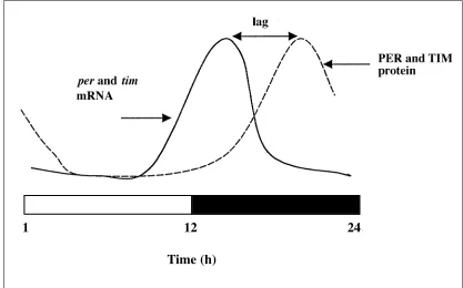
The discovery of these clock mutants lay the foundations for the molecular analysis of circadian rhythms which was to occur some 15 years later. perwas cloned, but translation of the primary amino acid sequence, which gave rise to a large putative protein of more than 1200 residues, gave few clues as to PER’s function. Progress was made only after the sub-sequent discovery that levels of the PER protein and mRNA cycled in the fly’s brain, with the transcript peaking early in the night phase (of a 12-h light – 12-h dark cycle, LD12:12), whereas the protein peaked late at night (Fig. 1). These mol-ecular rhythms were also maintained in constant darkness
Fig. 1 perandtim
mRNA and proteins cycle during a LD12:12 cycle or in constant darkness
1
12
24
with a period of 24 h (DD). This suggested that as the PER protein rises, it feeds back and shuts down its own mRNA production, giving rise to a negative feedback loop. Further evidence for this was provided by the permolecular cycles in the short 19-hour per mutant, which cycled with a corre-sponding 19-hour cycle. As the only difference between the normal wild-type PER protein and the mutant protein was a single amino acid change, this meant that the mutant protein had somehow fed back and influenced the mutant mRNA rhythm. The delay between mRNA and protein cycles would thus provide the necessary condition for the negative feedback to work, as without the delay, the protein would simply shut down transcription almost immediately, and cycling would damp.
Immunohistochemical studies using anti-PER antibodies also revealed that late at night, the PER protein was seen to move from the cytoplasm to the nucleus in the so-called lat-eral neurons in which PER was expressed. Experiments involving some sophisticated genetic trickery had shown that unless PER is expressed in these cells, the fly’s sleep-wake cycle will be arrhythmic, so consequently, these neu-rons appear to represent the fly’s behavioural pacemaker. The apparent nuclear role for PER in these neurons suggest-ed that PER may be a transcription factor, which acts to influence its own mRNA expression by binding to its own promoter region. If this is how it worked, then the PER pro-tein should carry a recognisable DNA-binding motif, which it does not.
In 1993 however, sequence analysis of PER and several other proteins, both in flies and mammals, revealed a mar-ginal similarity in a 270 residue region that was termed PAS. In the past two years it has become clear that PAS is a recog-nisable motif found in many signalling molecules, including transcription factors. PAS acts as a dimerisation domain, and can mediate various types of protein-protein interac-tions, which perform a wide variety of funcinterac-tions, from light sensing in bacteria, to potassium channel deactivation in mammals [10]. The primary sequence similarity between PAS domains is very poor, yet the structural integrity of dif-ferent PAS domains is remarkably conserved at the three-dimensional (3D) level [10]. The PER PAS domain would therefore be expected to physically interact with another molecule, and one of these binding partners is the product of the timeless (tim) gene. The first mutation identified in tim
also gives arrhythmic behaviour, suggesting that tim is also an important clock gene. TIM protein and mRNA cycle in the same brain cells, and in approximately the same phase, as do the perproducts (Fig. 1). Thus not only is timin a neg-ative loop with itself, but it is also a cardinal component of the per loop. Without TIM, PER cannot get into the nucle-us, and vice versa.
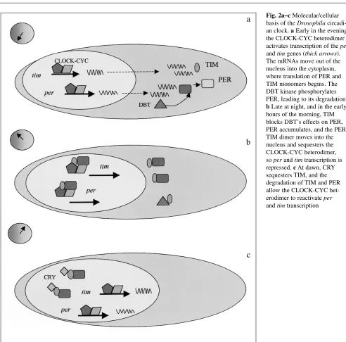
Late at night, as TIM and PER protein levels are on the rise, the two products dimerise via the PER PAS domain and
translocate to the nucleus (Fig. 2). Inside the nucleus are the protein products of the genes Clock and cycle (cyc), both of which have bHLH (basic helix-loop-helix) and PAS regions. CLOCK and CYC can associate with each other via their PAS domains, and their bHLH motifs further allow them to bind to specific DNA sequences called E-boxes, which are found in the perand timpromoters. The CLOCK-CYC com-plex is therefore the positive element in the feedback loop and activates perand tim transcription during the day and early part of the night (Fig. 2). As the PER-TIM dimer enters the nucleus, it sequesters the CLOCK-CYC dimer, and repress-es perand timtranscription. The PER-TIM dimer therefore represents the negative element of the feedback loop. As PER and TIM degrade during the day phase, the CLOCK-CYC dimer is freed to reactivate perand timtranscription. In LD cycles, this process by which perand tim transcrip-tion is derepressed is also aided by another molecule, CRYPTOCHROME (CRY). CRY changes its conformation when stimulated by light, and binds to TIM, sequestering the PER-TIM dimer from the CLOCK-CYC complex, which then reactivates per/timtranscription (Fig. 2) [9]. CRY’s light sensitivity thus appears to enhance the amplitude of the molecular oscillation in LD cycles.
The feedback loop therefore has positive (CLOCK and CYC) and negative (PER and TIM) components but relies on a delay between the translation of the proteins PER and TIM, and their negative feedback on their own promoters. This critical delay involves DOUBLETIME (DBT), a casein kinase, which phosphorylates PER monomers as they are produced in the cytoplasm in the early part of the night phase (Fig. 2). This earmarks PER monomers for degrada-tion, and contributes to the delay between the observation of the peak levels of the permRNA and the subsequent peak of the PER protein cycle (Fig 1). As TIM levels rise later in the evening, they somehow block the action of DBT on PER (Fig. 2), so PER monomers reach a level where they can dimerize with TIM, and so the PER-TIM complex moves into the nucleus [11, 12]. Mutations in doubletime which either shorten or lengthen the behavioural cycle have been isolated, but more severe mutations are lethal. This is not surprising as the kinase is likely to be involved in many other biological functions, and is not simply clock-specific. However, mutations in Clock and cyc are arrhythmic and non-lethal [13, 14], and a mutation in cry predictably leads to poorer circadian behavioural responses to light [15].
the fly, workers tried to find a similar gene in the mouse, but all failed, and it was thought in many quarters that perhaps the fly clock mechanism would be specific to insects. One should never make up one’s mind based on negative evi-dence, and this was underlined relatively recently, when mouse and human pergenes were finally identified. In fact it turned out that the mouse has three pergenes (mper1, 2
and 3). Many genes in mammals are duplicated, the most famous being the HOX gene complex, which has four copies, compared to Drosophila’s one [16]. These mper
transcripts cycle in various parts of the mouse brain but in a different phase to that seen in the fly. Crucially, the mper
genes are expresssed in the suprachiasmatic nucleus (SCN) of the hypothalamus, which has been known for many years to represent the circadian pacemaker of the mammal [6].
The mouse also has a Clockgene, which was defined by mutagenesis. The homozygous Clock mouse mutant has an arrhythmic circadian behavioural phenotype, and has a slightly longer period than normal in the heterozygote. There are also two murine Cry genes. Mutations in each produce slight changes in circadian behaviour, but a double mutant is arrhythmic, suggesting that mCRYs have a rather more direct function on the clock than Drosophila dCRY [17-19]. There is also a cychomologue in the mouse (called
Fig. 2a–c Molecular/cellular basis of the Drosophila circadi-an clock. aEarly in the evening the CLOCK-CYC heterodimer activates transcription of the per
and timgenes (thick arrows). The mRNAs move out of the nucleus into the cytoplasm, where translation of PER and TIM monomers begins. The DBT kinase phosphorylates PER, leading to its degradation. bLate at night, and in the early hours of the morning, TIM blocks DBT’s effects on PER, PER accumulates, and the PER-TIM dimer moves into the nucleus and sequesters the CLOCK-CYC heterodimer, soperand timtranscription is repressed. cAt dawn, CRY sequesters TIM, and the degradation of TIM and PER allow the CLOCK-CYC het-erodimer to reactivateper
and timtranscription
a
b
bmal1), a DBT homologue (casein kinase 1ε), and a tim
homologue, mTim.Mutations in the latter three mammalian genes have not been reported, and the role of mTim particu-larly is unclear at the moment.
Thus some of the central components of the mammalian clock have been identified and not surprisingly, the duplica-tion of clock genes in the mouse means that the different mPER or mCRY products will have slightly different func-tions from each other, providing a rather more complex reg-ulatory network. These same clock components may gener-ate circadian rhythmicity in the fly or the mouse, but the way in which these molecules are regulated will have altered through evolution. Even in insects, the regulation of PER and TIM in the brain of the giant silkmoth is quite dra-matically different from that of the fruitfly [8]. No doubt, more clock components will be identified in the future, but my guess is that there will not be so many more involved in generating the feedback loop. There will certainly be many clock-controlled genes (ccg’s)which connect the clock to the circadian phenotype, and some of these are already known in the mammal and the fly.
One example of the regulation of a ccg, concerns a recent study of the neuropeptide arginine vasopressin gene in the mouse, whose mRNA cycles with a circadian period in the mouse brain [20]. The gene’s promoter has an E-box, to which CLOCK-BMAL1 heterodimers bind and activate
vasopressintranscription. Addition of the mPER proteins or mTIM gives a modest repression of transcription, but the mCRY1 or mCRY2 products produce a far more powerful transcriptional inhibition, suggesting that mCRYs are part of the negative loop [19]. The mCRY proteins also dimerize with mPER proteins, revealing how mCRYs influence the negative regulation of the mper genes, and also explaining why mCRY double mutants are arrhythmic [18,19]. The role of the mCRYs thus contrasts with the role of dCRY in
Drosophila, which seems to act predominantly as a circadian photoreceptor [9]. The regulation of the vasopressin ccg can therefore be directly controlled by the central clock compo-nents mPER, mTIM, mCLOCK, BMAL1 (CYC) and the mCRYs. However it is unlikely that allccg’s will be regulat-ed in this way, because any mRNA which cycles with a dif-ferent phase tomper genes may require other intermediates interfacing between the cycling actions of the core clock genes, and the transcription factors which ultimately control the ccg.
It is clear from this brief overview of molecular chrono-biology that the basis of the clockworks in the mammal are beginning to be understood (thanks to the fly). This knowl-edge has broad implications for developing treatments for some of the clock problems that bedevil shift workers (>25% of the Western working population), insomniacs, and seasonal depressives, to name but a few.
Melatonin, headache, and the clock
Another output from the mammalian circadian clock is melatonin, which cycles with high levels at night, low lev-els during the day, and is produced predominantly from the pineal [21]. As there are a number of reports on the benefi-cial effects of melatonin administration for headache [22-24], perhaps examining the role of biological rhythms in headache may lead to new insights in the genesis of such disorders. There certainly are good reasons to suspect that the clock is involved in the circadian and seasonal patterns observed in cluster headache (CH) [23, 24], as well as a rare benign syndrome called hypnic or ‘alarm clock’ headache, which also shows monotonous temporal regularity [25]. In addition, positron emission tomography (PET) scans have implicated the hypothalamus in the onset of CH, and the pos-sibility that CH is due to a neuro-vascular disorder has been discussed [26]. The region of the hypothalamus that seems to be the focus of CH lies uncomfortably close to the SCN.
Melatonin is a master hormone which controls the pitu-itary-hypothalamic-adrenal axis, and regulates genes which stimulate the immune system [27]. Melatonin stimulates T-helper 1 (Th1) cell cytokines, among them interferon-γ (INF-γ) and interleukin-2 (IL-2) [28–31]. IL-2 is reported to be at lower levels in CH sufferers [32], fitting with the observation that plasma melatonin levels are also too low during a cluster period [33, 34]. At night, a disruption of the Th1/melatonin signaling, caused by lower-than-normal melatonin levels, could signal the beginning of a CH attack. If this were the case however, then CH sufferers should show symptoms of a less-than-perfectly functioning circadi-an clock. In fact, there are reports of clock defects in such CH patients [22, 34], but disentangling cause from effect is not easy. Does a dysfunctional clock cause the disorder, or does the disorder cause the apparently defective clock? After all, a CH sufferer will be exhausted, sleep-deprived and distressed during the cluster period, leading to a number of correlated circadian changes. The simple procedure of waking up during a cluster episode and putting on the lights will itself reduce melatonin levels.
References
1. Konopka RJ, Benzer S (1971) Clock mutants of Drosophila melanogaster. Proc Natl Acad Sci USA 68:2112–2116 2. Weiner J (1999) Time, Love, Memory.
Faber & Faber, London
3. Dunlap JC (1999) Molecular bases for circadian clocks. Cell 96:271–290 4. Sassone-Corsi P (1998) Molecular
clocks: mastering time by gene regula-tion. Nature 392:871–874
5. Dunlap JC (1998) An end in the begin-ning. Science 280:1548–1549
6. Wilsbacher LD, Takahashi JS (1998) Circadian rhythms: molecular basis of the clock. Curr Opin Genet Dev 595–602
7. Young MW (1998) The molecular con-trol of circadian behavioral rhythms and their entrainment in Drosophila. Annu Rev Biochem 67:135–152 8. Rosato E, Piccin A, Kyriacou CP
(1997) Molecular analysis of circadian behaviour. Bioessays 19:1075–1082 9. Ceriani MF, Darlington TK, Staknis D,
Mas P, Petti AA, Weitz CJ, Kay SA (1999) Light-dependent sequestration of TIMELESS by CRYPTOCHROME. Science 285:553–556
10. Taylor BL, Zhulin IB (1999) PAS domains: internal sensors of oxygen, redox potential, and light. Microbiol Mol Biol Rev 63:479–506
11. Kloss AB, Price JL et al (1998) The
Drosophilaclock gene double-time
encodes a protein closely related to human casein kinase I. Cell 94:97–107 12. Price JL, Blau J, Rothenfluh A et al
(1998) double-timeis a novel
Drosophilaclock gene that regulates Period protein accumulation. Cell 94:83–95
13. Allada R, White NE, So WV, Hall JC, Rosbash M (1998) A mutant
Drosophilahomolog of mammalian
Clockdisrupts circadian rhythms and transcription of periodand timeless. Cell 93:791–804
14. Rutila JE, Suri V, Le M, So WV, Rosbash M et al (1998) Cycle is a sec-ond bHLH-PAS clock protein essential for circadian rhythmicity and transcrip-tion of Drosophila periodand timeless. Cell 93:805–814
15. Stanewsky R, Kaneko M, Emery P, Beretta B, Wager-Smith K, Kay S, Rosbash M, Hall J (1998) The cryb mutation identifies cryptochrome as a circadian photoreceptor in Drosophila. Cell 95:681–692
16. Carroll SB (1995) Homeotic genes and the evolution of arthropods and chor-dates. Nature 376:479–485
17. Thresher RJ, Vitaterna MH et al (1998) Role of mouse cryptochrome blue light photoreceptor in circadian photo-responses. Science 282:1490–1494 18. Horst van der GTJ, Muijtjens M et al
(1999) Mammalian Cry1 and Cry2 are essential for maintenance of circadian rhythms. Nature 398:627–630 19. Kume K, Zylka MJ et al (1999)
MCRY1 and MCRY2 are essential components of the negative limb of the circadian clock feedback loop. Cell 96:193–205
20. Jin X, Shearman LP, Weaver DR, Zylka MJ, De Vrees GJ, Reppert SM (1999) A molecular mechanism regu-lating rhythmic output from the suprachiasmatic circadian clock. Cell 96:57–68
21. Illnerova H, Sumova A (1997) Photic entrainment of the mammalian rhythm in melatonin production. J Biol Rhythms 12:547–555
22. Leone M, Bussone G (1998) Melatonin in cluster headache - Rationale for use and possible therapeutic potential. CNS Drugs 9:7–16
23. Leone M, Lucini V, Damico D, Moschiano F, Maltempo C, Fraschini F, Bussone G (1995) 24-Hour mela-tonin and cortisol plasma levels in rela-tion to timing of cluster headache. Cephalalgia 15:224–229
24. Leone M, D’Amico D, Moschiano F, Fraschini F, Bussone G (1996) Melatonin versus placebo in the pro-phylaxis of cluster headache: A dou-ble-blind pilot study with parallel groups. Cephalalgia 16:494–496 25. Dodick DW, Mosek AC, Campbell JK
(1998) The hypnic (“alarm clock”) headache syndrome. Cephalalgia 18:52–156
26. May A, Bahra A, Buchel C, Frackowiak RSJ, Goadsby PJ (1998) Hypothalamic activation in cluster headache attacks. Lancet 352:275–278 27. Dawson D, Heuvel CR van den (1998) Integrating the actions of melatonin on human physiology. Ann Med
30:95–102
28. Constantinesu CS, Hilliard B, Ventura E, Rostami A (1997) Luzindole, a melatonin receptor antagonist sup-presses experimental autoimmune encephalitis. Pathobiology 65:190–194 29. Segal BM, Dwyer BK, Shevach EM
(1998) An interleukin (IL)-10/IL-12 immunoregulatory circuit controls sus-ceptibility to autoimmune disease. J Exp Med 187:536–546
30. Heurtier AH, Boitard C (1997) T-cell regulation in murine and human autoimmune diabetes: the role of TH1 and TH2 cells. Diabetes Metab 23:377–385
31. Garcia-Maurino S, Gonzalez-Haba MG, Calvo JR, Rafii-ElIdrissi M, Sanchez-Margalet V, Goberna R, Guerrero JM (1997) Melatonin enhances IL-2, IL-6, and IFN-gamma production by human circulating CD4(+) cells - A possible nuclear receptor-mediated mechanism involv-ing T helper type 1 lymphocytes and monocytes. J Immunol 159:574–581 32. Shimomura T, Araga S, Esumi E,
Takahashi K (1991) Decreased serum interleukin-2 level in patients with chronic headache. Headache 31:310–313
33. Waldenlind E, Ekbom K, Wetterberg L, Fanciullacci M, Marabini S (1994) Lowered circannual urinary melatonin concentrations in episodic cluster headache. Cephalalgia 14:199–204 34. Strittmatter M, Hamann G, Blaes F,
Fischer C, Grauer M, Hoffmann KH, Schimrigk K (1997) Alterations of the hypothalamic-pituitary-adrenal axis and chronobiological disturbance in cluster headache. Fortschritte der Neurologie Psychiatrie 65:1–7 35. Nagtegaal JE, Smits MG, Swart ACW,