www.fm.viamedica.pl
Address for correspondence: M. Üzel, MD, Saglik Hizmetleri MYO, Kocamustafapasa, 34098, Istanbul, Turkey, tel: +90 212 4143057, fax: +90 212 4143059, e-mail: muzel@istanbul.edu.tr
An anatomical study of the origins
of the lateral circumflex femoral artery
in the Turkish population
M. Üzel
1, E. Tanyeli
2, M. Yildirim
21Vocational School of Health Services, Istanbul University, Istanbul, Turkey
2Department of Anatomy, Cerrahpasa Medical Faculty, Istanbul University, Istanbul, Turkey
[Received 23 June 2008; Accepted 27 August 2008]
The aim of this study was to investigate the origins of the lateral circumflex femoral artery (LCFA) in the Turkish population. For this purpose, we investi-gated 110 inguinal regions of 56 cadavers between 1997 and 2007. The LCFA was found to be branched from the deep femoral artery (DFA) in 85 (77.3%) cases and from the femoral artery (FA) in 21 (19.1%) cases. In two (1.8%) cases the ascending and the descending branches of the LCFA branched separately from the DFA and FA. There was a common trunk of the DFA and the LCFA in one (0.9%) case, and a common trunk of the DFA, LCFA and the medial circumflex femoral artery (MCFA) (trifurcation) in another (0.9%). We also measured the distance between the mid-inguinal point (MIP) and the origin of the LCFA. For LCFAs branching from the DFA the mean distance between these points was 4.8 ± 1.2 cm, while for LCFAs branching from the FA the mean distance was 3.4 ± 0.9 cm. We discuss the clinical importance of the artery and compare the results with the literature. (Folia Morphol 2008;
67: 226–230)
Key words: lateral circumflex femoral artery, anatomy, variation
INTRODUCTION
The lateral circumflex femoral artery (LCFA) is commonly a branch of the deep femoral artery (DFA) or sometimes of the femoral artery (FA) and passes between divisions of the femoral nerve posterior to the sartorius and rectus femoris muscles. After pass-ing behind these structures, it divides into its as-cending, transverse and descending branches. It supplies blood to the head and neck of the femur, greater trochanter, the vastus lateralis and the knee [13]. It has many implications in clinical practice. The branches of the LCFA are used in an anterolateral thigh flap [15], aortopopliteal bypass [7, 14], coronary ar-tery bypass grafting (CABG) [6] and extracranial– –intracranial (EC–IC) bypass surgery [4]. The descending
branch of the artery can act as a collateral [8]. The ascending branch of the artery can be used as a sup-ply for vascularised iliac transplantation [16].
MATERIAL AND METHODS
RESULTS
During the dissections we observed that two of the 112 FAs had been destroyed, and so these were excluded from our study. In 85 (77.3%) sides the LCFA branched from the DFA, and in 21 (19.1%) from the FA proximal to the origin of DFA. In two cases the ascending and descending branches of the LCFA branched separately. In the first case (0.9%) the as-cending branch of the LCFA branched from the FA proximal to the origin of the DFA and the descend-ing branch from the FA distal to the origin of the DFA (Fig. 1). In the second case (0.9%) the ascend-ing and descendascend-ing branches branched from the DFA and FA respectively (Fig. 2). There was a common trunk for the DFA and LCFA in one (0.9%) case, in which both arteries branched from the same stem (Fig. 3). In another case (0.9%) there was a com-mon trunk of the DFA, LCFA and the medial circum-flex femoral artery (MCFA) (trifurcation) (Fig. 4). In addition, we also measured the distance between the mid-inguinal point (MIP) and the origin of the LCFA. The mean distance between these points was 4.8 ± 1.2 cm where the LCFA branched from the DFA, and 3.4 ± 0.9 cm where the LCFA branched from the FA. The mean diameter of the LCFA at its origin was 4.7 mm in our study. There were no sta-tistical differences with respect to sex or side in the branching patterns, distances and diameters.
DISCUSSION
Because of the increasing importance of LCFA in clinical practice numerous studies may be found of
its anatomy, both recent and old. The LCFA itself is used in aortopopliteal bypass [7, 14] and can be affected during total hip replacement surgery [2].
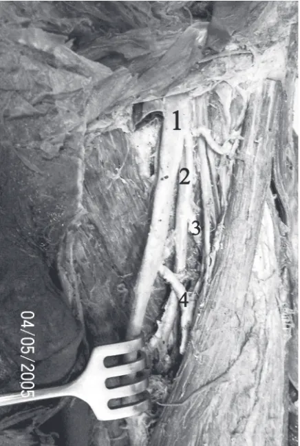
Figure 2. The left inguinal region of the case in which the ascending and descending branches of the lateral circumflex femoral artery (LCFA) branched from the deep femoral artery (DFA) and the femoral artery (FA) respectively; 1 — FA; 2 — DFA; 3 — ascending branch of LCFA; 4 — descending branch of LCFA.
Figure 3. The case of a common trunk of the deep femoral artery (DFA) and the lateral circumflex femoral artery (LCFA) on the right side; 1 — FA; 2 — DFA; 3 — LCFA.
Its branches have clinical implications as well. Its ascending branch can be used as a supply for vas-cularised iliac transplantation [16], while its de-scending branch can act as a collateral in obstruct-ed superficial FA [8] and can be usobstruct-ed in CABG [6]; perforators are important in anteroletaral thigh flap for reconstructive surgery [9, 15].
In his comprehensive study of the arterial sys-tem Adachi [1] reported the incidence of the LCFA branching from the DFA as 78.2% and from the FA as 18.3% (Table 1). He gave the proportion of the descending branch of the LCFA originating from the femoral artery as 2.7%.
In the 1980s, Lippert and Pabst [10] and Berg-man et al. [5] published their works on arterial
varia-tion in humans. Lippert and Pabst [10] gave the pro-portion of the LCFA branching from the DFA as 76% (type a + b in Lippert and Pabst’s classification), and from the FA as 19% (type c + d). They also reported the proportion with the descending branch of the LCFA originating from the FA to be 3% (type e).
Similar results were mentioned in the work of Bergman et al. [5] with proportions of 76.8% and 18% respectively. These authors gave the occurrence of both ascending and descending branches of the LCFA originating from the FA as 0.5%, with the de-scending branch of the LCFA originating from the FA in 3.2% of cases.
One of the studies most often referred to on the anatomy of the FA is that of Siddharth et al. [12]. In this the incidence of the LCFA branching from the DFA was reported to be 71% and from the FA 16%. The ratio of the descending branch of the LCFA ori-ginating from the FA was given as 3%. The study also reports the DFA, LCFA and MCFA as having a com-mon origin in 5% of cases, which was not mentioned in other studies. A further peculiarity of their study is that they measured the distance between the MIP and the branching point of the LCFA, the mean dis-tance being given as 5.9 cm.
In their angiographic study on 188 lower limbs Massoud and Fletcher [11] investigated the anato-my of the DFA and classified it according to Lippert and Pabst’s method [10]. The frequency of the LCFA branching from the DFA was found to be 81%, and from the FA 2.8%.
In an angiographic study on the ramification patterns of the FA in the Turkish population, the
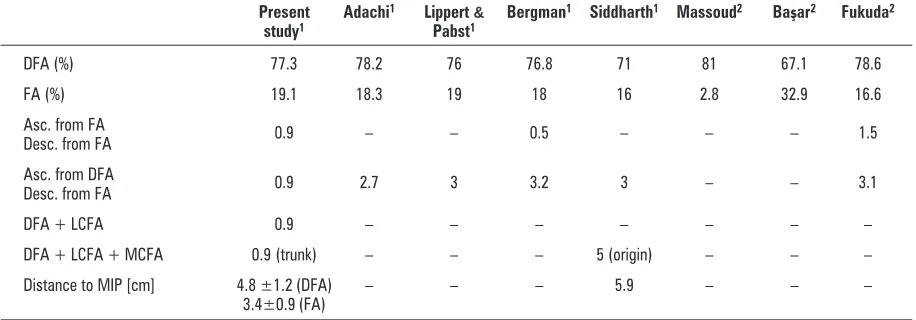
Table 1. Percentages for the origins of the lateral circumflex femoral artery (LCFA) in different studies; a comparison of
the results of our study with those of other studies in the literature
Present Adachi1 Lippert & Bergman1 Siddharth1 Massoud2 Başar2 Fukuda2
study1 Pabst1
DFA (%) 77.3 78.2 76 76.8 71 81 67.1 78.6
FA (%) 19.1 18.3 19 18 16 2.8 32.9 16.6
Asc. from FA
Desc. from FA 0.9 – – 0.5 – – – 1.5
Asc. from DFA
Desc. from FA 0.9 2.7 3 3.2 3 – – 3.1
DFA + LCFA 0.9 – – – – – – –
DFA + LCFA + MCFA 0.9 (trunk) – – – 5 (origin) – – –
Distance to MIP [cm] 4.8 ±1.2 (DFA) – – – 5.9 – – – 3.4±0.9 (FA)
DFA — deep femoral artery; FA — femoral artery; MIP — the mid-inguinal point; MCFA — medial circumflex femoral artery; 1cadaveric study, 2angiographic study
LCFA was found to branch from the DFA in 67.1%, and from the FA in 32.9% of cases [3].
More recently Fukuda et al. [6] published their angiographic study on the LCFA. Anatomical vari-ants of the LFCA were assessed on femoral arterio-grams obtained before CABG in 131 adult patients. In most of the cases examined the LCFA branched from the DFA (78.6%), while in 16.6% of cases the LCFA branched from the FA. In 3.1% of cases they found that ascending and the descending branches of the LCFA branched separately from the DFA and FA respectively. In 1.5% of their cases the ascending and descending branches of the LCFA originated separately from the FA.
In our study we found that the ascending and descending branches of the LCFA originated sepa-rately from the FA in 0.9% of cases. The same pat-tern was mentioned in the studies by Bergman et al. [5] and Fukuda et al. [6] as occurring in 0.5% and 1.5%, respectively (Table 1). Our result is equal to the mean of the results of these two studies. The descending branch of the LCFA originated separately from the FA in 0.9% of our cases, while the mean percentage of the same pattern in other studies is 3%. This difference may be attributable to the charac-teristics of our population. The most remarkable pattern in our study is the common origin of the DFA and LCFA (0.9%), and we were unable to trace this pattern in the literature. In our study we found a common trunk for the DFA, LCFA and MCFA in 0.9% of cases. Siddharth et al. mentioned a similar pattern in their study but with a slight difference; in our study these three arteries originated from a com-mon trunk (2 cm in length) from the FA, but in their study these arteries had a common origin on the FA (5%). In our study we also measured the distance between the origin of the LCFA and the MIP and found it to be 4.8 ± 1.2 cm for LCFAs originating from the DFA and 3.4 ± 0.9 cm for LCFAs originat-ing from the FA. Siddharth et al. also measured this distance and found it to be 5.9 cm.
Cadaveric and angiographic studies concerning the LCFA may be found in the literature. The angiographic studies are valuable, but their results may be inconsis-tent with the results of the cadaveric studies because of difficulties in defining some branches on arterio-grams. The literature includes an angiographic study of the LCFA in the Turkish population [3], but we were unable to find any cadaveric studies on the same pop-ulation. Our intention in the present study was to fill this gap. Our results for the normal origin of the LCFA from the DFA are fairly consistent with the results of
other cadaveric studies in different populations [1, 3, 5, 6, 10–12]. The percentage of cases in our study with a normal origin of the LCFA (77.3%) is higher than in the angiographic study in the same population (67.1%) [3]. In our study the distance between the branching point of the LCFA and the MIP was also measured. We could find only the study by Siddharth et al. on this issue (Table 1). In our cases where the LCFA branched directly from the FA the mean distance (3.4 ± 0.9 cm) was smaller than where the LCFA branched from the DFA (4.8 ± 1.2 cm); both of these are smaller than in the results of Siddharth et al. [12]. The distance be-tween the MIP and the origin of the LCFA may be of importance in surgical or angiographic interventions, and so these distances may be useful for health pro-fessionals dealing with this artery.
We have been investigating the DFA and its branches in our department for 11 years. During this time we have examined 56 cadavers. With regard to the LCFA and its branches our study is the largest cadaver study in our country. We have also found some branching configurations, such as a common trunk for the DFA and LCFA and for the DFA, LCFA and MCFA (trifurcation), which to our knowledge have not yet been referred to anywhere in the literature. In our study we both investigated the patterns and measured the distance of branching of the LCFA in the Turkish population. We compared our results with other studies, reporting patterns which had not pre-viously been described. We have therefore considered it useful to publish these findings and share them with our colleagues.
REFERENCES
1. Adachi B (1928) Das arteriensystem der Japaner, Band II. Verlag der Kaiserlich, Kyoto, pp. 151.
2. Aust JC, Bredenberg CE, Murray DG (1981) Mechanisms of arterial injuries associated with total hip replace-ment. Arch Surg Mar, 116: 345–349.
3. Başar R, Sargon MF, Cumhur M, Bayramoglu A, Demiryürek D (2002) Distinct intergender difference in the femoral artery ramification pattern found in the Turkish population: angiographic study. Anat Sci Int, 77: 250–253.
4. Başkaya MK, Kiehn MW, Ahmed AS, Ateş Ö, Niemann DB (2008) Alternative vascular graft for extracranial-intrac-ranial bypass surgery: descending branch of the lateral circumflex femoral artery. Neurosurg Focus, 24: 1–7.
6. Fukuda H, Ashida M, Ishii R, Abe S, Ibukuro (2005) Anatomical variants of the lateral femoral circumflex artery: an angiographic study. Surg Radiol Anat, 27: 260–264.
7. Gradman WS (1992) Bypass to the lateral circumflex femoral artery. Ann Vasc Surg, 6: 344–346.
8. Hage JJ, Woerdeman LA (2004) Lower limb necrosis after use of the anterolateral thigh free flap: is preoperative angiography indicated? Ann Plast Surg, 52: 315–318. 9. Koshima I, Fukuda H, Utunomiya R, Soeda S (1989) The
anterolateral thigh flap; variations in its vascular pedi-cle. Br J Plast Surg, 42: 260–262.
10. Lippert H, Pabst R (1985) Arterial variations in man: classification and frequency. JF Bergman Verlag, München, pp. 61.
11. Massoud TF, Fletcher EWL (1997) Anatomical variants of the profunda femoris artery: an angiographic study. Surg Radiol Anat, 19: 99–103.
12. Siddharth P, Smith NL, Mason RA, Giron F (1985) Varia-tional anatomy of the deep femoral artery. Anat Rec, 212: 206–209.
13. Standring S ed. (2005) Gray’s anatomy. 39th ed. Else-vier Churchill Livingston, Edinburgh, pp. 1452. 14. Sugawara Y, Sato O, Miyata T, Kimura H, Namba T,
Makuuchi M (1998) Utilization of the lateral circum-flex femoral artery as a midway outflow for aorto-popliteal grafting: report of a case. Surg Today, 28: 967–970.
15. Valdatta L, Tuinder S, Buoro M, Thione A, Faga A, Putz R (2002) Lateral circumflex femoral arterial system and perforators of the anterolateral thigh flap: an anato-mic study. Ann Plast Surg, 49: 145–150.