www.fm.viamedica.pl
Address for correspondence: J. Hreczecha, Department of Clinical Anatomy, Medical University of Gdańsk, Dębinki 1, 80–211 Gdańsk, Poland, tel./fax: +48 58 3491420, e-mail: [email protected]
The morphometry of the accessory leaflets
of the tricuspid valve in a four cuspidal model
M. Skwarek
1, J. Hreczecha
2, M. Dudziak
3, J. Jerzemowski
4, B. Wilk
1, M. Grzybiak
31Department of Sports Medicine, the Jędrzej Śniadecki Academy of Physical Education and Sport, Gdańsk, Poland 2Department of Clinical Anatomy, Medical University, Gdańsk, Poland
3Non-invasive Cardiovascular Diagnostic Unit, Institute of Cardiology, Medical University, Gdańsk, Poland 4Department of Anatomy and Anthropology, the Jędrzej Śniadecki Academy of Physical Education and Sport,
Gdańsk, Poland
[Received 13 March 2007; Revised 3 August 2007; Accepted 15 August 2007]
The tricuspid valve is of great importance because of the progress made in operative techniques and invasive cardiology accidents. This structure is more differentiated during evolution than the mitral valve. Accessory leaflets, their frequency and role are still controversial, despite the fact that they have been known from the beginning of the 20th century. The number of leaflets in the
tricuspid valve grows in an evolutionary line, but the rules governing their appearance are still not known. The samples were taken from a group of 107 human adult hearts. The four-cuspidal form of the tricuspid valve was used as the simplest model to show the appearance of accessory leaflets for anatomical and statistical examination. On the basis of the results of this study we conclude that the separation of accessory leaflets is a complex process.
Key words: tricuspid valve, morphometry, right atrioventricular orifice
INTRODUCTION
Understanding of the tricuspid valve remains im-portant because of progress in cardiac surgery, includ-ing the partial transfer of the posterior leaflet of the tricuspid valve for repair of the mitral valve [11, 13], tricuspid replacement [4, 5], complications after heart transplantation [7], invasive cardiology [17] and valvu-loplasty [24]. Many congenital malformations involve the tricuspid valve [10, 22, 23, 25, 33]. Complications of the right atrioventricular valve because of infection may be an indication for surgical treatment [21, 35].
The tricuspid valve is more differentiated during evolution than the mitral valve [1, 16, 31, 32]. Ata-vistic features and atypical forms of the tricuspid valve and the distribution of its tendinous chords (chordae tendineae cordis) and their connection with
the papillary muscles occur in a small percentage of human hearts [9, 20, 26–33]. The number of cusps in the tricuspid valve increases in an evolutionary line [31, 32], but the rules of division of the main leaflets are unknown. The four-cuspidal form of the tricus-pid valve was used as the simplest model for ana-tomical and statistical examination of the origin of accessory leaflets.
The objectives of the present study were to ex-amine the following:
— the attachment length of the main leaflets (an-terior, posterior and septal);
MATERIAL AND METHODS
The examinations were carried out on 107 hu-man hearts of adults (30 women and 77 men), rang-ing in age from 18 to 90 years (mean age 41.66 ± ± 15.87 years), who had died because of non-vas-cular disease and who did not display congenital malformations or pathological changes. The hearts were formalin-fixed. Dissection was performed ac-cording to standard techniques: from the superior vena cava and along the sharp margin of the right ventricle. A group of 45 tricuspid valves, classified according to an earlier scheme [15, 28] as Type 2, was identified. This group contained 11 female and 34 male hearts, ranging in age from 18 to 90 years (mean age 39.63 ± 15.14 years). On the basis of the location of the accessory leaflets, subtypes of Type 2 were identified:
— subtype 2A: an accessory leaflet (Cac) between the posterior cusp (CP) and the septal cusp (CS), a group of 24 hearts;
— subtype 2B: Cac between the anterior cusp (CA) and the CS, a group of 10 hearts;
— subtype 2C: Cac the CA and the CP, a group of 11 hearts.
Afterwards, using a flexible millimetre ruler, the following measurements were made [9] (Fig. 1): — the attachment length of the main leaflets:
ante-rior [1], posteante-rior [2] and septal [3];
— the attachment length of the accessory leaflets in particular subtypes: 2A [4], 2B [5], 2C [6]; — the length of the tricuspid attachment in
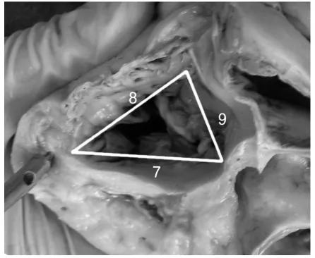
partic-ular walls of the right ventricle: anterior [7], pos-terior [8], and septal [9].
Measurements 1, 2, 3 and 5 for an example of valve type 2B are shown in Figure 2, and measure-ments 7, 8 and 9 are shown in Figure 3.
The results obtained were statistically analysed by Pearson’s analysis and one way analysis of vari-ance (ANOVA; p < 0.05).
RESULTS
The length of the anterior wall of the right ven-tricle (dimension 7) was 34 ± 6.9 mm, the posterior wall (dimension 8) was 31 ± 9 mm and the septal
Figure 1. Scheme of location of accessory cusps in subtypes of Type 2.
Figure 2. Example of measurements of the attachment leaflets in
valve Type 2B. The attachment length of the main leaflets — an-terior (CA-cut and shown in 2 parts), posan-terior (CP), septal (CS) and accessory leaflet (CaC).
Figure 3. Example of measurements of the tricuspid attachment
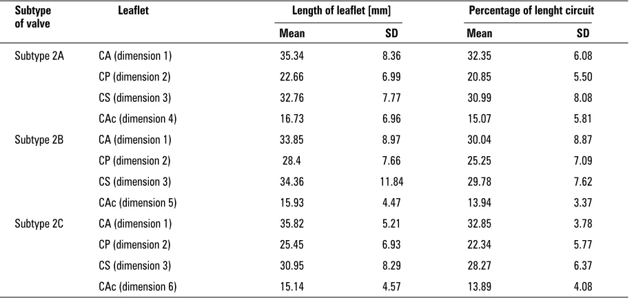
wall (dimension 9) was 29.32 ± 4.74 mm. Table 1 presents the mean values and SD of the length and percentage of lenght circuit for the main and acces-sory leaflets in particular subtypes of the four-cusp-idal form of the tricuspid valve.
Table 2 presents the statistical results for the cor-relation between the length of the main and acces-sory cusps shown in the column heading and the length of particular walls of the right ventricle in particular subtypes of type 2.
Strong negative correlations (Pearson: –0.549) were noticed between CP and CS and between CS and Cac in subtype 2A.
In subtype 2B a high positive correlation was observed between CS and Cac, while lower correla-tions were noticeable between these and CA, these being positive between CA and CS and negative be-tween CA and Cac.
A small negative correlation between CA and CP was observed in subtype 2C.
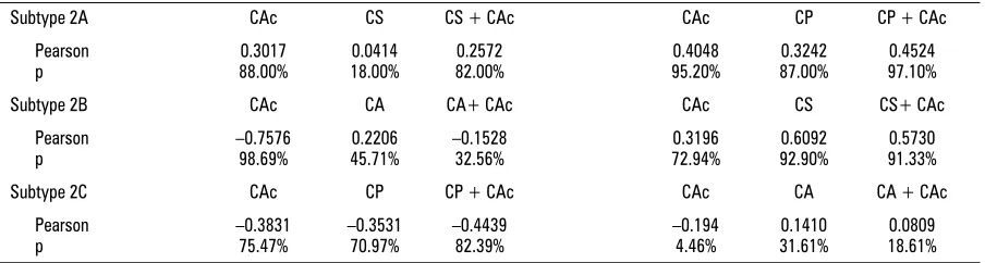
Table 3 presents the statistical results for the ra-tio of the sum of the attachment lengths for the main and accessory leaflets shown in the column heading and the length of the tricuspid attachment in particular walls of the right ventricle. On the basis of the statistics shown in Table 2 we reach the fol-lowing conclusions:
— subtype 2A: the correlation between the acces-sory leaflet and the septal part of the tricuspid valve is stronger than that between the septal leaflet and the septal part of the attachment; — subtype 2B; there is a strong negative
correla-tion between the anterior part of the attachment and the accessory leaflet;
— subtype 2C: no statistically significant correlation was observable.
Table 1. Values and SD of length and percentage of lenght circuit for main and accessory leaflets
Subtype Leaflet Length of leaflet [mm] Percentage of lenght circuit of valve
Mean SD Mean SD
Subtype 2A CA (dimension 1) 35.34 8.36 32.35 6.08
CP (dimension 2) 22.66 6.99 20.85 5.50
CS (dimension 3) 32.76 7.77 30.99 8.08
CAc (dimension 4) 16.73 6.96 15.07 5.81
Subtype 2B CA (dimension 1) 33.85 8.97 30.04 8.87
CP (dimension 2) 28.4 7.66 25.25 7.09
CS (dimension 3) 34.36 11.84 29.78 7.62
CAc (dimension 5) 15.93 4.47 13.94 3.37
Subtype 2C CA (dimension 1) 35.82 5.21 32.85 3.78
CP (dimension 2) 25.45 6.93 22.34 5.77
CS (dimension 3) 30.95 8.29 28.27 6.37
CAc (dimension 6) 15.14 4.57 13.89 4.08
Table 2. Correlation between length of accessory leaflets and main leaflets adhering to them
Subtype 2A CS CAc CP CAc CS CP
Pearson –0.2009 0.2940 –0.5495
p 70.00% 77.58% 85.00%
Subtype 2B CA CAc CS CAc CA CS
Pearson –0.1900 0.6358 0.2511
p 54.55% 82.79% 60.50%
Subtype 2C CA CAc CP CAc CA CP
Pearson 0.3238 0.3313 –0.3533
The correlations between the anterior, posterior and septal parts of the attachment of the tricuspid valve and accessory leaflets were calculated. The sta-tistics obtained showed no clear correlation which could explain which leaflets divided into main and accessory cusps. The correlations are comparable and the thesis that accessory cusps originate from dif-ferent main leaflets may be accepted.
DISCUSSION
The tricuspid valve is a heterogeneous structure. The leaflets of the tricuspid valve develop from en-docardial cushions and the myocardium, which comes from two sources, namely the tricuspid gully complex and the supraventricular crest [15]. The number of leaflets in the tricuspid valve increases during evolution [6, 19, 31, 32], but the rules of this process are unknown. Atavistic features and atypi-cal forms of the tricuspid valve and the distribution of the tendinous chords and their connection with the papillary muscles occur in a small percentage of human hearts [9, 20, 26, 27, 29, 31, 32, 34]. Bi-di-visible leaflets are also observed in some primates [31, 32]. On the basis of this, the thesis was put for-ward that the accessory leaflet had separated from the main leaflet.
We based our thesis on our findings, which showed that, if accessory cusps are separated direct-ly from the main leaflets, there is a correlation be-tween the length of the accessory and maternal main leaflets and the attachment length of the analogical wall of the right ventricle, and also that there is no correlation between the length of an accessory and another main leaflet adhering to it and the wall of the right ventricle.
On the basis of the results of this study we con-clude that the separation of accessory leaflets is a complex process and that it is impossible to make explicit connections with the main leaflets from which
the accessory ones have separated. The explanation of the mechanism of the separation of accessory leaf-lets may be linked to the question of the different sources of the tissues which form the cusps and their non- synchronised phylo- and ontogenetic develop-ment [1, 2, 6, 9, 19]. The role of differences in tension in particular parts of the tricuspid valve during leaflet formation is unknown [14, 18].
The mechanism of the separation of the accesso-ry cusps is a complex process and demands further study of groups of human foetal hearts and the hearts of other primates. This will be the subject of our next study.
REFERENCES
1. Anderson RH, Benson R, Wilcox MD (2000) Reply. Ann Thorac Surg, 69: 1990.
2. Anderson RH, Webb S, Brown NA, Lamers W, Moor-man A (2003) Development of the heart: (2) Septation of the atriums and ventricles. Heart, 98: 949–958. 3. Benninghoff A (1933) Herz. In: Goppert E (ed)
Hand-buch der wergleichenden Anatomie der wirbeltiere. Vol. VI. Urban and Schwarzenberg, Berlin–Wien: 346–389.
4. Cardarelli MG, Gammie JS, Brown JM, Poston RS, Pier-son RN 3rd, Griffith BP (2005) A novel approach to tri-cuspid valve replacement: the upside down stentless aortic bioprosthesis. Ann Thorac Surg, 80: 507–510. 5. Carrier M, Hebert Y, Pellerin M, Bouchard D, Perrault LP,
Cartier R, Basmajian A, Page P, Poirier NC (2003) Tricuspid valve replacement: an analysis of 25 years of experience at a single center. Ann. Thorac. Surg, 75: 47–50. 6. Cayré R, Valencia-Mayoral P, Coffe-Ramirez V,
Sánchez-Gómez C, Angelini P, De la Cruz MV (1993) The right atrioventricular apparatus in the chick heart. Acta Anat, 148: 27–33.
7. Crumbley AJ, Van Bakel AB (1994) Tricuspid valve re-pair for biopsy-induced regurgitation after cardiac transplantation. Ann Thorac Surg, 58: 1156–1160. 8. Dimas VV, Grifka RG, Fraser CD Jr (2004) Combined
tricuspid valvuloplasty and superior cavopulmonary anastomosis for repair of traumatic tricuspid valve in-jury. Tex Heart Inst J, 31: 418–20. Comment in: Tex Heart Inst J (2005) 32: 114; author reply 114. Table 3. Correlation between accessory leaflets and main leaflets adhering to them
Subtype 2A CAc CS CS + CAc CAc CP CP + CAc
Pearson 0.3017 0.0414 0.2572 0.4048 0.3242 0.4524
p 88.00% 18.00% 82.00% 95.20% 87.00% 97.10%
Subtype 2B CAc CA CA+ CAc CAc CS CS+ CAc
Pearson –0.7576 0.2206 –0.1528 0.3196 0.6092 0.5730
p 98.69% 45.71% 32.56% 72.94% 92.90% 91.33%
Subtype 2C CAc CP CP + CAc CAc CA CA + CAc
Pearson –0.3831 –0.3531 –0.4439 –0.194 0.1410 0.0809
9. Dudziak M (1984): Budowa pierścienia włóknistego zastawki przedsionkowo-komorowej prawej serca u człowieka w rozwoju osobniczym. Rozprawa doktors-ka. Gdańsk.
10. Hartyanszky I, Prodan Z, Kiraly L, Mihalyi S, Bodor G, Tamas C, Lozsadi K (2005) Challenges in the surgical management of hearts with functional single ventri-cle. Orv Hetil, 33: 1721–26.
11. Hvass U, Juliard JM, Assayag P, Laperche T, Pansard Y, Chatel D (1996) Tricuspid autograft for mitral-valve repair. Lancet, 347: 659–661.
12. Jastrzębski C (1926) O zmienności kształtu zastawki trójdzielnej serca i o otworach wrodzonych w jej płatkach. Kosmos, Seria A. Biologia, 51: 191–198. 13. Khoury GE, d’Udekem Y, Noirhomme P, Verhelst R,
Rubay J, Dion R (2000) Transfer of the posterior leaflet of the tricuspid valve to the mitral valve. J Heart Valve Dis, 9: 350–352.
14. Kilner PJ, Yang GZ, Wilkes AJ, Mohiaddin RH, Firmin DN, Yacoub MH (2000) Asymmetric redirection of flow through the heart. Nature, 6779: 759–761.
15. Kosiński A, Kuta W, Grzybiak M, Ciszkowicz M, Ka-miński R(2000) Morfologia zastawki trójdzielnej w ser-cu człowieka dorosłego i innych naczelnych. Przegląd Medyczny, 2: 80.
16. Lamers WH, Virágh S, Wessels A, Moorman AFM, Anderson RH (1995) Formation of the tricuspid valve in the human heart. Circulation 91, 111–121. 17. Langberg JJ, Man KC, Vorperian VR, Williamson B.,
Kalbfleisch SJ, Strickberger SA, Hummel JD, Morady F (1993) Recognition and catheter ablation of sub-epicardial accessory pathways. JACC 22: 1100– –1104.
18. Lomholt M, Nielsen SL, Hansen SB, Andersen NT, Hasenkam JM (2002) Differential tension between sec-ondary and primary mitral chordae in an acute in vivo porcine model. J Heart Valve Dis, 11: 337–345. 19. Lu Y, James TN, Boottsma M, Terasaki T (1993)
Histo-logical organization of the right and left atrioventric-ular valves of the chicken heart and their relationship to the atrioventricular Purkinje ring and the middle bundle branch. Anat Rec, 235: 74–86.
20. Łukaszewska-Otto H (1970) Zmienność budowy zastawki przedsionkowo-komorowej prawej u człowieka. Rozprawa habilitacyjna. Akademia Medyc-zna w Warszawie, Warsaw.
21. Miki K, Maekura R, Higara T, Hirotani A, Hashimoto H, Kitada S, Miki M, Yoshimura K, Naka N, Motone M, Fujikawa T, Takashima S, Kitazume R, Kanzaki H, Na-katani S, Watanuki H, Tagusari O, Kobayashi J, Ito M
(2005) Infective tricuspid valve endocarditis with pul-monary emboli caused by Campylobacter fetus after tooth extraction. Intern Med, 10: 1055–1059. 22. Oppido G, Napoleone CP, Ragni L, Turci S, Loforte A,
Angeli E, Gargiulo G (2006) Double orifice tricuspid valve in an infant with tetralogy of Fallot Ann Thorac Surg, 81: 1121–1123.
23. Radermecker MA, Somerville J, Li W, Anderson RH, de Leval MR (2001) Double orifice right atrioventricular valve in atrioventricular septal defect: morphology and extension of the concept of fusion of leaflets. Ann Thorac Surg, 7: 358–360.
24. Sharieff S, Sagir T, Shah-e-Zaman K (2005) Concurrent percutaneous valvuloplasty of mitral and tricuspid valve stenoses. J Invasive Cardiol, 17: 340–342.
25. Siebert FM, Lorenzo FJM, Rojas TM, Becerra PE, Mar-tinez EF, Dans CA (1981) Tricuspid atresia with hypo-plasia of aortic isthmus in absence of transposition of great vessels. An Esp Pediatr, 14: 117–121.
26. Skwarek M, Dudziak M, Hreczecha J, Grzybiak M (2006) The connection between the papillary muscles and leaf-lets of the tricuspid valve. Folia Morphol, 65: 322–328. 27. Skwarek M, Dudziak M, Hreczecha J, Grzybiak M (2006) The morphology of right atrioventricular valve in the human adult heart. Folia Morphol, 65: 105–113. 28. Skwarek M, Hreczecha J, Grzybiak M, Kosiński A (2005)
Unusual anatomical features of the right atrioventric-ular valve. Folia Morphol, 64: 183–187.
29. Skwarek M, Hreczecha J, Grzybiak M, Kosiński A (2004) Remarks on the morphology of the papillary muscles of the right ventricle. Folia Morphol, 64: 176–182. 30. Sutton JP III, Yen Ho S, Vogel M, Anderson RH (1995)
Is the morphologically right atrioventricular valve tri-cuspid? J Heart Valve Dis 4: 571–575.
31. Szostakiewicz-Sawicka H, Grzybiak M (1981) Zgodność rozwoju osobniczego niektórych cech budowy serca z przypuszczalnym kierunkiem ich rozwoju w antropo-genezie. Morfologia, podręczniki, skrypty AWF. Seria: Monografie, 199: 9–16.
32. Szostakiewicz-Sawicka H (1967) Zastawka przedsion-kowo-komorowa prawa u naczelnych. Rozprawa ha-bilitacyjna. Acta Biol Med Soc Sc Gedan, 11: 545–589. 33. Victor S, Nayak VM (2000) Tricuspid valve is bicuspid.
Ann Thorac Surg, 69: 1989–1990.
34. Wafae N, Hayashi H, Gerola LR, Vieira MC (1990) Ana-tomical study of the human tricuspid valve. Surg Radi-ol Anat, 12: 37–34.