Mercury free dentistry
Naziya Butt1, Sangeeta Talwar2,
Sarika Chaudhry3, Ruchika Roongta Nawal4
ABSTRACT:
Dental amalgam has been a highly successful, cost-effective, restorative material. In the past 20 years, significant research has been conducted on the health effects of mercury. Mercury toxicity has become a compelling rationale for replacing amalgam restorations with tooth-coloured materials. However, no causal link between mercury in restorations and systemic disease has been proven, despite the billions of restorations placed over 160 years. Nonetheless, it is impossible to prove that amalgam is entirely safe and its continued use implies acceptance of possible risks. The development of competing restorative materials continues apace and, in those countries where the incidence and severity of caries is decreasing, a progressive reduction in the use of amalgam is inevitable. Resin-based composite, glass ionomer cements, cast gold and ceramic and/or processed composites inlay and onlay have become predictably successful in the restoration of posterior teeth. However, none of the currently available restorative materials can fulfil all of the requirements of a 'perfect restorative materials'. This article will review the health hazards of mercury and the possible amalgam substitutes in clinical situations.
Key words: Amalgam, mercury, composite resins, ceramic,
toxicity
doi: 10.5866/2013.521186
1PG Student 2Professor & HOD 3Associate Professor 4Assistant Professor
Department of Conservative dentistry & Endodontics Maulana Azad Institute of Dental Sciences
MAMC Complex, Bahadur Shah Zafar Marg, New Delhi-110002, India
Article Info:
Received: January 9, 2013
Review Completed: February 11, 2013 Accepted: March 10, 2013
Available Online: July, 2013 (www.nacd.in) © NAD, 2013 - All rights reserved
Email for correspondence: [email protected]
Quick Response Code
I
NDIANJ
OURNALOFD
ENTALA
DVANCEMENTSJ o u r n a l h o m e p a g e : w w w. n a c d . i n
INTRODUCTION
posterior restorations have increased in use significantly over the past several years.5 A survey accomplished previously showed 22% or more of intra-coronal restorations were tooth-coloured.5 However, when considering these restorations by category, 94% of these restorations were direct-placement resins, 4% were indirect-direct-placement resins, and only 2% were indirect ceramic.5 Over the past 20 years or so, various anti-mercury groups have fought to effect a ban on the use of dental amalgam. As no fully biocompatible material exists, this would appear to be a short-sighted objective.
HISTORY
Dental amalgam apparently was first used by the Chinese.6 The invention of a “silver dough” in China is mentioned in a manuscript of the Tang dynasty, the “Materia Medica” by SU KUNG in the year 659 A.D. The English chemist Charles Bell in 1819 invented a kind of silver amalgam. Originally it was named “Bell’s putty” and later on “Mineral succedaneum”, meaning “mineral substitute”.6 In the early 1830s the family Crawcour in London advertised that they filled teeth with the “Royal Mineral Succedaneum in two minutes without any pain, inconvenience or pressure”. In 1833 two of the Crawcour brothers brought amalgam fillings to America. Skilled in the use of cohesive gold the American dentists stamped the use of amalgam as quackery.6
The First Amalgam War
In 1845, American Society of Dental Surgeons condemned the use of all filling material other than gold as toxic and requested members to sign a pledge refusing to use amalgam. However, this policy was reconsidered in 1850, and the use of amalgam was promoted by the work of J Foster Flagg and the final stamp of approval for its clinical use came from G V Black.7 By combining the principles of cavity design, extension of the cavity into “immune” areas and the development of an alloy with the composition of 68.5% silver, 25.5% tin, 5% gold, 1% zinc, Black advanced amalgams into modern times.
The Second Amalgam War
In 1926, the German chemist Alfred Stock wrote an article, “Die Gefahrlichkeit des Quecksilberdampfes und der Amalgame” (“The danger of mercury vapour and amalgams”).8 Dr. Stock himself was exposed to significant Hg vapours and recognized its danger. In 1930, a commission
issued a report that validated the safety of newer amalgam that need not be heated and it replaced the older formulation.9
The Third Amalgam War
In 1985, Dr. Huggins10 published a book that detailed his belief about Hg toxicity. He mentioned that Hg released from amalgam restorations caused a wide variety of neurological, CVS, immunological, collagen, emotional and allergic disorders. In 1995, a survey reported that 8.7% of dentists wanted to ban Amalgam use and 14.3% were undecided about its safety.11 American Council on Science and Health, a consumer education and advocacy group has determined that allegations against amalgam constitute one of the greatest unfounded health scares of recent times.12
DENTAL AMALGAM
The dental industry uses about 75 tons of mercury to place approximately a half-billion amalgam restorations per year.13 Dental amalgam consists, essentially, of mercury combined with a powdered silver-tin alloy.The reaction between mercury and alloy which follows mixing is termed an amalgamation reaction. The amalgamation is a chemical process unique to elemental mercury, in which another metal forms a semisolid alloy “amalgam” with mercury. Mercury dissolves in the solid metal, forming a solid solution. The process is reversible, so that mercury can be released from these alloys by heating. Amalgams, although solid, show a significant vapor pressure and solubility of mercury.14
(lathe-cut) particles made from low-copper Ag-Sn alloys. The ‘‘highcopper single-composition” powders was developed by Asgar and Reichman16 in 1975. Particles in this high-copper single-composition powder are ternary Ag-Sn-Cu (13 wt% Cu) alloy. Perhaps the most important advantage offered by amalgam is its greater clinical longevity than tooth-coloured materials, notably when placed in large cavities subject to occlusal forces. The approximate median expectations for clinical use of amalgam and competing materials is listed in Table 1.
MERCURY AND ITS BIOCOMPATIBILITY ISSUES
Globally. around 10,000 tons of mercury are produced yearly for anthropogenic use. It has been estimated that 3-4% is used in dentistry.19 Mercury (Hg) is globally recognized as a toxic substance with numerous national and international efforts to phase out its use, the most recent being the initiative of the United Nations Environment Programme20 on a global phase out strategy, for which negotiations began in June 2010. The one lingering exception to this phase out is dental amalgam. Although now banned in Sweden21 and Norway,22 dental amalgam is still a restorative material of choice for the majority of US general dentists for repair of dental caries (cavities).23
Mercury is generally found in three forms:4
Elemental mercury (Hg0)
Inorganic mercury compounds
(mercurous-Hg2++ and mercuric-Hg2+)
Organic mercury compounds (primarily methyl mercury -MeHg compounds)
Each form possesses its own characteristic toxicokinetics and human health effects. Elemental Hg volatilizes at room temperature and human exposure is primarily through inhalation of the vapor. Hg vapor is lipid soluble and easily crosses alveolar membranes of the lungs; it is taken up by red blood cells and transported to the central nervous system. Absorption of inorganic Hg (also known as ionic Hg) by the gastrointestinal tract in humans is relatively limited and approximates 7% of the ingested dose.24 Kidney tissue contains the highest concentration of Hg after exposure to inorganic salts and elemental Hg. It has been demonstrated that elemental Hg in human saliva can be oxidized to ionic Hg, which may be protective since ionic Hg is a less toxic species.25 Organic Hg is
the most important form in terms of toxicity to humans.4 The serious health consequences of MeHg exposure was dramatically illustrated in 1953, when an epidemic of MeHg poisoning occurred in humans from the consumption of fish in villages around Minamata Bay, Japan. The resulting medical disorders associated with this epidemic became known as “Minamata disease”.26 The high-dose chronic and acute MeHg poisoning resulted in many deaths and other effects, which included mental retardation, cerebral palsy, deafness, blindness, and dysarthria, especially in children exposed in utero.
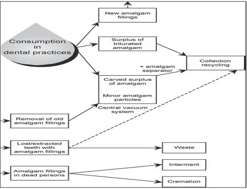
As a natural element mercury is ubiquitous in the environment.27 As long as amalgam fillings are produced inrestorative dentistry and patients have amalgamfillings in their teeth, the dental profession has an obligation to minimize or, preferably totally to eliminate release of mercury to the environment. The mercury cycle in dentistry is illustrated in Fig. 1.28
Estimates of mercury released and absorbed initially varied markedly, but are generally accepted to lie between 2-5μg/day for the average adult.29 A method to evaluate occupational exposure is to measure the air mercury level at the workplace. WHO has decided an exposure limit of 50μg Hg/m3 air (TWA: Time weighted average) corresponding to an estimated urine concentration of about 80μg Hg/l which with today’s knowledge of mercury toxicology seems to be too high. Some countries have therefore adopted a lower concentration of 25 or 30 μg Hg/m3 as the upper limit.28 The lowest dose of mercury that illicits a toxic reaction is 3 to 7 μg/kg body weight.28 Paresthesia (tingling of extremities) occurs at about 500 μg/kg of body weight, followed by ataxia at 1000 μg/kg of body weight, joint pain at 2000 μg/kg of body weight, and hearing loss and death at 4000 μg/kg of body weight. Therefore these values are much greater in magnitude than the exposure to mercury from amalgam or from a normal diet.
BEST MANAGEMENT PRACTICES (BMP) FOR AMALGAM WASTE
ALTERNATIVES TO DENTAL AMALGAM
The demand for tooth-colored restorations has grown considerably during the last decadebecause of concerns about the esthetics and biocompatibility of dental amalgam.5 Amalgam may be replaced by three different categories of filling materials or restorations (Fig. 2), defined as standards I, II and III (Lutz, Krejci & Besek, 1997).31
However, a cost/benefit analysis is essential if a tooth-colored material is considered as a substitute. Two types of restorative materials are commonly used in dentistry; they are designated depending on whether they can be applied directly to the tooth or require fabrication of the restoration in the dental laboratory. Dental materials are used for direct restoration of a tooth in order to save its function while indirect materials include pre-formed metal crowns, dental porcelain, and cast restorations.32 It can be categorised as:
1. Direct Restorative Dental Materials
a. Composites (Direct/Indirect)
b. Glass ionomers
c. Resin ionomers (Compomers/Giomers)
2. Indirect Restorative Dental Materials
a. All ceramic
b. Porcelain fused to metal
c. Gold alloys ( high noble)
d. Base metal alloys
Advances in resin-based adhesives and restorative materials, as well as increased patient demand for esthetic restorations, have stimulated an increase in the use of resin-based composites in posterior teeth.33 Gold alloys have been used as a standard of care for indirect restorative services.34 Their characteristics such as low restoration wear and low wear of antagonistic teeth have been unavailable in other restorative materials. Indirect composite materials have been occasionally used as an alternative to dental porcelain when use of the porcelain is contraindicated.35 Although dental porcelains have an advantage of chemical inertness, they are at times not the material of choice because they possess inherent problems including brittle characteristic and abrasiveness to antagonistic dentition.36 The indications for use of these
restorative materials span from small cavities to extensive loss of tooth substance. Materials are employed for cavities in primary teeth; for cavities in permanent teeth, ranging from “minimal interventions” to the need for extensive replacements and/or build-procedures; replacement or repair of failed or less satisfactory restorations, or materials are used in people with compromised health and having dental caries on certain locations, e.g. root caries.37
Restoration longevity
The longevity of different materials is not easily established because the data depends on a multitude of factors, where material selection is just one. Annual failure rates of different restorative materials are given in Table 4, with glass ionomers having the highest failure rate of 7.6%.37 The most prevalent reasons for failure of fillings are secondary caries and fracture.37,38
Biological considerations
A balanced discussion of the biocompatibility of dental amalgam requires consideration of the relative biocompatibility of other restorative materials that potentially could serve as alternatives to amalgam. Amalgam has been associated with general health concerns, while local oral effects from different restorative materials are reported.40 Many of the biocompatibility considerations pertaining to dental restorative materials are sized in Table 5. All materials in current use are considered acceptable, in terms of their biocompatibility with local tissues, when properly handled and placed. Adverse systemic reactions are believed to be rare and self-limiting and tend to be of an allergenic nature.
Despite the innovations in biocompatibility, strength, marginal adaptation, and optical qualities of dental materials, the prognosis of esthetic restorations appears to hinge predominantly on choice of material, precise technique, and patient selection. In the face of rapid technological advances, evidence-based research offers a powerful tool to dental practitioners to assess the risk/benefit calculus of various tooth-colored restorations and provide appropriate information to patients.
CONCLUSIONS
Dental amalgam has been the main direct
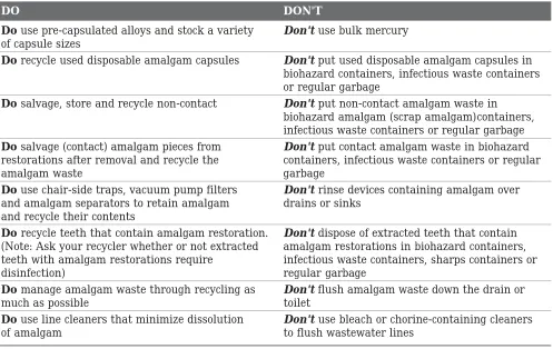
TABLE 2: Best Management Practices for dental offices using amalgam30
DO DON'T
Do use pre-capsulated alloys and stock a variety Don't use bulk mercury
of capsule sizes
Do recycle used disposable amalgam capsules Don't put used disposable amalgam capsules in
biohazard containers, infectious waste containers or regular garbage
Do salvage, store and recycle non-contact Don't put non-contact amalgam waste in
biohazard amalgam (scrap amalgam)containers, infectious waste containers or regular garbage Do salvage (contact) amalgam pieces from Don't put contact amalgam waste in biohazard
restorations after removal and recycle the containers, infectious waste containers or regular
amalgam waste garbage
Do use chair-side traps, vacuum pump filters Don't rinse devices containing amalgam over
and amalgam separators to retain amalgam drains or sinks and recycle their contents
Do recycle teeth that contain amalgam restoration. Don't dispose of extracted teeth that contain
(Note: Ask your recycler whether or not extracted amalgam restorations in biohazard containers, teeth with amalgam restorations require infectious waste containers, sharps containers or
disinfection) regular garbage
Do manage amalgam waste through recycling as Don't flush amalgam waste down the drain or
much as possible toilet
Do use line cleaners that minimize dissolution Don't use bleach or chorine-containing cleaners
of amalgam to flush wastewater lines
TABLE 1: Longevity of dental restorations17,18
Restoration Median clinical performance (years)
Amalgam 8-12
Direct Composite 6-8
Glass Ionomer Cement 5
Gold foil 10
Gold alloy inlay/crown 12-18
Ceramic 6-10
direct restorative materials like composite resins and glass ionomers and several indirect restorative materials are available for use, although at much higher cost.
The issue of mercury and dental amalgam in dentistry resolves around the proposition that mercury leaching out of dental amalgam fillings may have an adverse effect on health. At high doses mercury is recognised as a neurotoxin capable of producing a variety of neurobehavioural effects.
Altered approaches to cavity preparation, including a philosophy of minimum tooth removal, and the availability of alternative materials are leading to a further movement away from dental amalgam as a direct restorative material.
Non-contact (scrap) amalgam·
Place non-contact, scrap amalgam in a wide-mouthed container that is marked “Non-contact Amalgam Waste for Recycling”.·
Make sure the container lid is well sealed.
When the container is full, send it to a recycler.
Amalgam capsules
Stock amalgam capsules in a variety of sizes.
After mixing amalgam, place the empty capsules in a wide-mouthed, airtight container that is marked “Amalgam Capsules Waste for Recycling”.
Capsules that cannot be emptied should likewise be placed in a wide-mouthed airtight container that is marked “Amalgam Capsules Waste for Recycling”.
Make sure the container lid is well sealed.
When the container is full, send it to a recycler.
Disposal chair-side traps
When the chair-side unit to expose the trap.
Remove the trap and place it directly into a wide-mouthed, airtight container that is marked “Contact Amalgam Waste for Recycling”.
Make sure the container lid is well sealed.
When the container is full, send it to a recycler.
Traps from dental units dedicated strictly to hygiene may be placed in with the regular garbage.
Reusable chair-side traps
Open the chair-side unit to expose the trap.
Remove the trap and empty the contents into a wide-mouthed, airtight container that is marked “Contact Amalgam Waste for Recycling”.
Make sure the container lid is well sealed.
When the container is full, send it to a recycler.
Replace the trap into the chair-side unit (Do not rinse the trap under running water as this could introduce dental amalgam into the waste stream).
Vacuum pump filters·
Change the filter according to the manufacturer’s recommended schedule.
Note: The following instructions assume that your recycler will accept whole filters; some recyclers require different handling of this material, so check with your recycler first.
Remove the filter.
Put the lid on the filter and place the sealed container in the box in which it was originally shipped. When the box is full, the filters should be recycled.
Amalgam separators
Select an amalgam separator that complies with ISO 11143.
Follow the manufacturer’s recommendations for maintenance and recycling producers.
Line cleaners
TABLE 4: Annual failure rates of dental restorations37,39
Material Age at replacement Annual failure rate
Resin-based composites 8 years 2.3%
Poly-acid modified composites 7 years 3.5%
Resin-modified glass ionomers 2 years 3.1%
Glass ionomers 4 years 7.6%
Amalgam 10 years 2.2%
Ceramic 9 years 3.9%
Gold inlay 20 years 2.4%
TABLE 5: Biocompatibility considerations of various dental restorative materials40,41
Restorative Biocompatibility Consideration
Material
Dental No adverse pulpal responses from mercury
Amalgam: Corrosion may limit marginal leakage, but in the long-term may lead to breakdown of marginal integrity, especially with low-copper amalgams
Lichenoid reasons reported
Thermal conduction to pulp
Mercury allergy (6%)
Resin-Base Documented estrogenicity issue
Composites Very little research on systemic biocompatibility
Allergic to resin composite ingredients (8%)
Incomplete polymerization leading to degradation, teaching, and imperfect bonding
Predisposed to polymerization shrinkage
Associated with adverse local pulpal and dentin reactions, development of recurrent caries, and pain
Higher proportion of streptococcus mutans leading to secondary caries
Glass lonomer Few documented systemic adverse effects
Cements Early pulpal reactions, although less than with cements or composite resins, and with rapid recovery
Hydraulic pressure and etching during placement may irritate the pulp
Good adhesion, minimal leakage at margins, high biocompatibility
Leaching of component materials offers opportunity for slow release of fluoride
Gold Foil and Inert; sensitivities are rare
Cast Alloys Potential pulpal reactions due to condensation
Gold contact allergy (23%)
Ceramics Inert material
No long-term data on biocompatibilility
Fig. 1: Mercury cycle in dentistry (adapted from Horsted-Bindslev 2004)28
Fig. 2: Classification and characteristics for posterior fillings and restorations in operative dentistry.31 12345678901234567890123
12345678901234567890123 12345678901234567890123 12345678901234567890123 12345678901234567890123 12345678901234567890123 12345678901234567890123 12345678901234567890123 12345678901234567890123
12345678901234567890123 12345678901234567890123 12345678901234567890123 12345678901234567890123 12345678901234567890123 12345678901234567890123 12345678901234567890123 12345678901234567890123 12345678901234567890123 12345678901234567890123 12345678901234567890123 12345678901234567890123 12345678901234567890123 12345678901234567890123 12345678901234567890123 12345678901234567890123 12345678901234567890123 12345678901234567890123 12345678901234567890123
Classification Characteristics of posterior fillings and restorations
STANDARD TOOTH PLUS PLUS
PREVENTION FUNCTION AESTHETICS
temporary
I filling
amalgam filling
II cast gold inlay/onlay
gold foil filling
compomere: for primary dentition
adhesively placed composite filling (incremental technique)
III lab-made composite inlay/onlay
REFERENCES
1. Molin C. Amalgam - fact and fiction. Scand J Dent Res 1992;100:66-73.
2. Eley BM, Cox SW. The release, absorption and possible health effects of mercury from dental amalgam: a review of recent findings. Br Dent J 1993;20:355-362.
3. Brownawell AM, Berent S, Brent RL, Bruckner JV, Doull J, Gershwin EM et al. The potential adverse health effects of dental amalgam. Toxicol Rev 2005;24:1-10.
4. Syversen T, Kaur P. The toxicology of mercury and its compounds. J Trace Elem Med Biol 2012;26:215-226. 5. Christensen GJ. Current use of tooth-colored inlays, onlays,
and direct-placement resins. J Esthet Dent 1998;10: 290-295
6. Charles AD. The story of dental amalgam. Bull Hist Dent 1982;30:2-7.
7. Flagg JF. The amalgam question. Dent Cosmos 1882;24:237-242.
8. Stock A. Die Gefahrlichkeit des Quecksilberdampfes und der Amalgame. Med Klin 1926;22:1209-1212,1250-1252. 9. Harndt E. Clinical examination results of research on the
amalgam-mercury question. Deutsche Zahnarztliche Wochenschrift 1930;33:564-575.
10. Huggins HA, Huggins SA. It’s all in your head: Diseases caused by silver-mercury restorations. Solona Beach, Calif.: APW; 1985.
11. Product Use Survey, 1995. Clin Res Associates Newsletter. 1995;19:4.
12. Lieberman AJ, Kwon SC. Facts versus fears: A review of the greatest unfounded health scares of times. 3rd ed. New York. American Council on Science and Health; 1998. 13. Osborne JW. Safety of dental amalgam. J Esthet Restor
Dent 2004;16:377-388.
14. Rodríguez O, Padilla I, Tayibi H, López-Delgado A. Concerns on liquid mercury and mercury-containing wastes: a review of the treatment technologies for the safe storage. J Environ Manage 2012 Jun;101:197-205. 15. American Dental Association (ADA), J Am Dent Assoc
1977;95:1171.
16. Asgar K, Reichman S. Special Metals Corporation assignee. Dental amalgam. US patent 3,871,875; March 1975. 17. Mjor IA, Jokstad A, Qvist V. Longevity of posterior
restorations. Int Dent J 1990;40:11-17.
18. Hickel R, Manhart J. Longevity of restorations in posterior teeth and reasons for failure. J Adhes Dent 2001;3:45-64. 19. Arenholt-Bindslev D. Dental amalgam-environmental
aspects. Adv Dent Res 1992;6:125-130.
20. UNEP (United Nations Environment Program). Press release: historic treaty to tackle toxic heavy metal mercury gets green light. Nairobi, Kenya: UNEP Secretariat; 2009. 21. Sweden Ministry of Environment. Press release: government bans all use of mercury in Sweden. Government offices of Sweden; 2009.
22. Norway Ministry of Environment. Press release: Minister of the Environment and International Development Erik Solheim: bans mercury in products. Dated December 21, 2007.
23. Public Health Service (USA). Dental amalgam: A scientific review and recommended public health service strategy for research, education and regulation. Committee to Co-ordinate Environmental Health and Related Programs. Final report (January 1993).
24. Mertz-Fairhurst EJ, Curtis JW Jr, Ergle JW, Rueggeberg FA, Adair SM. Ultraconservative and cariostatic sealed restorations: Results at year 10. J Am Dent Assoc 1998;129:55-66.
25. Liang L, Brooks RJ. Mercury reactions in the human mouth with dental amalgams. Water Air and Soil Pollut 1995;80:103-107.
26. Guzzi G, La Porta CA Molecular mechanisms triggered by mercury. Toxicology 2008;244:1-12.
27. Zahir F, Rizwi SJ, Haq SK, Khan RH. Low dose mercury toxicity and human health . Environ Toxicol Pharmacol 2005;20:351-360.
28. Hörsted-Bindslev P. Amalgam toxicity-environmental and occupational hazards. J Dent 2004;32:359-365.
29. Spencer AJ. Dental amalgam and mercury in dentistry Aust Dent J 2000;45:224-234.
30. Future Use of Materials for Dental Restoration. WHO HQ, Geneva, Switzerland : Final Report (November 2009). 31. Lutz FU, Krejci I, Besek M. Operative dentistry: the
missing clinical standards. Pract Periodontics Aesthet Dent 1997;9:541-548.
32. ADA Council on Scientific Affairs. Direct and indirect restorative materials. J Am Dent Assoc 2003;134:463-472. 33. Zimmerli B, Strub M, Jeger F, Stadler O, Lussi A. Composite materials: Composition, properties and clinical applications A literature review. Schweiz Monatsschr Zahnmed 2010;120:972-986.
34. Naert I. Materials in Fixed Prosthodontics for Indirect Dental Restorations Comprehensive Biomaterials 2011;6:353-365.
35. Suzuki S, Nagai E, Taira Y, Minesaki Y. In vitro wear of indirect composite restoratives. J Prosthet Dent 2002;88:431-436.
36. Sadowsky SJ. An overview of treatment considerations for esthetic restorations: a review of the literature. J Prosthet Dent 2006;96:433-442.
37. Manhart J, Chen H, Hamm G, Hickel R. Buonocore Memorial Lecture. Review of the clinical survival of direct and indirect restorations in posterior teeth of the permanent dentition. Oper Dent 2004;29:481-508. 38. Mjör IA, Moorhead JE, Dahl JE. Reasons for replacement
of restorations in permanent teeth in general dental practice. Int Dent J 2000;50:361-366.
39. Espelid I, Tveit AB, Tornes KH, Alvheim H. Clinical behaviour of glass ionomer restorations in primary teeth. J Dent 1999;27:437-442.
40. Cobos-Fuentes MJ, Martinez-Sahuquillo-Marquez A, Gallardo-Castillo I, Armas-Padron JR, Moreno-Fernandez A, Bullon-Fernandez P. Oral lichenoid lesions related to contact with dental materials: A literature review. Med Oral Patol Oral Cir Bucal 2009; 14:e514-520.