Periodontal Microsurgery: A Changing Perspective
Satyanarayana D1, Vikram Reddy G2, Raja Babu P3
Department of Periodontics & Implantology Kamineni Institute of Dental Sciences Narketpally, Nalgonda Dist. AP.
Email for correspondence: [email protected] INTRODUCTION:
Recent developments in medicine have shown that magnification and microsurgery can greatly improve clinical practice. Over the past several years, a therapeutic revolution has taken place in general surgery requiring retraining tens of thousands of surgeons and the retooling their operating rooms. This startling change has come about due to the acceptance of microscopic surgical therapy. These procedures were a natural evolution of microsurgical advances that took place in the early 1970s and culminated in modern medical microsurgery. Microsurgery today is applied to a variety of medical operations ranging from limb replantation to coronary artery bypass procedures.1
Periodontal microsurgery is defined as refinements in existing basic surgical techniques that are made possible by the use of the surgical microscope and subsequent improved visual acuity. Microsurgery was broadly defined as surgery performed under magnification provided by the microscope. In 1980, Serafin describe microsurgery as a methodology - a modification and refinement of existing surgical techniques using magnification to improve visualization - that had implications and applications to all specialties.2
Periodontal plastic microsurgery incorporates the use of a surgical dissecting microscope in an attempt to increase visibility, minimize trauma and enhance surgical results. Miniature instruments such as micro scalpels and micro sutures have developed with hope to assist the surgeon and minimize tissue injury. Surgical magnification can allow the operator
Article Info
Received: July 18, 2011
Review Completed:August, 19, 2011 Accepted: September, 20, 2011 Available Online: January, 2012 © NAD, 2011 - All rights reserved
R
EVIEWABSTRACT:
Periodontal microsurgery is a refinement in existing basic surgical techniques that uses surgical microscopes and loupes and subsequent improvement in vision. Apothekar and Jako first introduced surgical microscopes to dentistry in 1978. Magnification systems in surgery have revolutionized surgical treatment in recent era. Delivering treatment care through magnification requires understanding of optical principles of various magnification instruments. Microscopes provide magnification above 10X, Dental optical loupes provide economic and mobile options to Periodontists. These loupes follow Keplerian optical principles.
The advantages include better diagnosis and assessment of root surface, less tissue trauma and improved cosmetic result. On the contrary disadvantages include further training and high cost of instrumentation, which can translate into higher treatment costs. Although clinical studies and evidence is lacking further research is needed, so that better care is delivered through magnification with less morbidity and improved cosmetic result.
Key words: Periodontal therapy, Microsurgery, Magnification
J o u r n a l h o m e p a g e : w w w. n a c d . i n
Professor1
Senior Lecturer2
Professor & HOD3
doi: 10.5866/3.4.698
to see things that are not distinguishable with the naked eye.
Historically microsurgery in general is not an independent discipline, but a technique that can be applied to different surgical disciplines. It is based on the fact that the human hand, by appropriate training, is capable of performing finer movements than the naked eye is able to control.1
HISTORICAL PERSPECTIVE
First report of microsurgery was available in nineteenth century when a microscope was developed for using ophthalmology.3
In 1694, Amsterdam merchant Anton van Leeuwenhookconstructed the first compound lens microscope. Magnification for microsurgical procedures was introduced to medicine during the late nineteenth century.
In 1921, Carl Nylen, who is considered the father of microsurgery, first used a binocular microscope for ear surgery.3
First surgical operation with microscope was performed in Sweden to correct otosclerotic deafness (Nylen 1924).
In 1950’s the first surgical microscope, OPMI1, with a coaxial lighting system and option for stereoscopic view, was invented and commercialized by the Carl Zeiss company.
The micro vessel surgery revolutionized plastic and transplantation surgery was mainly developed by neurosurgeons (Jacobsen & Suarez1960, Donaghy & Yesargil 1967).
Apotheker and Jako first introduced the microscope to dentistry in 1978.
MAGNIFICATION SYSTEMS:
Magnification Systems are of a variety of simple and complex magnifications are available to dentists, ranging from simple loupes to prism telescopic loupes and surgical microscopes. Each magnification system has its specific advantages and limitations. Although magnification improves the accuracy of clinical and diagnostic skills, it requires an understanding of optical principles that govern all
magnification systems. The assumption that “more magnification is better ‘’must always be weighed against the decrease in field of view and depth of focus that can occur as magnification increases. This is a problem the most common with dental loupes than with operating microscopes.
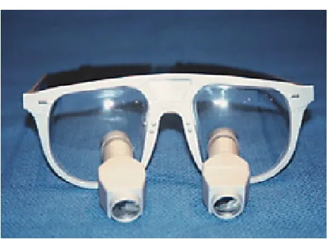
Dental loupes are the most common system of optical magnification used in Periodontics. Loupes are fundamentally dual monocular telescopes with side-by-side lenses convergent to focus on the operative field. The magnified image formed has stereoscopic properties by virtue of their convergence. A convergent lens optical system is called a Keplerian optical system.5
Although dental loupes are widely used, they have disadvantages compared with the microscope. The clinician’s eyes must converge to view the operative field. This can result in eyestrain, fatigue, and even pathologic vision changes, especially after prolonged use.
Three types of Keplerian loupes are typically used in Periodontics: simple or single-element loupes, compound loupes, and prism telescopic loupes. Each type may differ widely in optical sophistication and individual design.
Simple loupes consist of a pair of single meniscus lenses. Simple loupes are primitive magnifiers with limited capabilities. Each lens is limited to only two refracting surfaces. Their magnification can only increase by increasing lens diameter and thickness. Size and weight constraints make simple loupes impractical for magnification beyond 1.5X. Another disadvantage of simple loupes is that they are greatly affected by spherical and chromatic aberration. This distorts the image shape and color of objects being viewed.
Compound loupes use multi element lenses with intervening air spaces to gain additional refracting surfaces. This allows increased magnification with more favorable working distance and depth of field. Magnification of compound loupes can be increased by lengthening the distance between lenses, thereby avoiding excessive light.
In addition to offering improved optical performance, compound lenses can be achromatic. This is an optical feature that clinicians should always choose when selecting magnifying loupes. Achromatic lenses consist of two glass lenses, joined
together with clear resin .The specific density of each lens counteracts the chromatic aberration of its paired lens to produce a color-correct image. However multi element compound loupes become optically inefficient at magnifications above 3X.5
Prism telescopic loupes arethe most advanced
loupe optical magnification currently available is the prism telescopic loupe. Such loupes employ Schmidt or “rooftop” prisms to lengthen the light path through a series of switch back mirrors between the lenses. This arrangement folds the light so that the barrel of the loupes can be shortened. Prism loupes produce better magnification, wider depths of field, longer working distances, and larger fields of view than other types of loupes. The increased weight of prism telescope loupes with magnification above 4X makes headband mounting more comfortable and stable than eye glass frame mounting. Recent innovations in prism telescopic loupes include coaxial fiberoptic lighting incorporated in the lens elements to improve illumination.
Fig 5: Prismatic loupe optical diagram
Fig 6: Prismatic loupe
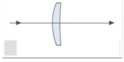
Fig 2: Simple loupe
Fig4: Compound loupes
BASIC PRINCIPLES OF THE SURGICAL MICROSCOPE
A microscope is nothing more than a monocular or binocular with a close-up lens. A binocular is simply mounted side-by-side for stereoscopic vision. In the binocular concept, the length of the telescope becomes condensed by the use of prisms.
The components of microscope are the basic stereo microscope, the binocular head, and the objective lens. This microscope, however, contains two additional elements: a magnification changer and an illuminator which beams the light in through the objective lens. This type of illumination is desirable because the line of illumination is very close to the viewer’s line of vision. Therefore, the surgical field will be illuminated and free of shadows. Shadows would result if the line of illumination was at a large angle of incidence from the viewing axis.
Benefits of microscopes in dentistry8
The operating microscope offers three distinct advantages to the clinician: illumination, magnification, and increased precision in the delivery of surgical skills. Collectively, these advantages are referred to as the microsurgical triad.
ADVANTAGES AND DISADVANTAGES OF SURGICAL MICROSCOPE
Advantages
1. Less tissue trauma 2. Less mobility 3. Less patient anxiety 4. Atraumatic tissue management5. Accurate primary wound closure. 6. Increased diagnostic skills.7. Minimally invasive 8. Improved cosmetic results 9. Increased surgical quality
10. Increased effectiveness of root debridement results in greater predictability of
a) Regeneration procedures,
b) Cosmetic procedures.
11. Improved documentation e.g. video, slide, digital.
Disadvantages
1. Educational requirements A) Surgical technique B) Understanding of optics
2. Long adjustment period for clinical proficiency
3. Initial increased surgical time
4. High patient cost5. Limited surgical access
BENEFITS OF MICROSCOPES IN PERIODONTICS1
The surgical operating microscope, like all magnification, enhances visual acuity. This leads to:
1. Increased precision in delivery of surgical skills, which results in more accurate incisions via smaller instrumentation, less trauma, and quicker postoperative healing.
2. Precise repositioning of tissues with smaller needles and sutures.
3. Improved view of root surfaces, which permits more definitive removal of calculus and improved smoothness of the root.
MICROSURGICAL INSTRUMENTS
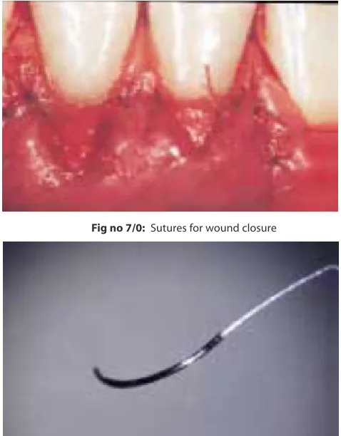
With addition to the use of magnification and reliance on atraumatic technique, microsurgery requires specially constructed instruments designed specifically to minimize trauma. An important characteristic of microsurgical instruments is their ability to create clean incisions that prepare wounds for healing by primary intention. Microsurgical incisions are established at a 90-degree angle to the surface using ophthalmic microsurgical scalpels. Microscopy permits easy identification of ragged wound edges for trimming and freshening. For primary wound closure, micro sutures in the range of 6-0 to 9-0 are needed to approximate the wound edges accurately. Microsurgical wound apposition minimizes gaps or voids at the wound edges. This encourages rapid healing with less postoperative pain.
SUTURE MATERIALS1,6
Thethree principal goals of surgery areeliminating dead space, closing withsufficient but appropriate tension,and immobilizing the wound.
trauma as possible while eliminating dead space and preventing movement of the wound.
Microsurgery has increased the periodontist’s options for appropriately sized needles and sutures. Needles vary in size, shape, and curvature, but most needles used in dentistry are 3/8 curvature.
Periodontists frequently use a reverse cutting needle of a significant size (16 to 19 mm). Although larger needles are sometimes indicated for Periodontics, several needles allow more precise approximation of tissue edges. One such needle is a spatula needle, which is 6.6 mm in length and has a curvature of 140 degrees.
Designed for ophthalmic surgery, the needle track is shallow and the needle purchase point is precise. These characteristics allow extremely accurate apposition, closure, and immobilization of the connective tissue graft.
Several other needles with sizes ranging from 6.6 to 19 mm can be used in Periodontics. The availability of smaller needles can affect the choice of suture. An accepted surgical practice is to select the smallest suture that will adequately hold the mending tissue. This practice minimizes the opening made by the needle and minimizes the trauma through the tissues.
Although 4-0 or 5-0 sutures are typically used in Periodontics, in periodontal microsurgery 6-0 and 7-0 sutures are appropriate.
Non-absorbable and absorbable sutures can be multifilament or monofilament in design. Although absorption rates vary significantly, surgical gut (plain and chromic), polyglactin 910, poliglecaprone 25, and polydioxanone are four absorbable sutures indicated for use in periodontal surgery. Non-absorbable sutures such as silk, nylon and polyester sutures are available for the surgeon who does not mind removing sutures.
Effectively using smaller needles and sutures requires magnification.The surgical operating microscope allows the clinician to use smaller sutures and needles and results in minimal dead space, closure with sufficient but appropriate tension, and immobilization of the wound.
Fig no 7/0: Sutures for wound closure
Fig no 8: A spatula shaped needle with 7/0
APPLICATIONS IN PERIODONTAL FLAP SURGERIES4,13:
Originally from general surgical principles, periodontal flap procedures have been used since the latter part of the 19th century. Flap reflection, as a basic rule, is to gain exposure of the underlying tissues for whatever surgical procedure the surgeon has in mind. Flaps must be adequate to the clinical situation being treated, and the use of surgical microscope has introduced the reality of considerably less invasive surgical incisions and flap reflections in Periodontics. Successful periodontal flap surgery depends on the surgeon’s ability;
1) To diagnose the problem correctly.
2) To plan the appropriate procedures.
4) To reflect gently and a traumatically, the flaps for passive procedure access and treatment.
5) To suture properly the flap in the most advantageous position for the defined results and patient comfort at the completion of the procedure.
DISCUSSION
As recent developments in medicine have shown, magnification and microsurgery can greatly impact clinical practices. Over the past several years, a therapeutic revolution has taken place in general surgery requiring the retaining of tens of thousands of surgeons and the retooling of their operating rooms. This startling change has come about due to the acceptance of microscopic and endoscopic surgical therapy, particularly laparoscopic removal of gall bladder and arthroscopic repair of the knee. These procedures were a natural evolution of microsurgical advances that took place in the early 1970s and culminated in modern medical microsurgery.
Microsurgery today is applied to a variety of medical operations ranging from limbs replantation to coronary artery bypass procedures and in dentistry, the general dental practitioners consider microsurgery is suitable for all clinical procedures except orthodontics and prosthodontics. In common usage, microsurgery refers to a refinement in surgical technique by which normal vision is enhanced through magnification.
An important factor in recent public and professional acceptance of microsurgery is the significant decrease in morbidity. The reduced trauma and relative painlessness that microsurgery offers, is an appealing alternative to major surgery. Patients may not fully understand the medical reasons for their therapy, but they are firmly grounded in a belief that medicine and technology should advance in their behalf. They expect sound advice and careful treatment and readily appreciate advances that give more predictable, more cosmetic and safer results, to say nothing of lessening their inconvenience, anxiety and discomfort.
There is every reason to believe that the public experts will welcome and eventually demand
advancement in the technology and procedures available for treating periodontal diseases. A case in point is the growth and acceptance of technical diagnostics, non-surgical mechanical therapies and pharmaceutical therapies in Periodontics. Despite these advances in Periodontics over the past decade, much of the everydaypractice of Periodontics still involves surgically treating periodontal anatomy altered by trauma or disease. Aside from many potential promotional or marketing advantages, periodontal microsurgery offers an improvement in predictability, cosmetic results and patient comfort level over conventional periodontal surgical procedures.2This is especially true for regenerative procedures that apply materials and techniques that are difficult to use successfully and predictability within the confines of normal vision.10
directly guides the hand through its entire range of motion using “visual sensory feedback” to accomplish mid-course corrections. Under magnification, not only are such cognitive perceptual skills readily learnable, but it is also possible to retain the hand and arm muscles to move in much smaller and more accurate incremental motion sequences. Provided the reprogrammed sequences are maintained by occasional practice sessions. Although, such cognitive retaining and muscle fiber reconditioning cannot be accomplished without practice using a microscope, once established the new skills become an indispensable resource available to execute the fine motor movement required in microsurgical technique.2
In a fully developed microsurgical periodontal practice, perhaps 70-80% of typical periodontal microsurgical procedures could be performed with the surgical microscope at 10-20X. The remainder of the procedure could be accomplished with loops under 6-8X using enhanced motor skills learned and conditioned during microsurgery training sessions. Such enhanced motor skills operating on the outer borders of distinct visual acuity have been termed metascopic motor skills.2
In spite of their significant cost, the relatively long learning curve associated with their use, frustrations during use, their occasional need for being replaced and peculiar appearance to patients, magnifying loops assist all types of clinical dentists in producing higher quality dentistry. Seeing better also meads decreasing operating time. Properly fitted loupes also can improve posture during operating and reduce muscle pain in the shoulders, neck and back. Working under magnification is useful and clinicians should give strong consideration to adopting the concept.
CONCLUSION
The surgical operating microscope provides a microsurgical triad of illumination, magnification, and an environment in which surgical skills can be refined. Incorporation of smaller instrumentation, sutures, and needles into this environment should allow clinicians to increase the precision of their surgical skills.
Important reason why microsurgery is likely to gain more rapid acceptance among periodontists is unrelated to improved outcome or lessened morbidity of the procedure. The endpoint visual appearance of the typical microsurgical procedure is simply far superior to the end-point appearance of conventional surgery.
Education through movement concentrates the mind and raises the neurobiology of learning to new levels of performance and new possibilities for achievement. As we progress into the twentyfirst century, such learning methods will come to occupy an increasingly important role in training periodontists for microsurgery as it moves into the mainstream of periodontal therapy.
Although clinical studies are lacking and research is needed, the increase in visual acuity provided by the surgical operating microscope should enhance the periodontist’s delivery of surgical skills.
REFERENCES
1. Belcher A. perspective on periodontal microsurgery.Int J Periodontics Restorative Dent. 2001;21:191-196.
2. Serafin D. Microsurgery : Past, present and future. Plastreconstrsurg 1980;66:781.
3. Shenelec DA and Tibbett LS. A perspective on the future of periodontal microsurgery. Periodontol 2000 1994; 11: 58-64.
4. Tibbetts LS and Shenelec DA. Periodontal microsurgery. Dental Clinics of North America 1998; 42(2): 339-359. 5. www.surgicalmicroscopes.com accessed on 7/8/2007,
11pm.
6. Miller B J. Focus on loupes. Br Dent J 1998; 185(10): 504-508.
7. Burkhardt R, Lang NP. Coverage of localized gingival recessions: comparison of micro and macrosurgical techniques. J ClinPeriodontol 2005; 32: 287-293.
8. Shenelec DA.Periodontal microsurgery. J Esthet Rest Dent.2003; 15: 402-408.
9. Lindhe J, Nyman S. Long-term maintenance of patients treated for advanced periodontal disease. J ClinPeriodontol 1984;11:504-514.