84
Copyright © 2018. IJEMR. All Rights Reserved.
Volume-8, Issue-3, June 2018
International Journal of Engineering and Management Research
Page Number: 84-89
DOI: doi.org/10.31033/ijemr.8.3.12
Microwave Assist Green Synthesis of Silver Nano Particles Using
Rhynchostylisretusa(L.) Blume
Leaf Extract and Its Antioxidants Activity
Jahnabi Sarma1 and Dr. A M Dutta2
1Ph.D. Scholar, Department Of Chemistry, Assam Down Town University, Guwahati, INDIA 2HOD, Department Of Chemistry, Assam Down Town University, Guwahati, INDIA
1Corresponding Author: [email protected]
ABSTRACT
A novel green approach for the synthesis and stabilization of silver nanoparticles (AgNPs) using water extract of Rhynchostylisretusa(L.) Blume leaf has been developed. As obtained, the nanoparticles are characterized by UV-visible (UV-Vis), FTIR, X-ray diffraction (XRD) and SEM analysis. The crystalline nature of the AgNPs is confirmed by the prominent peaks in the XRD pattern. FTIR spectra suggest that the possible biomolecules are responsible for the efficient stabilization of the sample. The prepared nanoparticle shows good antioxidant activity.
Keywords--- Novel Green Synthesis, Silver Nanoparticles,
Rhynchostylisretusa (L.) Blume, Antioxidant activity
I.
INTRODUCTION
The discipline of nanotechnology is swiftly evolving as an interdisciplinary science, interfacing chemical, medical, environmental and physical sciences not leaving behind diverse engineering fields, with myriad of applications in the development of biosensors and biomedical devices, alternative energy generation and environmental restoration [1]. Various nanostructures such as thin films, nanospheres, nanorods, and a variety of nanoparticles (both metallic and non-metallic) are increasingly contributing to several innovative applications. Green nanotechnology encourages not only fundamental but also goal-oriented research in both the academic and industrial fields for the design and development of Green Nanoparticles (GNPs) [2]. Green nanoparticles have already been used in the design of smart electronic devices, life-saving nano-pharmaceuticals, and in substitute green energy production devices as well. [3]
The synthesis and characterization of noble metal nanoparticles such as silver, gold and platinum are an
emerging field of research due to their important applications in the fields of biotechnology, bioengineering, textile engineering, water treatment, metal-based consumer products and other areas, electronic, magnetic, optoelectronics, and information storage.
II.
MATERIALS AND METHOD
2.1. Chemicals
Fresh leaf of Rhynchostylisretusa(L.) Blume collected from golaghat Assam, India. All the chemicals except DPPH were obtained from Hi Media Laboratories, DPPH is from Sigma-Aldrich. Ultra purified water was used for experiment.
2.2 Preparation of the extract for synthesis of silver nanoparticles
www.ijemr.net
ISSN (ONLINE): 2250-0758, ISSN (PRINT): 2394-6962
85
Copyright © 2018. IJEMR. All Rights Reserved.
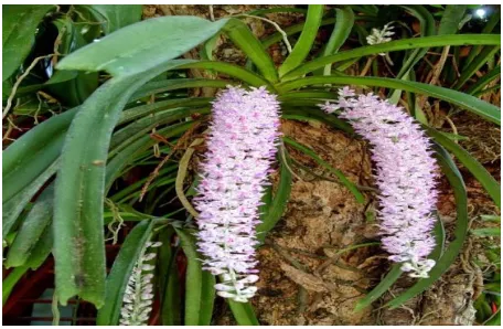
Fig1: Picture of Foxtail orchid or kopouphool
2.3 Preparation of silver nanoparticles
For efficient synthesis of silver nanoparticles, effect of boiling time and effect of extract amount to be added to 0.3 Mm AgNO3solution were varied and the best one was selected. AgNP was prepared in three ratios, Extract: AgNO3 solution, these are 3:5, 3:10 and 3:15 and subjected to microwave at 100% power of 800 W, frequency 2450 MHz just for 5 Sec. The best result was obtained in the 3:15 ratio.
2.4 Free radical scavenging ability on 2,2-didhenyl-2-picrylhydrazyl (DPPH)
To find the antioxidant activity by DPPH method, each extract (5-20 mg/ml) in water and ethanol was mixed with 1 ml of methanolic solution contain DPPH radicals (0.2 mM). The mixture was shaken vigorously in vortex mixture and left to stand for 30 mins in dark before measuring the absorbance at 517 nm against a blank []. Then the scavenging ability was recorded by using the following formula. [4][5]
I (%)= 100 x (A blank – A Sample/ A blank) Where I (%) is the inhibition present, A blank is the absorbance of the control reaction (containing all reagents except the test compound) and A sample is the absorbance of the test compound.
III.
CHARACTERIZATION OF THE
SYNTHESIZEDSILVER NANOPARTICLES
The Ag NPs obtained using silver nitrate and Rhynchostylisretusa(L.) Blume, leaves extract was characterized by using UV-VIS spectra, FTIR, XRD and SEM-EDX.
The reduction of pure Ag+ ions was monitored by UV-Visible spectrophotometer (SPECORD 50 PLUS). Scanning electron microscopy (SEM) & EDX analysis of
synthesized AgNPs was done using a Make: Zeiss, Model:-Sigma 300. XRD patterns were recorded using Rigaku X-ray diffractometer (Model: ULTIMA IV, Rigaku, Japan) with a scanning rate of 3◦ min-1 and 2θ value ranging from 5 to min-100◦ using Cu K (= 1.54056 A) as the X-ray source and operates at a generator voltage of 40 kV and current 40 mA, respectively. Fourier transform infrared (FT-IR) spectra were recorded using IR Affinity, Shimadzu, Japan FTIR spectrophotometer equipped with a Shimadzu DRS-8000 DRIFT accessory and IR solution software with 4 cm-1 spectral resolution.
IV.
RESULTS AND DISCUSSION
4.1 UV-Vis spectrophotometry analysis
The sample was observed under UV-Visspectrophotometer (SPECORD 50 PLUS) for its maximum absorbance and wavelength to confirm the reduction of Silver nitrate [2]. UV spectra of AgNP were taken at different time interval are shown below:
Fig 2: UV Spectra of Silver Nano Particle
4.2 Fourier-Transform Infrared Spectroscopy (FT-IR) Infrared Spectroscopy gives information on the vibrational and rotational modes of motion of a molecule and hence an important technique for identification and characterization of a substance. The particles were analyzed under FT-IR for the size conformation.
86
Copyright © 2018. IJEMR. All Rights Reserved.
Fig 3: IR spectra of Silver Nano particle
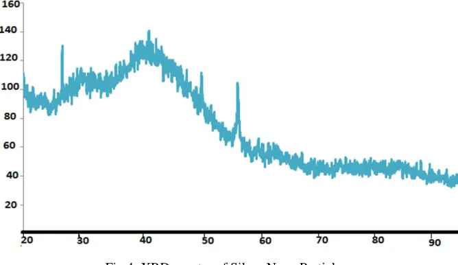
4.3 XRD Analysis
XRD spectrum showed distinct diffraction peaks around 38Ο, which are indexed by the (100) of the cubic face centered silver. These sharp Bragg peaks might have resulted due to capping agent stabilizing the nanoparticle. Intense Bragg reflections suggest that strong X-ray scattering centers in the crystalline phase and could be due to capping agents. Independent crystallization of the capping agents was ruled out due to the process of centrifugation and redispersion of the pellet in Millipore
water after nanoparticles formation as a part of purification process. Therefore, XRD results also suggested that the crystallization of the bio-organic phase occurs on the surface of the silver nanoparticles or vice versa. Generally, the broadening of peaks in the XRD patterns of solids is attributed to particle size effects. Broader peaks signify smaller particle size and reflect the effects due to experimental conditions on the nucleation and growth of the crystal nuclei. [7]
Fig 4: XRD spectra of Silver Nano Particle
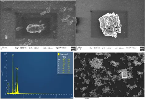
4.4 SEM Analysis
The SEM image of silver nanoparticle synthesized is shown below. The Ag NPs are monodispersed with star & cuboids’ shaped. The average size of the particles is
www.ijemr.net
ISSN (ONLINE): 2250-0758, ISSN (PRINT): 2394-6962
87
Copyright © 2018. IJEMR. All Rights Reserved.
Fig 5: SEM-EDX Analysis of Silver nanoparticle
V.
ANTIOXIDANTS ACTIVITY OF
AGNP AND THE PLANT EXTRACT
The freshly prepared DPPH solution exhibited a deep purple colour with maximum absorbance at 517 nm.
The diaappriance of purple colour may be due to presence of antioxidant activities of prepared silver Nano particle. The free radical scavenging is an increase with increase in the concentration. It was observed from the fig below that the standard of Gallic acids showed almost similar antioxidant activity AgNP. [9]
Table 1:- Gallic standard reading by DPPH method
Concentration (µl)
%scavenging antioxidant
activity for Gallic acid
%scavenging antioxidant activity for AgNP
%scavenging antioxidant
activity for plant extracts
50 22.46 16.38 9.62
100 29.96 23.01 16.02
150 43.46 34.65 27.04
88
Copyright © 2018. IJEMR. All Rights Reserved.
Fig 6: Antioxidant activity of AgNP & Plant extract wrt Gallic Acid
VI.
CONCLUSION
The rapid biological synthesis of silver nanoparticles using Rhynchostylisretusa(L.) Blume leaves extract provides environmental friendly, simple and efficient route for synthesis of benign nanoparticles. The synthesized nanoparticles were of spherical and sheet shaped and the estimated sizes were 160-180 nm. The size was bigger as the nanoparticles were surrounded by a thin layer of proteins and metabolites such as terpenoids having functional groups of amines, alcohols, ketones, aldehydes, etc., which were found from the characterization using UV-vis spectrophotometer, SEM-EDX, XRD and FTIR techniques. From the technological point of view these obtained silver nanoparticles have potential applications in the biomedical field [10] and this simple procedure has several advantages such as cost-effectiveness, compatibility for medical and pharmaceutical applications as well as large scale commercial production [11].
ACKNOWLEDGEMENT
The authors are grateful to the Assam Down Town University and CSIR-NEIST for instrumental support in the characterization part of The Research work.
REFERENCES
[1] Chinnasamy C, Tamilselvam P, V. Karthik, & B. Karthick. (2017, April). Optimization and characterization studies on green synthesis of silver nanoparticles using Response Surface Methodology. Advances in natural and
applied sciences, 11(4), 214-221.
[2] Balavandy, S.K., et al. (2014). Stirring time effect of silver nanoparticles prepared in glutathione mediated by green method. Chemistry Central Journal, 8(1), 11. [3] Mohammad A, Khalilzadeh, & Mina Borzoo. (2016). Green synthesis of silver nanoparticles using onion extract and their application for the preparation of a modified electrode for determination of ascorbic acid. Journal of food and drug analysis, 24(4), 796-803.
[4] G. Clarke, K.N. Ting, C. Wiart, & J. Fry. (2013). High correlation of 2, 2-diphenyl-1-picrylhydrazyl (DPPH) radical scavenging, ferric reducing activity potential and total phenolics content indicates redundancy in use of all three assays to screen for antioxidant activity of extracts of plants from the Malaysian rainforest. Antioxidants(Basel), 2(1), 1-10.
[5] Ajithadas Aruna, Ramraj Nandhini, Venkatachalam Karthikeyan, & Pandi Bose. (2014). Synthesis and characterization of silver nanoparticles of insulin plant (costus pictus d. don) leaves. Asian Journal of Biomedical
and Pharmaceutical Sciences, 4(34), 1-6.
[6] Genevieve A. Kahrilas, Laura M. Wally, Sarah J. Fredrick, Michael Hiskey, Amy L. Prieto, & Janel E. Owens. (2014). Microwave-assisted green synthesis of silver nanoparticles using orange peel extract. ACS
Journals, 2(3), 367–376.
[7] Karunakar Rao Kudle,Manisha R. Donda, Jahnavi Alwala, Rama Koyyati, Veerababu Nagati, Ramchander Merugu, Y.Prashanthi, & M.P.Pratap Rudra. (2012). Biofabrication of silver nanoparticles using Cuminum cyminum through microwave irradiation. International
www.ijemr.net
ISSN (ONLINE): 2250-0758, ISSN (PRINT): 2394-6962
89
Copyright © 2018. IJEMR. All Rights Reserved.
[8] Naheed Ahmad & Seema Sharma. (2012). Green synthesis of silver nanoparticles using extracts of ananas comosus. Green and Sustainable Chemistry, 2, 141-147. [9] Karunakar Rao Kudle, Manisha R. Donda, Ramchander Merugu, Y.Prashanthi, & M.P.Pratap Rudra. (2013). Microwave assisted green synthesis of silver nanoparticles using Stigmaphyllon littorale leaves,their characterization and anti-microbial activity. International Journal of
Nanomaterials and Biostructures, 3(1), 13-16.
[10] Morones JR, Elechiguerra JL, Camacho A, Holt K, Kouri JB, Ramírez JT, & Yacaman MJ. (2005). The bactericidal effect of silver nanoparticles. Nanotechnology, 16(10), 2346–2349.
[11] Thirumurugan A, Neethu Anns Tomy, Hema Priyanka Kumar, & Prakash P. (2011). Biological synthesis of silver nanoparticles by Lantana camara leaf extracts. International
Journal of Nanomaterials and Biostructures, 1(2), 22-24.
[12] Thirumurugan A, Neethu Anns Tomy, R Jai Ganesh, & S Gobikrishnan. (2010). Biological reduction of silver nanoparticles using plant leaf extracts and its effect on increased antimicrobial activity against clinically isolated organism. Scholars Research Library Der Pharma