Cytotoxic and Apoptogenic Activity of Bryonia
aspera Extract on Pre-B Acute Lymphoblastic
Leukemia Cell Lines
Sorur Yazdanpanah1, Somayeh Esmaeili2, Davood Bashash1, Nasrin Dehghan Nayeri1, Mohammad Esfini Farahani1, Ahmad Gharehbaghian1
1Department of Laboratory Hematology and Blood Bank, School of Allied Medical Sciences, Shahid Beheshti University of Medical Sciences, Tehran, Iran
2
Traditional Medicine and Materia Medica Research Center (TMRC), Shahid Beheshti University of Medical Sciences, Tehran, Iran
Corresponding Author: Ahmad Gharehbaghian, Department of Laboratory Hematology and Blood Bank, School of Allied Medical Sciences, Shahid Beheshti University of Medical Sciences, Tehran, Iran
Tel: +98 912 320 43 79
Email: gharehbaghian@sbmu.ac.ir
Received: 11, Oct, 2017 Accepted: 01, Feb, 2018
ABSTRACT
Background: The natural products and conventional chemotherapeutic drug combinations are believed to increase cure rates of anticancer treatment while reducing its toxicity. The current study investigates cytotoxic and apoptogenic effects of methanolic extract of Beryonia aspera, and also synergistic effects of this extract and Prednisolone on acute lymphoblastic leukemia cell lines.
Materials and Methods: The under study populations were NALM-6 and REH cell lines. Cells were treated by
Prednisolone and B. aspera extract alone and in combination. The effect of the drugs on survival and apoptosis were examined using MTT and flow cytometry, respectively. Moreover, the effects of the drugs on the mRNA expression levels of Bax and Bcl-2 were studied using RQ-PCR. Finally, both the transcriptional and enzymatic activity of caspase-3 were investigated by caspase-3 assay kit.
Results: The B. aspera extract induced cell growth inhibition and triggered apoptosis in a dose- and time-dependent manner. Real-time PCR analysis of apoptotic target genes revealed that this agent shifted the ratio of the death promoter to death repressor genes via alteration of Bax and Bcl-2 expression levels. These changes resulted in caspase-3 activation, which led to DNA fragmentation and subsequent apoptosis. Our study has also demonstrated that the combined treatment of B. aspera extract with Prednisolone did not induce greater cytotoxic effect as compared to treatment series using either Prednisolone alone.
Conclusion: Our study demonstrated that the B. aspera extract has anti-leukemic properties on BCP-ALL cell lines and could be regarded as a promising agent for the treatment of ALL.
Keywords: Acute lymphoblastic leukemia, Bryonia aspera, Prednisolone, Apoptosis, Combination therapy
INTRODUCTION
B-cell precursor acute lymphoblastic leukemia (BCP-ALL) is the most common type of malignancy in children1,2. Glucocorticoids have been widely used as effective therapeutic agents for these
204
International Journal of Hematology Oncology and Stem Cell Research ijhoscr.tums.ac.ir
International Journal of Hematology Oncology and Stem Cell Research ijhoscr.tums.ac.ir
These agents have many side effects such as immunodeficiency, hyperglycemia, increased skin fragility, easy bruising, steroid-induced osteoporosis, etc5. Dramatic advancement in an effective treatment for childhood ALL has led to an overall cure rate of more than 80%, providing opportunities for innovative combined-modality strategies that would increase the efficacy of treatment while reducing the toxic side-effects of current intensive regimens1. Since the medicinal herbs due to the natural origin are more compatible than chemical drugs with living organisms and cause fewer side effects, thus, as a potential resource of new supplements for chemotherapy drugs, are paid special attention6. According to the World Health Organization (WHO), approximately 60% of anti-tumor drugs are derived directly or indirectly from medicinal plants 7. The selection of plants that may contain new biological agents is done, using different strategies. In the ethnomedical strategy, belief is given to oral or written information on medicinal use of the plant, and accordingly the plant is collected and evaluated 8,9. Bryonia aspera Steven ex Ledeb. is a species of Bryonia which belongs to the family of Cucurbitaceae, and is locally known as “Andaz”. Biological values of cucurbit plants were early recognized by traditional medicine. They were used actively as traditional herbs for treating many types of diseases and indicated to have anti-inflammatory, anti-tumor, hepatoprotective and immunomodulatory properties. Ethnopharmacological information indicates that the B. aspera is used in the Turkmen Sahra region, at the north of Iran, to treat cancer, gastric, hepatic and heart disorders and rheumatism9,10. Additionally, this plant has been reported to possess cytotoxic effects on breast cancer9, cervix adenocarcinoma and head and neck squamous cell carcinoma cell lines11.
The present study was conducted to investigate the therapeutic effect of methanolic extract of aerial parts of B. aspera, and also synergistic cytotoxic effects of this extract and Prednisolone on
glucocorticoid non-sensitive (REH)12, and partially sensitive (NALM-6)13 acute lymphoblastic leukemia cell lines.
MATERIALS AND METHODS Plant material
The aerial parts of B. aspera were collected from the entrance of Pelor, Tehran province, Iran, and were identified by Mr. Moazeni, Traditional Medicine & Materia Medica Research Center, Shahid Beheshti University of Medical Sciences, Tehran, Iran. Voucher specimen of the plant (TMRC 3637) has been deposited in the herbarium of the Traditional Medicine & Materia Medica Research Center.
Preparation of extract
The aerial parts of plant were air dried at room temperature, powdered and then extracted with methanol by maceration and with constant shaking for 24 h. The plant extract was then filtered and the solvent was evaporated under vacuum by means of a rotary evaporator and stored at 4°C before evaluating biological activities.
Cell culture and B. aspera extract /Prednisolone treatment
amounts of the Prednisolone to attain concentration of 500 nM, 1, 10, 50, and 100 M.
MTT assay
The MTT assay was used to evaluate the cytotoxicity14. For suspension cells (NALM-6 and REH), cells were briefly cultured at 5000/well in 96-well plate and incubated with desired concentrations of B. aspera extract, Prednisolone and the combination of B. aspera extract and Prednisolone for 24 and 48 h. After removing the media, cells were further incubated with MTT solution (5 mg/ml in PBS) at 37ºC for 3 h and the untreated cells were defined as the control group. The resulting formazan was solubilized with Dimethyl sulfoxide (DMSO) and the absorption was measured at 570 nm (620 nm as a reference) in ELISA reader and IC50 was calculated as the concentration of fractions and compounds causing a 50% inhibition of cell viability.
For adherent cells (MDBK), 10000 cells were cultured into a 96-well plate and 24 h later cells were washed and maintained with different concentrations of B. aspera extract for 24 and 48 h, at 37°C under 5% CO2 atmosphere. After removing the media, cells were further incubated with MTT solution (5 mg/ml in PBS) at 37ºC for 3 h and the untreated cells were defined as the control group. The resulting Formazan was solubilized with DMSO and the absorption was measured at 570 nm (620 nm as a reference) in ELISA reader.
Phosphatidylserine externalization (Annexin-V assay)
The apoptosis induced by B. aspera extract were analyzed by Annexin-V/PI double staining kit (eBioscience) based on the manufacturer’s instructions. Briefly, leukemic cells were cultured into 12-well cell culture plates and treated with different concentrations of B. aspera for 48 h and were then collected. Afterwards, 200 l of a binding buffer and then 5 l of Annexin-V were added to the cell suspension and were incubated for 10
minutes. The cells were rinsed and 200 l of the binding buffer was added. PI was added before reading the values with flow cytometry. Annexin-V positive and PI-negative cells were considered to be in early apoptotic phase and cells with Annexin-V and PI positive were considered to undergo late apoptosis or necrosis.
RNA purification, reverse transcription, and real-time PCR amplification
Total RNA was isolated 48 h after treatment with B. aspera extract using RNX plus (SinaClon, Iran). The reverse transcription (RT) reaction was performed using the revertAid First Strand cDNA Synthesis Kit (Takara BIO). The cDNA prepared was subjected to qRT-PCR on a light cycler instrument (Roche). Thermal cycling conditions included activation step for 30 s at 95°C followed by 45 cycles, including a denaturation step for 5 s at 95°C and a combined annealing/extension step for 20 s at 60°C. A melting curve analysis was applied to verify the specificity of the products, and the values for the relative quantification were calculated based on 2-ΔΔCt relative expression formula. Nucleotide sequences of the primers used for qRT-PCR are shown in Table 1.
Gene Forward primer (5’-3’) Reverse primer (3’-5’) Size
(bp)
HPRT TGGACAGGACTGAACGTCTTG CCAGCAGGTCAGCAAAGAATTTA 111
Bax CGAGAGGTCTTTTTCCGAGTG GTGGGCGTCCCAAAGTAGG 242
Bcl-2 CGGTGGGGTCATGTGTGTG CGGTTCAGGTACTCAGTCATCC 90
HPRT (hypoxanthine phosphoribosyltransferase), Bax (Bcl-2 associated protein), Bcl-2 (B-cell lymphoma)
Caspase-3 enzymatic activity
206
International Journal of Hematology Oncology and Stem Cell Research ijhoscr.tums.ac.ir
International Journal of Hematology Oncology and Stem Cell Research ijhoscr.tums.ac.ir
pellets were lysed and the lysates were centrifuged at 20,000×g for 10 min. In a total volume of 100 µl, 5µg of the supernatant was incubated with 85 µl of assay buffer plus 10 µl of caspase-3 substrate (Ac-DEVD-pNA) in a 96-well plate at 37ºC for 2 h. The cleavage of the peptide by caspase releases the chromophore pNA, which was quantified spectrophotometrically at a wavelength of 405 nm. The results were expressed as fold increase in caspase activity of apoptotic cells in the treated cells over that of untreated cells.
Statistical analysis
Experimental data are expressed by mean ± standard deviation of three independent assays. All tests were done in duplicate or triplicate. Statistical significance was calculated using paired two-tailed Student’s t-tests. Statistically different values were defined significant at *P <0.05, ** P < 0.01, and ***P <0.001.
RESULTS
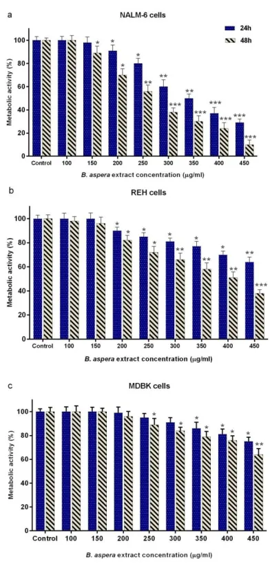
Reduction of cellular metabolic activity in BCP-ALL cell lines by B. aspera extract
The cytotoxic effects of different concentrations of
B. aspera extract (100-450 μg/ml) on metabolic activity of NALM-6 and REH cells after 24 and 48 h treatment were evaluated with the MTT assay. As indicated in Figure 1 a and b, the extract of B. aspera reduced the survival of NALM-6 and REH cells in a dose- and time-dependent manner. In this study, CompuSyn software was used to calculate IC50. The IC50 values of B. aspera extract in NALM-6 and REH cell lines after 48 h incubation were approximately 282 and 380 μg/ml, respectively. In addition, the MDBK cells were used as the normal cell line to examine the effect of this extract on normal cells. The IC50 value of the B. aspera extract on MDBK cells was approximately 467 μg/ml after 48 h incubation (figure 1c), suggesting the lower cytotoxic effect of this extract on MDBK cells than on acute lymphoblastic leukemia cell lines.
Figure 1. Cytotoxic effect of B. aspera extract on NALM-6, REH and MDBK cell lines.The cells were treated with different concentration of
B. aspera extract and their metabolic activities were measured after 24 and 48 h incubation, using the MTT assay. Values are given as mean ±SD
of three independent experiments. *P < 0.05, **P < 0.01, and ***P < 0.001 represent considerable alterations from untreated control.
Induction of apoptosis in BCP-ALL cell lines by B. aspera extract
To validate whether cell survival reduction was due to apoptosis induction, NALM-6 and REH cells were treated with intended concentrations of B. aspera
manner. As shown in Figure 2, 200, 250 and 300 μg/ml doses of B. aspera extract caused 16.5, 21.6 and 26% early apoptosis on NALM-6 cells, and similar apoptogenic effects were also apparent on REH cell line as indicated in Figure 3, such that 200, 300 and 400 μg/ml doses of B. aspera extract caused 12.6, 17.5 and 23.7% early apoptosis on these cells.
Figure 2. Results from the induction of apoptosis by the B. aspera
extract on NALM-6 cell line.The NALM-6 cells were exposed to intended concentrations of the B. aspera extract and then the induction
of apoptosis after 48 h incubation was evaluated. Flow cytometry images, Q1, Q2, Q3 and Q4 represent early apoptosis, late apoptosis,
live cells, and necrotic cells, respectively.
Figure 3. Results from the induction of apoptosis by the B. aspera
extract on REH cell line.The REH cells were exposed to intended concentrations of the B. aspera extract and then the induction of apoptosis after 48 h incubation was evaluated. Flow cytometry images,
Q1, Q2, Q3 and Q4 represent early apoptosis, late apoptosis, live cells, and necrotic cells, respectively.
Increased transcription of Bax and reduced transcription of Bcl-2 in BCP-ALL cell lines by B. aspera extract
The Bax (pro-apoptotic) and Bcl-2 (anti-apoptotic) proteins are the most important members of Bcl-2
anti-208
International Journal of Hematology Oncology and Stem Cell Research ijhoscr.tums.ac.ir
International Journal of Hematology Oncology and Stem Cell Research ijhoscr.tums.ac.ir
apoptotic proteins in favor of pro-apoptosis factors in BCP-ALL cell lines.
Figure 4. Expression of Bax and Bcl-2 genes in NALM-6 and REH cells treated with desirable concentration of B. aspera extract. (a) The NALM-6 and REH cells were exposed to the desirable concentration of
the B. aspera extract, and the expression of Bax and Bcl-2 genes was examined after 48 h incubation using real-time RT-PCR. (b) Bax/Bcl-2
ration. Values are given as mean ±SD of three independent experiments. *P < 0.05 and **P < 0.01 represent considerable
alterations from untreated control.
Increased transcription and enzymatic activity of caspase-3 in BCP-ALL cell lines by B. aspera extract To confirm the apoptotic effects of B. aspera
extract, the enzymatic activity of caspase-3 was also quantified. To this end, the cells were treated with desirable concentrations of the extract for 48 h, and the enzymatic activity changes of caspase-3 was evaluated, using caspase-3 assay kit (Colometric). Results indicated that the B. aspera extract increased the enzymatic activity of caspase-3 in BCP-ALL cells treated with this extract (Figure 5).
Figure 5. Caspase-3 activity in NALM-6 and REH cells treated by B. aspera extract.The NALM-6 and REH cells were exposed to the desirable concentration of the B. aspera extract, and the activity of caspase-3 enzyme was examined after 48 h incubation. Values are given
as mean ±SD of three independent experiments. *P < 0.05 and **P < 0.01 represent considerable alterations from untreated control.
The lack of synergistic effect in using B. aspera extract in combination with Prednisolone in BCP-ALL cell lines
with CompuSyn software. The obtained CI was almost equal to 1, suggesting that B. aspera extract did not induce greater cytotoxic effect as compared to using either drug alone and had additive effect.
Figure 6. Cytotoxic effect of Prednisolone on NALM-6 and REH cell lines. The cells were treated with different concentration of Prednisolone and
their metabolic activities were measured after 24 and 48 h incubation, using the MTT assay. Values are given as mean ±SD of three independent experiments. *P < 0.05 and **P < 0.01 represent
considerable alterations from untreated control.
DISCUSSION
Plants have provided a prime source of highly effective drugs for the treatment of many diseases17. Nowadays, many clinically approved or under trial anticancer medications are derived from nature18. In this study, we observed that the extract of aerial parts of B. aspera plant induces a significant dose- and time-dependent cytotoxic effect on the acute lymphoblastic leukemia cell lines, namely NALM-6 and REH. Interestingly, we found lesser cytotoxic effect by different doses of this extract on non-cancerous cell line (MDBK), which is consistent with a study by Sahranavard et al. who reported that the extract from the root of B.
aspera has a cytotoxic effect on the breast cancer cell line (MCF-7) and they found a weaker cytotoxic effect on MDBK cells9. Additionally, in this study, the synergistic effects of B. aspera extract were investigated in combination with Prednisolone, the most used glucocorticoid19, and no synergistic effect was seen in BCP-ALL cell lines (CI1).
In the next stage, the apoptotic effects of B. aspera
extract on two BCP-ALL cell lines were evaluated. Apoptosis plays an axial role in cancer and its induction in cancer cells is essential to achieve a successful treatment20. Our results showed that the
B. aspera extract causes cell death in BCP-ALL cell lines (NALM-6 and REH) through apoptosis induction, which increases in dose-dependent manner. In line with our findings, Pourgonabadi et al. reported that the B. aspera root extract triggers apoptosis in HN-5 (head and neck squamous cell carcinoma) and Hela (cervix adenocarcinoma) cell lines11. Apoptosis is highly regulated and conserved cellular process21 controlled by regulators, which have either an inhibitory effect on programmed cell death (anti-apoptotic) or block the protective effect of inhibitors (pro-apoptotic)22. Among the intracellular factors, the balance between Bax
(powerful activator of apoptosis) and Bcl-2 (anti-apoptotic counterpart of Bax) is known as the most significant parameter in cell survival15. Therefore, the mRNA expression levels of Bax and Bcl-2 were investigated to evaluate the effect of the B. aspera
extract on the induction of cell death. Our gene expression study demonstrated that the extract of
210
International Journal of Hematology Oncology and Stem Cell Research ijhoscr.tums.ac.ir
International Journal of Hematology Oncology and Stem Cell Research ijhoscr.tums.ac.ir
through increasing the activity of caspase-3 and caspase-8 in MCF-7 (human breast adenocarcinoma) and SiHa (human squamous cell carcinoma; cervix) cell lines24. In general, it can be concluded that the extract of B. aspera causes the inhibition of Bcl-2 and dimerization of Bax that trigger the release of cytochrome c from the mitochondria and activation of caspase-3, and this phenomenon finally induces cell death.
In summary, our study demonstrates that the B. aspera extract can induce apoptosis in BCP-ALL cell lines. However, further investigations, including clinical trials and a detailed understanding of the B. aspera extract underlying mechanism of action are warranted to determine the efficacy of this natural agent.
CONCLUSION
By and large, our present findings provide evidence that the B. aspera extract has anti-leukemic properties, and the mechanism of action is likely to be dependent on transcriptional regulation of apoptotic signaling proteins. Thus, B. aspera
could be considered as promising source of developing novel therapeutics against acute lymphoblastic leukemia.
ACKNOWLEDGMENT
The authors would like to gratitude the Shahid Beheshti University of Medical Sciences for supporting of this study.
CONFLICT OF INTEREST
The authors report no conflicts of interest.
REFERENCES
1. Safa M, Tavasoli B, Manafi R, et al. Indole-3-carbinol suppresses NF-κB activity and stimulates the p53 pathway in pre-B acute lymphoblastic leukemia cells. Tumor Biol. 2015; 36(5):3919-30.
2.Bhojwani D,Yang JJ, Pui CH. Biology of childhood acute lymphoblastic leukemia. Pediatr Clin North Am. 2015; 62(1):47-60.
3. Rahman M, Hirabayashi Y, Ishii T, et al. Prednisolone sodium succinate down-regulates BSAP/Pax5 and causes a growth arrest in the Nalm6 pre-B cell line.Tohoku J Exp Med. 2001;193(3):237-44.
4. Lambrou GI, Vlahopoulos S, Papathanasiou C, et al. Prednisolone exerts late mitogenic and biphasic effects
on resistant acute lymphoblastic leukemia cells: Relation to early gene expression. Leuk Res. 2009; 33(12):1684-95. 5. Schäcke H, Döcke W-D, Asadullah K. Mechanisms involved in the side effects of glucocorticoids. Pharmacol Ther. 2002;96(1):23-43.
6. Ghaderi S, Falahati hosein abad A, Sarailoo MH, et al. Investigation of the components and antibacterial effects of three plant's essential oil Coriandrum sativum, Achilleh millefolium, Anethum graveolens in vitro. J Shahrekord Univ Med Sci. 2012; 14(5): 74-82
7. Robinson MM, Zhang X. The world medicines situation 2011. 3rd edition. Genava. World Health Organization. 2011. 1-4.
8. Cragg GM, Newman DJ, Snader KM. Natural products in drug discovery and development. Journal of natural products. 1997 Jan 22; 60(1):52-60.
9. Sahranavard S, Naghibi F, Ghffari S. Cytotoxic activity of extracts and pure compounds of Bryonia aspera. Int J Pharm Pharmaceut Sci. 2012;4(3):541-3.
10. Esmaeili S, Naghibi F, Mosaddegh M, et al. Screening of antiplasmodial properties among some traditionally used Iranian plants. J Ethnopharmacol. 2009; 121(3):400-4
11. Pourgonabadi S, Amiri MS, Mousavi SH. Cytotoxic and apoptogenic effects of Bryonia aspera root extract against Hela and HN-5 cancer cell lines. Avicenna J Phytomed. 2017; 7(1):66-72.
12. Jiang N, Kham SK, Koh GS, et al. Identification of prognostic protein biomarkers in childhood acute lymphoblastic leukemia (ALL). J Proteomics. 2011; 74(6):843-57
13. Millar BC, Bell JB, Hobbs S, et al. Lymphotoxic activity of methyl prednisolone in vitro—I: Comparative toxicity of methyl prednisolone in human cell lines of B and T origin.Biochem Pharmacol. 1987;36(6):831-7.
14. Carmichael J, DeGraff WG, Gazdar AF, et al. Evaluation of a tetrazolium-based semiautomated colorimetric assay: assessment of chemosensitivity testing. Cancer Res. 1987; 47(4):936-42.
15. Elmore S. Apoptosis: a review of programmed cell death. Toxicol Pathol. 2007;35(4):495-516.
16. Bashash D, Ghaffari SH, Zaker F, et al. BIBR 1532 increases arsenic trioxide-mediated apoptosis in acute promyelocytic leukemia cells: therapeutic potential for APL. Anti-Cancer Agents Med Chem. 2013; 13(7):1115-25.
17. Shoeb M. Anticancer agents from medicinal plants. Bangladesh J pharmacol. 2006; 1(2):35-41.
19. Tissing WJ, den Boer ML, Meijerink JP, et al. Genomewide identification of prednisolone-responsive genes in acute lymphoblastic leukemia cells. Blood. 2007;109(9):3929-35.
20. Bunz F. Cell death and cancer therapy. Current Opinion in Pharmacology. 2001;1(4):337-41.
21. Kerr J. A histochemical study of hypertrophy and ischaemic injury of rat liver with special reference to changes in lysosomes.J Pathol Bacteriol. 1965;90(2):419-35.
22. Wang K, Yin XM, Chao DT, et al. BID: a novel BH3 domain-only death agonist. Genes Dev. 1996; 10(22):2859-69.
23. Benarba B, Meddah B, Aoues A. Bryonia dioica aqueous extract induces apoptosis through mitochondrial intrinsic pathway in BL41 Burkitt's lymphoma cells. J Ethnopharmacol. 2012; 141(1):510-6.