Alper Biler
1, A–F, Sait Yucebilgin
2, A–F, Fatih Sendag
2, A–F, Levent Akman
2, A–F,
Ali Akdemir
2, A–F, Utku Ates
3, A–C, E, F, Yigit Uyanikgil
3, A–C, E, F,
Ozlem Yilmaz-Dilsiz
3, A–C, E, F, Ebru Sezer
4, A–C, E, FThe Effects of Different Intraabdominal
Pressure Protocols in Laparoscopic Procedures
on Oxidative Stress Markers and Morphology
in Rat Ovaries
1 Fatsa Gynecology and Maternity Training and Research Hospital of TC Ministry of Health, Ordu, Turkey 2 Department of Obstetrics and Gynecology, Faculty of Medicine, Ege University, Bornova, İzmir, Turkey 3 Department of Histology and Embryology, Faculty of Medicine, Ege University, Bornova, İzmir, Turkey 4 Department of Biochemistry, Faculty of Medicine, Ege University, Bornova, İzmir, Turkey
A – research concept and design; B – collection and/or assembly of data; C – data analysis and interpretation;
D – writing the article; E – critical revision of the article; F – final approval of article; G – other
Abstract
Background. To determine the effects of different intraabdominal pressure (IAP) on the ovaries in a laparoscopic rat model.
Objectives. The aim of the study was to determine the effects on the ovaries of different intraabdominal pressures (IAP) in laparoscopic surgery in a rat model.
Material and Methods. Thirty-two post-pubertal nonpregnant Sprague-Dawley rats were divided randomly into four groups. In the control group, no intraabdominal pressure (IAP) was applied. In Group Pp10 and Group Pp15, an IAP of 10 and 15 mm Hg, respectively, were applied by carbon dioxide insufflation for 60 min, and a 30-min desufflation was carried out. In Group IPp15, a 15 mm Hg IAP was applied for 10 min, and then CO2 was desufflated for 10 min.
After this ischemic preconditioning, IAP was established at 15 mm Hg for 60 min, after which CO2 was desufflated for
30 min. Erythrocyte and ovarian tissue malondialdehyde (MDA) and histopathologic damage scores were evaluated.
Results. In Groups Pp10 and Pp15, ovarian tissue MDA values were significantly increased compared to the con-trol group. In Groups Pp10 and Pp15, erythrocyte MDA values were significantly increased when compared to Group IPp15 and the control group. Ovarian histopatological assesment scores were significantly higher in Group Pp15 than in Groups Pp10 and IPp15.
Conclusions. Pneumoperitoneum causes injuries to abdominal organ such as the ovaries. The ischemic precon-ditioningmethodismore effective in reducing oxidative stress due to laparoscopic pneumoperitoneum than low-pressure pneumoperitoneum methods (Adv Clin Exp Med 2014, 23, 6, 885–892).
Key words: ischemic preconditioning,pneumoperitoneum, oxidative stress, reperfusion, laparoscopy.
Adv Clin Exp Med 2014, 23, 6, 885–892 ISSN 1899–5276
ORIGINAL PAPERS
© Copyright by Wroclaw Medical University
As skills and knowledge in diagnostic lapa-roscopy have improved and more advanced sur-gical instruments have been developed, the use of laparoscopic techniques has become much more common and has earned the admiration of the majority of surgeons. More complex pro-cedures are being performed via laparoscopy, including traditional abdominal hysterectomy.As laparoscopic surgery has developed and surgeons
have gained more experience, the procedure has been extended to the pregnant population. The most common reported laparoscopic operations during pregnancy are cholecystectomy, adnexial surgery, appendectomy and management of het-erotopic pregnancy [1].
not a harmless procedure. In some laparoscopic procedures, an intraabdominal pressure (IAP) of 18–25 mm Hg is needed [2, 3], but these pres-sure levels cannot be accessed in routine laparos-copy, as most clinical procedures are performed at 10–15 mm Hg [4]. This pressure range, although it is higher than the normal portal systemic pressure (7–10 mm Hg), maintains a balance between estab-lishing working space with adequate visualization and the undesireable effects of increased IAP [5–9].
The increasing experience of large number of trials has revealed the side effects of PN [10]. These trials have mostly focused on blood flow changes in the intraabdominal organs. PN leads to a 10–80% reduction in the rate of blood flow to the in-traabominal organs, but it has been reported to re-turn to the normal range after desufflation [10]. In terms of the hypoperfusion and subsequent rep-erfusion periods, both clinical and experimen-tal studies show that laparoscopic procedures re-sult in a typical model of ischemia and reperfusion (I/R) injury in the organs [11–13]. After desuffla-tion, visceral perfusion returns to normal, but oxi-dative stress remains in the tissues. The use of min-imal pressure to get adequate visualization is more recommended than the use of constant pressure in laparoscopic procedures [9].
The ischemic preconditioning method is anoth-er way to reduce I/R injury. The ischemic precondi-tioning method is defined as a brief insufflation fol-lowed by brief desufflation at the beginning of the procedure. During the ischemic preconditioning method, some cellular proteins are secreted in order to increase tissue resistance to I/R injury [14, 15].
The aim of this study was to determine the ef-fects of different intraabdominal pressure models and the ischemic preconditioning model on oxida-tive stress markers and morphology in laparoscop-ic rat ovary surgery.
Material and Methods
The Animals
and Experimental Design
The study protocol complies with the European Community guidelines for the use of experimental animals. All the experiments were approved by the local Animal Care Ethics Committee at Ege Uni-versity, İzmir, Turkey. The study involved 32 non-pregnant Sprague-Dawley rats weighing 264 ± 28 g, housed in suitable environmental conditions (12 h daylight at 25 ± 2ºC temperature with sequential dark and light cycles) and fed ad libitum with rat chow and tap water. The rats were randomly di-vided into 4 groups: Group Pp10 (n : 8), in which
pneumoperitoneum (PN) was achived at 10 mm Hg; Group Pp15 (n:8), in which PN was achived at 15 mm Hg; Group IPp15, in which the ischemic preconditioning model was used; and the control group, which only underwent general anesthesia.
Briefly, each rat was anesthetized with an in-tramuscular injection of a mixture of Ketamine (60 mg/kg, Alfamine®, Ege Vet, Izmir, Turkey)
and Xylazine (10 mg/kg, Alfazyne®, Ege Vet, Izmir,
Turkey). Using sterile techniques, an 18-gauge an-giocatheter was placed in the abdominal cavity from the caudal of the sternum. Possible gas escape from the abdomen was blocked with sutures close to the angiocatheter entry point. A CO2
insuffla-tor (Nortech, Model No:3-315-00) was attached to the angiocatheter with the help of cannula. In the control group, the angiocatheter was inserted into the abdominal cavity but there was no gas insuffla-tion. In Group Pp10 and Group Pp15, PN was per-formed at 10 mm Hg and 15 mm Hg, respectively, by an automatic insufflator for 60 min, and then desufflation was performed for 30 min. In Group IPp15, after the ischemic preconditioning proce-dure, PN was achived at 15 mm Hg by an automat-ic insufflator for 60 min and desufflation was car-ried out for 30 min. The ischemic preconditioning procedure consisted of performing PN at 15 mm Hg for a duration of 10 min and desufflation for 10 min, as decribed above. In the control group the angiocatheters were removed after 90 min and midline laparatomies were performed; in the other 3 groups this was done after a 30-min desufflation period. Both ovaries were removed from the sub-jects immediately after the laparatomy. Afterwards, an intracardiac blood sample was drawn into tubes with K-EDTA using 22-gauge × 1 needles. These laparotomy procedures took approximately 2 min. After the sampling, all the subjects were sacrified with intracardiac potassium injections.
After the fat and connecting tissue were cleaned from the samples, half of the samples were washed with Ringer’s lactate solution at ice temperature and stored at –80ºC for the biochemical assesments. The other half of the samples were fixed within a form-aldehyde (10%) solution and stored at room tempa-ture for 24 h for the histological assesments. Hemo-lysates were prepared from the blood samples and stored at –80ºC for the biochemical studies.
Biochemical Analysis
Preparation
of Tissue Homogenates
and Analytical Methods
The samples were homogenized at a propor-tion of 1 : 10 (w/v) in cold phosphate-buffer (0.5 M, pH = 7.0). The homogenates were centrifuged for 5 min at 700 × g at 4oC to sediment cellular debris.
The TBARS contents in the supernatants were de-termined immediately.
Quantification of MDA
MDA levels were determined by colorimetric as-say. After dilution in the required amount of tissue homogenates, TBARS levels were determined accord-ing to the protocol described by Sozmen et al [16], by incubation with MDA solution (0.12 M TBA in 15% TCA and 1% HCl) for 30 min at 95°C, and were cal-culated as nmol/mg protein using a 1,1,3,3,tetrame-thoxypropane standard curve while the hemolysate MDA levels were expressed as nmol/g Hb.
Histological Procedures
The excised ovarian tissues were fixed by over-night immersion in 10% neutral buffered formal-dehyde (0.2M; pH = 7; Merck) for 24 h at 4ºC, then dehydrated, embedded in parafin wax and sec-tioned by microtome (Leica RM 2145). For the his-tological analyses, 3 μm thick serial sections were dewaxed, and rehydrated tissue slides were stained with hematoxylin eosin (HE) and mounted with entellan. All the sections were examined and pho-tographed using an Olympus C-5050 digital camera with Olympus BX51 microscope. The histological procedures applied in this study are conventional and well-established methods [17]. The chemicals were purchased from Sigma (St Louis, MO, USA) unless noted otherwise. The slides were immersed in the stain/buffer solutions at all times during the incubations.
Two investigators, blinded to the group dis-tinctions of the specimens, obtained 5 images from random fields under various objectives in 10 dif-ferent sections for determination and evaluation of the cellular components. No morphological evalu-ation was done on cells in areas with necrosis or with poor morphology, or on cells from the slide margins. In all cases the areas of interest were se-lected randomly.
In order to evaluate histological morpholog-ical alterations via light microscopy, the ovarian samples were analyzed according to a scoring sys-tem specifically developed for this study by the au-thors (UA, YU and OYD)
The criteria for ovarian injury were changes in the gerninative epithelium, increases in stromal connective tissue (cellular components and tissue skeleton) or medulla (increases in vascular struc-tures, increases in tunica-media vessel thickness), alterations in the morphology of primordial folli-cles, preanthral follifolli-cles, anthral follicles and ma-ture follicles, splitting of the granulosa cells and increases in interstitial spaces, vacuolisation of the oocytes, cellular alignment of the theca cells and increases in the vascularity and thickening of the
zona pellucid. Alterations in these structures were evaluated and graded semiquantitatively as follows: 0 for no difference, 1 for minor alterations, 2 for moderate alterations and 3 for severe alterations.
Statistical Analysis
The statistical analysis of the data was per-formed using the SPSS 11.0 software package for Windows (Statistical Package for Social Sciences, SPSS Inc., Chicago IL, USA). All the values were expressed as mean ± S.E.M. The Kruksal-Wallis analysis of variance and the Mann-Whitney U test were used for statistical comparison of the groups. A p-value of less than 0.05 was accepted as statisti-cally significant.
Results
A biochemical analysis of erythrocyte and tis-sue MDA levels for the ovaries was carried out.
Erythrocyte MDA Levels
Eythrocyte MDA values in Group Pp10 and Group Pp15 were significantly increased when compared to the control group (p = 0.004 and p = 0.004 respectively). There was no statistical-ly significant difference between Group IPp15 and the control group (p = 0.148). Eythrocyte MDA values in Group Pp10 and Group Pp15 were signif-icantly increased when compared to Group IPp15 (p = 0.006 and p = 0.004); but no statistically sig-nificant difference was noted between Group Pp10 and Group P15 (p = 0.873) (Fig. 1).
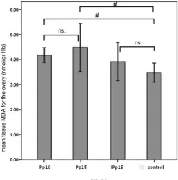
Ovarian MDA Levels
significant differences were found when compar-ing ovarian MDA values of Group Pp10, Group Pp15 and Group IPp15 (Fig. 2).
Histological Findings
for the Ovaries
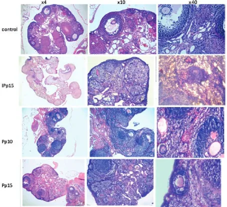
The histological examination scores and the results of the statistical analysis for the ovaries are shown in Fig. 3. The histological examination of the ovaries revealed normal ovarian morphology in the control group (Fig. 4); but two of the sub-jects displayed an increase in vascular structures
and vessel formation in the stroma and there was also a lack of thecal cell arrangement. In the ova-ries of Group IPp15, dispersed lymphatic dilata-tion and varicose enlargements of the vessels were observed; there was no evidence of capillary con-gestion. Most of the sections displayed many reg-gressed follicles. Zona pellucida formation was nor-mal in most of the follicles. Additionally, a loss of integrity between the tunica albuginea and the me-sothelium was noted (Fig. 4). The histological eval-uation scores of Group IPp15 were significantly higher than the control group’s score (p = 0.003).
In Group Pp10, zona pellucida formation was found to be normal in all samples except one. In
Fig. 1. Statistical analysis of erythrocyte MDA levels for all groups,
# p < 0.05; ns. – non-significant
Fig. 2. Statistical analysis of tissue MDA levels for the ovaries from all groups,
addition to medullar lymphatic dilatation findings in the stroma, medullar congestion and hemor-rhage were noted. However, there were no findings
of oocyte maturation defects in the follicles (Fig. 4). The histological scores of Group Pp10 were signif-icantly higher than the control group score and
Fig. 3. Histological examination scores for the ova-ries and the results of the statistical analysis,
# p < 0.05
the Group IPp15 score (p = 0.003 and p = 0.004, respectively).
In Group Pp15, there were no findings of fol-licular cycle arrest, but evident dilatation and con-gestion of medullar blood vessels were noted, as well as stromal lymphocytic infiltration. Oocyte maturation defects were noted within the follicles. Due to vessel congestion in close proximity with the follicles, deformation of the oocytes was deter-mined (Fig. 4). The histological scores of Group Pp15 were significantly higher than those of the control group, Group Pp10 and Group IPp15 (p = 0.003, p = 0.004 and p = 0.004, respectively).
Discussion
Laparoscopy has a both advantages and disad-vantages [2, 10]. After the development of mini-mally invasive laparoscopic surgical techniques, many trials were conducted to determine the side effects of PN. PN is performed for visualization, but it can cause ischemia and reperfusion (I/R) in-jury. The nature and severity of I/R injury depends on the time and level of IAP and the properties of the intraabdominal organs exposed to IAP.
Previously, the clinically permitted IAP level was 25 mm Hg at the beginnig of laparoscopy, and IAP levels not exceeding 18 mm Hg during the lap-aroscopic procedure [2]. Recently, this range has been reduced to 10–15 mm Hg [18]. But in some gynecological laparoscopic procedures, 20 mm Hg IAP is required. Abu-Rafea at el. indicated that a higher CO2 volume is required to estabilish
appro-priate pneumoperitoneum in tall, overweight and parous women at IAPs of 20–30 mm Hg at the be-ginning of laparoscopy [19]. Madl and Druml [20] previously reported that an IAP of no more than 10 mm Hg would not cause any major hazardous effects, but an IAP exceeding 12 mm Hg could re-sult in inflamation of the tissues, including process-es like leukocyte migration, a release of cytokinprocess-es, increased formation of reactive oxygen interme-diates, etc. An IAP between 18 and 20 mm Hg is described as intraabdominal hypertansion. An IAP over 20 mm Hg is an emergency situation, called abdominal compartment syndrome. In-traabdominal organ oxidative stress injuries oc-cur due to changes in systemic circulation and the direct effects of IAP on abdominal vessels [13]. There is usually a physiological interval when re-active oxygen radicals and inflammatory cytokines overlay the tissue after surgical trauma, until the organism succesfully eradicates them via multi-ple signalling cascades. But uncontrolled produc-tion and eliminaproduc-tion defects of these compounds may result in tissue necrosis and apoptosis [21].
Therefore, optimal care must be taken to limit the production of any post-inflammatory cytokines af-ter surgery. Eleftheriadis et al. [22] defined PN-re-lated hepatic oxidative stress for the first time in literature [23, 24], while Yılmaz et al. [25] firstly demostrated PN-related renal oxidative stress in rat models.
In the available literature many human and animal studies have investigated the consequenc-es of high IAP-related injuriconsequenc-es and the benefits of low IAP. Giraudo and et al. [26] performed a lap-aroscopic cholecystectomy using a gasless od, a 10 mm Hg method and a 14 mm Hg meth-od. Their study revealed higher transaminase values for the 14 mm Hg group, while the gasless group and 10 mm Hg group displayed almost the same values. Yılmaz et al. [25] reported statistical-ly different values of oxidative stress responses for 10 mm Hg and 15 mm Hg groups in a rat model; and Polat et al. [13] validated these results in their study with human subjects. Moreover, Guven and et al. [27] reported that pneumoperitoneum, even at normal IAP levels, leads to significant oxidative-stress-induced biochemical and histologic damage to the ovaries.
The ischemic preconditioning method is an-other way to decrease ischemic injury related to PN. Different time periods for the precondition-ing method have been reported in previous pub-lications. Five- or ten-minute-ischemia followed by a reperfusion of 5–10 min is most commonly used [28–32]. Cevrioglu and et al. [33] described the preconditioning method as PN at 15 mm Hg IAP for 10 min followed by immediate desuffla-tion for 10 min.
Increased IAP forms free oxygen radicals and ischemia in the peritoneum in relation to the time and pressure applied [14]. After reperfusion, the amount of free oxygen radicals initially increas-es, then the amount of antioxidant substances that help the elimination of free oxygen radicals de-creases [14].Meanwhile, several proteins, such as heat shock protein 70 (HSP70) from coronary en-dotelial cells, are secreted in order to protect cells from the hazardous effects of the free radicals and ischemic injury in the course of the ischemic pre-conditioning method.
In this experimental study, statistically signif-icant differences in the oxidative effects of PN on erythrocyte and ovary tissues were demonstrated biochemically. Although there was no difference between Groups Pp10 and Pp15, light microscop-ic analyses of the tissue histology for the ovaries revealed morphologically significant differenc-es between the control group and the other three groups. Group Pp15 showed the highest histologi-cal scores, and Group Pp10 had higher scores than Group IPp15.
This study has clearly demonstrated that the ischemic preconditioning method should be used to reduce I/R injuries, rather than other low-pressure
models. However, further detailed studies are need-ed to prove likewise effects of ischemic precondi-tioning on human beings. The analysis of PN via
histological data contributes to the available scien-tific literature on the repair process in I/R injury. The method used is elegant, but the mechanisms governing the repair process of I/R injury in rats are different from those governing human oxida-tive stress mechanisms. Thus it is possible to report that the repair processes after I/R injury are regulat-ed differently in rat subjects. In conclusion, the au-thors believe that this is still a challenging area of re-search, and that it would be helpful to explain basic cellular processes via novel molecular approaches.
Acknowledgements. The authors are grateful to Assoc. Prof. Dr. Timur Köse, PhD, from the Department of Biostatistics at the Ege University Faculty of Medicine, İzmir, Turkey, for his support, guidance and many useful discussions. The authors also wish to thank the technical staff of the Departments of Histology & Embryology and Animal Experimental Surgery for their kind assistance.
References
[1] Al-Fozan H and Tulandi T: Safety and risks of laparoscopy in pregnancy. Curr Opin Obstet Gynecol 2002, 14, 375–379.
[2] Sharma KC, Kabinoff G, Ducheine Y, Tierney J, Brandstetter RD: Laparoscopic surgery and its potential for medical complications. Heart Lung 1997, 26, 52–64.
[3] Sutton C: A practical approach to surgical laparoscopy. In: Endoscopic surgery for gynecologists. Eds.: Sutton C, Daimond M, W.B. Saunders, Philadelphia 1998, 2nd ed., 41–53.
[4] Goitein D, Papasavas P, Yeaney W, Gagne D, Hayetian F, Caushaj P: Microsphere intestinal blood flow analysis during pnömoperitonyum using carbon dioxide and helium. Surg Endosc 2005, 19, 541–545.
[5] Chiu AW, Chang LS, Birkett DH and Babayan RK: The impact of pnömoperitonyum and gassles laparoscopy on systemic and renal hemodynamics. J Am Coll Surg 1995, 181, 397–406.
[6] Eleftheriadis E and Kotzampasi K: Influence of pnömoperitonyum on the mesenteric circulation. In Rosenthal RJ, Friedman RL, and Philips EH (eds) The pathophysiology of pnömoperitonyum. Springer, New York, USA, 1998, 49–61.
[7] O’Malley C and Cunningham AJ: Physiologic changes during laparoscopy. Anesthesiol Clin North Am 2001, 19, 1–19.
[8] Richter S, Olinger A, Hildebrandt U, Menger MD and Vollmar B: Loss of physiologic hepatic blood flow control (‘hepatic arterial buffer response’) during CO2 pnömoperitonyum in the rat. Anesth Analg 2001, 93, 872–877.
[9] Neudecker J, Sauerland S, Neugebauer E, Bargamaschi R, Bonjer HJ, Cuschieri A: European Association for Endoscopic Surgery clinical practice guideline on the pnömoperitonyum for laparoscopic surgery. Surg Endosc 2002, 16, 1121–1143.
[10] Schäfer M, Krähenbühl L: Effects of laparoscopy on intra-abdominal blood flow. Surgery 2001, 129, 385–389.
[11] Bentes de Souza AM, Wang CC, Chu CY: The effect of intra-abdominal pressure on the generation of 8-iso pros-taglandin F2α during laparoscopy in rabbits. Hum Reprod 2003, 18, 2181–2188.
[12] Kaya Y, Coşkun T, Demir MA: Abdominal insufflation-deflation injury in small intestine in rabbits. Eur J Surg 2002, 168, 410–417.
[13] Polat C, Yilmaz S, Serteser M: The effect of different intra-abdominal pressures on lipid peroxidation and protein oxidation status during laparoscopic cholecystectomy. Surg Endose 2003, 17, 1719–1722.
[14] Li C and Jackson RM: Reactive species mechanisms of cellular hypoxia-reoxygenation injury. Am J Physiol Cell 2002, 282, 227–241.
[15] Ates E, Yilmaz S, Ihtiyar E: Preconditioning-like amelioration of erythropoietin against laparoscopy-induced oxidative injury. Surg Endosc 2006, 20, 815–819.
[16] Sozmen EY, Tanyalçın T, Kutay F: Ethanol induced oxidative stress and membrane injury in rats. Eur J Clin Chem Clin Biochem 1994, 32, 741–744.
[17] Sheehan DC, Hrapchak BB: Theory and Practice of Histotechnology. Battelle Memorial Institute, Ohio 1987.
[18] Schachtrupp A, Toens C, Hoer J, Klosterhalfen B, Lawong AG, Schumpelick V: A 24-h pnömoperitonyum leads to multiple organ impairment in a porcine model. J Surg Res 2002, 106, 37–45.
[19] Abu-Rafea B, Vilos GA, Vilosa AG: Effect of body habitus and parity insufflated CO2 volume at various
intraab-dominal pressures during laparoscopic Access in women. JMIG 2006, 13, 205–210.
[21] Sies H: Oxidative stress: Oxidants and antioxidants. Exp Physiol 1997, 182, 291–295.
[22] Eleftheriadis E, Kotzampassi K, Papanotas K, Heliadis N and Sarris K: Gut ischaemia, oxidative stress and bac-terial translocation in elevated abdominal pressure in rats. World J Surg 1996, 20, 11–16.
[23] Serracino-Inglott F, Habib NA, Mathie RT: Hepatic ischaemia-reperfusion injury. Am J Surg 2001, 181, 160–166.
[24] Lieber CS: Role of oxidative stres and antioxidant therapy in alcoholic and nonalcoholic liver diseases. Adv Pharmacol 1997, 38, 601–628.
[25] Yilmaz S, Polat C, Kahraman A, Koken T, Arikan Y, Dilek ON, Gokce O: The comparison of the oxidative stress effects of different gases and intra-abdominal pressures in an experimental rat model. J Laparoendos Adv Surg Techn 2004, 14, 165–168.
[26] Giraudo G, Brachet CR, Cacceta M, Morino M: Gasless laparoscopy could avoid alterations in hepatic function. Surg Endosc 2001, 15, 741–746.
[27] Guven S, Muci E, Unsal MA: The effects of carbon dioxide pneumoperitoneum on ovarian blood flow, oxidative stres markers, and morphology during laparoscopy: a rabbit model. Fertil Steril 2010, 93, 1327–1332.
[28] Imamoglu M, Cay A, Unsal MA: The effects of increased intraabdominal pressure on testicular blood flow, oxida-tive stres markers, and morphology. J Pediatr Surg 2006, 41, 1118–1124.
[29] Murry CE, Jenning RB, Reimer KA: Preconditioning with ischaemia: a delay of lethal cell injury in ischemic myocardium. Circulation 1986, 74, 1124–1136.
[30] Sola A, Hotter G, Prats N, Xaus C, Gelpi E, Rosello-Catafau J: Modification of oxidative stress in response to intestinal preconditioning. Transplantation 2000, 69, 767– 772.
[31] Gonj JP, Tu B, Wang W, Peng Y, Li SB, Yan LN: Protective effect of nitric oxide induced by ischemic precondi-tioning on reperfusion injury of rat liver graft. World J Gastroenterol 2004, 10, 73–76.
[32] Unsal MA, Imamoglu M, Kadioglu M: The acute alterations in biochemistry, morphology, and contractility of rat-isolated terminal ileum via increased intra-abdominal pressure. Pharmacol Res 2006, 53, 135–141.
[33] Cevrioglu AS, Yilmaz S, Koken T: Comparison of the effects of low intraabdominal pressure and ischaemic pre-conditioning on the generation of oxidative stres markers and inflammatory cytokines during laparoscopy in rats. Hum Reprod 2004, 19, 2144–2151.
Address for correspondence:
Levent Akman
Department of Obstetrics and Gynecology Faculty of Medicine
Ege University Bornova 35100 İzmir
Turkey
E-mail: [email protected] Tel.: +90 232 390 17 00
Conflict of interest: None declared