Reviewable v1 Research Article
Lyonet’s gland of the tomato fruitworm,
Helicoverpa zea
(Lepidoptera: Noctuidae)
Sulav Paudel, Istvan Miko , Edwin G Rajotte, Gary W Felton
‡ Pennsylvania State University, University Park, United States of America § University of New Hampshire, Durham, United States of America
Corresponding author: Sulav Paudel ([email protected])
Received: 03 May 2019 | Published: 03 Jun 2019
Citation: Paudel S, Miko I, Rajotte EG, Felton GW (2019) Lyonet’s gland of the tomato fruitworm, Helicoverpa zea (Lepidoptera: Noctuidae). Research Ideas and Outcomes 5: e35906. https://doi.org/10.3897/rio.5.e35906
Abstract
The Lyonet’s gland is a widespread accessory labial gland in Lepidoptera. Although its function is ambiguous, the Lyonet’s gland arguably plays an important role in silk production. Our knowledge of the Lyonet’s gland in heliothine species is extremely limited; it is reportedly missing from Helicoverpa armigera and Heliothis virescens, whereas it is reportedly reduced in size in Helicoverpa zea. Using confocal microscopy and brightfield imaging, we show that the Lyonet’s gland in Helicoverpa zea is present and the size is relatively enlarged relative to other lepidopterans. We also examined whether glucose oxidase, an abundant enzyme found in labial salivary gland is also present in the extracts of Lyonet’s gland, but we found no evidence of that. Based on the size and accessibility of the Lyonet’s gland, future studies should include transcriptomic and proteomics studies in H. zea to provide evidence for potential functions.
Keywords
Lyonet's gland, tomato fruitworm, confocal microscopy and brightfield imaging, glucose oxidase
‡ § ‡ ‡
Introduction
The Lyonet’s gland is an accessory gland located at the proximal region of the silk or labial gland (Waku and Sumimoto 1974, Sehnal and Akai 1990). The gland is also referred to as “Fillipi’s gland” (Waku and Sumimoto 1974) and was first described in 1760 (Machida 1965, Waku and Sumimoto 1974, Akai 1984). A gland with a similar location is also recorded from larvae of the other amphiesmenopteran order, Trichoptera (Glasgow 1936, Allegret and Denis 1963, Cianficconi and Moretti 2000l). The homology of these trichopteran glands with the lepidopteran Lyonet’s gland, however, is questionable, as these glands are absent from non-dytrisian lepidopteran lineages (Victoriano and Gregório 2004, Vegliante 2005). Several functions of the glands are suggested for by various authors. Helm (1876) and Wigglesworth (1972) suggested that the gland produces a cementing substance; while others hypothesized that the secretion serves as a lubricant and helps in the extrusion of silk (Day and Waterhouse 1953, Glasgow 1936). An ablation study, however, demonstrated no significant impact on silk quality following the removal of Lyonet’s gland (Machida 1965). Recent studies on the gland ultrastructure (Waku and Sumimoto 1974) and transcriptome (Wang et al. 2016) suggested the glands’ role in transporting small molecules to the labial gland duct. The role of Lyonet’s gland in silk production is also suggested by the fact that these glands are missing from some taxa that do not typically produce silk, e.g., Manduca sexta (Leslie and Robertson 1973).
Many heliothine moths (Lepidoptera: Noctuidae) are major insect pests in several crops worldwide, and disruption of their silk production could have potential as a management strategy. While there are several studies on the main silk glands (Akai et al. 2003, Sorensen et al. 2006, Li et al. 2010), very limited information is available about the Lyonet’s gland. Sorensen et al. 2006 and Chi et al. 1975 did not find the Lyonet’s gland in Helicoverpa armigera, H. zea and Heliothis virescens. This finding is surprising, as these taxa all produce silk. Only a single, superficial illustration of a putative Lyonet’s gland from MacGown and Sikorowski (1982) suggests its presence in heliothines where it is described as a small, bi-lobed dilution of the proximal region of the silk gland. Helm (1876) classified the Lyonet’s glands into three types based on their gross morphology. The first and second types have proximal canal-like components while in the third type, the leaf-like glandular lobes arises from the main silk gland without any gland canal.
Material and methods
Fifth instar Helicoverpa zea were dissected in 0.1 M phosphate buffer (pH 7.4). Lyonet’s glands were fixed in 2.5% glutaraldehyde in 0.1 M phosphate buffer, and 5% sucrose for 24 hours on room temperature, washed in phosphate buffer, transferred and imaged in glycerol on concavity microscope slides. The glands were imaged with an Olympus BX41 compound microscope equipped with a Cannon EOS 70D SLR digital camera and with an Olympus FV10i Confocal Laser Microscope using two excitation wavelengths: 473 nm, and 559 nm. Auto-fluorescence was detected using three channels with emission ranges of 490–590 nm (green), and 570–670 nm (red), respectively. Volume rendered micrographs and media files were generated with ImageJ (Schneider et al. 2012) using maximum intensity projection.
Glucose oxidase (GOX) activity in Lyonet's gland was assayed using six pairs of Lyonet's gland and labial salivary glands (+ve control) collected from 5th instar caterpillars. Glands were homogenized with phosphate buffer (0.1 M, pH 7.0), and the supernatant was then collected after centrifugation (4 °C, 7,500× g, 10 min) to quantify GOX enzyme activity following Eichenseer et al. (1999).
Results
Based on our observation, the Lyonet’s glands in Helicoverpa zea larvae were branched from the proximal region of the silk glands (Figs 1, 2). The gland lumen is substantially smaller than the silk gland lumen (Lgl, sgl: Fig. 3). It was composed of multiple elongate lobes of 30–500 micrometers each, whose surfaces are scattered with less fluorescing wavy areas. We were not able to differentiate cell borders on the lobes even using higher magnification (Fig. 4). Interestingly, GOX activity was not detected in the Lyonet's gland, whereas a significant amount was found in the labial salivary gland (df=1, F=326.38. p<0.001) (Fig. 5).
Figure 1.
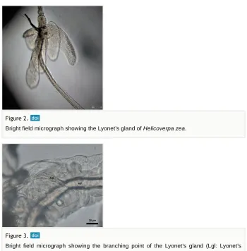
Figure 2.
Bright field micrograph showing the Lyonet’s gland of Helicoverpa zea.
Figure 3.
Bright field micrograph showing the branching point of the Lyonet’s gland (Lgl: Lyonet’s gland lumen) and main silk gland (sgl: Silk gland lumen) of Helicoverpa zea.
Figure 4.
CLSM volume rendered image showing granules on the surface of Lynoett’s gland of
Discussion
The structure of the Lyonet's gland in H. zea was different from what was illustrated by MacGown and Sikorowski 1982, with a relatively larger size while composed of multiple, leaf-like lobes. But it was similar to that described by in other noctuids with the lobes of the gland arising directly from the silk gland. The delicacy of the gland might be the explanation for these reports and it is also possible that specimen used by was partially destroyed and the illustration was mostly based on the proximal portion of the gland. We also experienced difficulties in keeping the Lyonet’s gland attached to the main salivary gland during our dissections but were always able to easily regain pieces of the gland.
The relative size of this gland in H. zea, which is 2–3 times larger than the same gland in Bombyx mori suggests that the gland plays an important role in the biology of H. zea. The labial glands have multiple function in Llepidoptera; in addition to silk formation saliva produced by the glands may be involved with digestion, detoxification, lubrication of the mouthparts, suppression of plant defenses, etc. (Rivera-Vega et al. 2017). What role the auxiliary Lyonet’s glands contribute to these functions is unknown. As there was no any indication of Glucose oxidase (GOX) in the gland, it may have other roles in the insects such as transport/secretion of small molecules involved in suppression of plant defenses. Based on the size and accessibility of the gland, future studies should include transcriptomic and proteomic studies in H. zea to provide clues regarding function. Examination of the gross morphology of other heliothines should also be considered, as authors who reported the absence of the Lyonet’s gland from H. zea also reported that this gland is absent from H. armigera and Heliothis virescencs (Sorensen et al. 2006, Chi et al. 1975).
Figure 5.
Acknowledgements
The current work is the result of the” Know Your Insect” 2017 fall graduate course of the Entomology Department at the Pennsylvania State University. We thank Adam Rork, Asifa Hameed, Po-An Lin, Ching-Wen Tan, and Maria Perezsandi for thoughtful discussions, literature search and assistance with dissections. SP is supported by the United States Agency for International Develpoment (USAID) Integrated Pest Management Innovation Lab (IPM IL) program.
Author contributions
SP and IM: Conceptualization, investigation, and writing
EGR and GWF: Supervision, review and editing
Conflicts of interest
There is no conflict of interest.
References
• Akai H (1984) The ultrastructure and functions of the silk gland cells of Bombyx mori . Insect Ultrastructure 323‑364. https://doi.org/10.1007/978-1-4613-2715-8_9
• Akai H, Nagashima T, Yamaguchi S (2003) Ultrastructures of silk glands and cocoon filaments from the Mexican silkmoth, Eucheira socialis . International Journal of Wild Silkmoth & Silk 8: 65‑72.
• Allegret P, Denis C (1963) Étude morphologique de l’appareil séricigene des larves de quelques espèces de Trichoptères et conséquences physiologiques immédiates. Bulletin de la Société Zoologique de France 88: 556‑558.
• Chi C, Drew W, Young J, Curd M (1975) Comparative morphology and histology of the larval digestive system of two genera of Noctuidae (Lepidoptera): Heliothis and Spodoptera
. Annals of the Entomological Society of America 68 (2): 371‑380. https://doi.org/10.1093/ aesa/68.2.371
• Cianficconi F, Moretti G (2000) Silk weave and silk glands in aquatic instars of two species of Helicopsyche von Siebold, 1856 (Trichoptera, Helicopsychidae). Aquatic Insects 22 (1): 58‑65. https://doi.org/10.1076/0165-0424(200001)22:1;1-z;ft058
• Day MF, Waterhouse DF (1953) The mechanism of digestion. In: Roeder KD (Ed.) Insect Physiology. John Wiley, New York, 311-330 pp.
• Eichenseer H, Mathews MC, Bi J, Murphy JB, Felton G (1999) Salivary glucose oxidase: Multifunctional roles for Helicoverpa zea? Archives of Insect Biochemistry and Physiology 42 (1): 99‑109.
• Glasgow JP (1936) Memoirs: Internal anatomy of a caddis (Hydropsyche colonica). Journal of Cell Science 2 (313): 151‑179.
• Helm E (1876) Über die Spinndrüsen der Lepidopteren. Wilhelm Engelmann
• Leslie RA, Robertson HA (1973) The structure of the salivary gland of the moth (Manduca sexta). Zeitschrift für Zellforschung und Mikroskopische Anatomie 146 (4): 553‑564. https:// doi.org/10.1007/bf02347183
• Li Q, Deng X, Yang W, Huang Z, Tettamanti G, Cao Y, Feng Q (2010) Autophagy, apoptosis, and ecdysis-related gene expression in the silk gland of the silkworm (Bombyx mori) during metamorphosis. Canadian Journal of Zoology 88 (12): 1169‑1178. https:// doi.org/10.1139/Z10-083
• MacGown MW, Sikorowski PP (1982) Anatomy of the digestive system of Heliothis zea
(Lepidoptera; Noctuidae) larvae. Bulletin - Mississippi Agricultural and Forestry Experiment Station 905: 1‑15.
• Machida Y (1965) Studies on the silk glands of the silkworm, Bombyx mori L. I.
Morphological and functional studies of Filippi’s glands in the silkworm. Science Bulletin of the Faculty of Agriculture, Kyushu University 22: 95‑108.
• Musser R, Hum-Musser S, Eichenseer H, Peiffer M, Ervin G, Murphy JB, Felton GW (2002) Caterpillar saliva beats plant defences. Nature 416 (6881): 599‑600. https://
doi.org/10.1038/416599a
• Musser RO, Cipollini DF, Hum-Musser SM, Williams SA, Brown JK, Felton GW (2005) Evidence that the caterpillar salivary enzyme glucose oxidase provides herbivore offense in solanaceous plants. Archives of Insect Biochemistry and Physiology 58 (2): 128‑137. https://doi.org/10.1002/arch.20039
• Rivera-Vega LJ, Acevedo FE, Felton GW (2017) Genomics of Lepidoptera saliva reveals function in herbivory. Current Opinion in Insect Science 19: 61‑69. https://doi.org/10.1016/ j.cois.2017.01.002
• Schneider CA, Rasband WS, Eliceiri KW (2012) NIH Image to ImageJ: 25 years of image analysis. Nature Methods 9 (7): 671‑675. https://doi.org/10.1038/nmeth.2089
• Sehnal F, Akai H (1990) Insect silk glands: their types, development and function, and effects of environmental factors and morphogenetic hormones on them. International Journal of Insect Morphology and Embryology 19 (2): 79‑132. https://
doi.org/10.1016/0020-7322(90)90022-h
• Sorensen GS, Cribb BW, Merritt D, Johnson M-L, Zalucki MP (2006) Structure and ultrastructure of the silk glands and spinneret of Helicoverpa armigera (Hübner) (Lepidoptera: Noctuidae). Arthropod Structure & Development 35 (1): 3‑13. https:// doi.org/10.1016/j.asd.2005.10.002
• Vegliante F (2005) Larval head anatomy of Heterogynis penella (Zygaenoidea, Heterogynidae), and a general discussion of caterpillar head structure (Insecta, Lepidoptera). Acta Zoologica 86 (3): 167‑194. https://doi.org/10.1111/
j.1463-6395.2005.00198.x
• Victoriano E, Gregório EA (2004) Ultrastructure of the Lyonet's glands in larvae of Diatraea saccharalis Fabricius (Lepidoptera: Pyralidae). Biocell 28 (2): 165‑9.
• Waku Y, Sumimoto K (1974) Ultrastructure of Lyonet's gland in the silkworm (Bombyx mori
L.). Journal of Morphology 142 (2): 165‑185. https://doi.org/10.1002/jmor.1051420205 • Wang X, Li Y, Peng L, Chen H, Xia Q, Zhao P (2016) Comparative transcriptome analysis
Insect Biochemistry and Molecular Biology 68: 89‑99. https://doi.org/10.1016/ j.ibmb.2015.11.003