Reconstructive lntracranial Vascular Surgery*
JOHN M. TEW, JR., M.D., F.A.C.S.
Instructor in Surgery, Division of Neurosurgery, University of
Cincinnati
Medical Center; Assistant Director, Neurosurgical
Training Program, Good Samaritan Hospital, Cincinnati, Ohio
It is scarcely less than two decades since the initial cautious explorations of reconstructive sur-gery for the prevention of cerebral infarction caused by extracranial vascular disease began. The next years recorded an accumulative experience in the field of vascular surgery. Early clinical investigations led to the discovery that extracranial vascular disease is a major cause of cerebral infarction or stroke. Estimates indicate, however, that only 30-40% of the patients with cerebrovascular insufficiency have significant extracranial occlusive disease. For this larger group of patients, previous surgical methods offer no hope. It is the purpose of this report to review the current status of our clinical explorations in the area of reconstructive intracranial vascular surgery.
This is not a new venture for as early as 1944, Henschen ( 1) transplanted a pedicle of temporalis muscle and artery over the surface of the brain in a type of encephalomyosynangiosis. This pro-cedure, performed in a patient with bilateral carotid stenosis, was similar in concept to the Vineberg-J ewett operation for revascularization of the myo-cardium, first published in 194 7 (2). The patient is reported to have improved, but angiographic confirmation of graft function was not accomplished. The procedure apparently was not explored further until the work of Donaghy and Yasargil ( 3) in
1966.
Direct reconstruction of the middle cerebral artery was successfully performed by Welch ( 4) in 1956, an amazing feat achieved without the aid of a surgical microscope and many other re-fined techniques which today are regarded as es-sential; subsequent reports by Shiliito ( 5), Scheibert
* Presented by Dr. Tew at the 27th Annual Stone. burner Lecture Series, February 7, 1974, at the Medical College of Virginia, Richmond.
MCV QUARTERLY 10(3): 139·145, 1974
(6), Driesen (7), and Chou (8) followed. Jacob-son, Donaghy, and colleagues (9) first reported that the microsurgical technique was of considerable value in performing this procedure. Subsequently several centers ( 10, 11) have reported their results with surgical treatment of occlusions of the middle cerebral artery. Yasargil (12) tabulated eleven cases in 1969, of which he operated on nine, more than one week following the onset of symptoms. One case, on which he operated withirt two hours of occlusion, failed to improve although the artery remained patent. A second case, on which he operated twelve hours following occlusion, developed a hemorrhagic infarction. All cases studied by postoperative an-giography were patent.
In our personal experience, we operated on only five cases two days-to-two weeks following occlusion. Patency was reestablished in each case although, in one case, the embolus had already passed on to a more distant site. One patient wors-ened as a result of the procedure probably due to hemorrhagic infarction and the other four irriproved, but their improvement was similar to that expected in the natural history of this disorder.
Recorded experience certainly indicates that reconstruction of flow through the embolically oc-cluded middle cerebral artery is technically feasible; yet, experimental evidence indicates that if restora-tion of flow is to be of value, it must be accom-plished in less than four-to-eight hours in order to reverse hypoxia. It must also be remembered that it is in this period that one incurs the greatest risk of hemorrhagic infarction if the flow is restored. Finally, it is rare, indeed, that the patient reaches the surgeon in the allotted time. In conclusion, it seems that this procedure, although technically suc-cessful, has few indications in practice.· Hopefully with the advent of less thrombogenic cardiac valves,
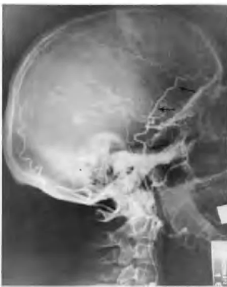
Fig. !-Preoperative angiogram demonstrates occlusion of internal carotid artery, collateral circulation through oph-thalmic artery, and the course of the superficial temporal artery (arrows).
the temptation to consider the procedure will dis-appear.
Having removed the acutely occluded middle cerebral artery from consideration for reconstruction, what other lesions and indications for intracranial vascular reconstruction exist? A recent study from UCLA (13) indicates that 19% of
a
series of pa-tients with symptoms of transient cerebral ischemia had a completely occluded internal carotid artery as a principle hemodynamic factor in their angio-graphic study. Other studies (14, 15) indicate that 30-35 % of the patients presenting with transient cerebral ischemia or mild infarction have obstruc-tive lesions inoperable by general vascular tech-niques. Accordingly, any effective surgical pro-cedure must either extract the obstructing lesion or bypass the area of obstruction. Since a major cause of cerebral insufficiency is unsuspected carotid oc-clusion, extending from the cervical to the cavernous portion, extraction of the thrombosis rarely is prac-tical. Bypass procedures then become important in the creation of compensatory vascular channels.What then are the indications for these vascular augmentation procedures? In a collaborative attempt to answer this question, investigators from four centers have pooled a hundred cases for retrospective analysis ( 16). As a result of this study, the follow-ing general indications are suggested:
Generalized Low Perfusion Syndromes
Symptoms:
1. impaired mentation 2. syncopal episodes
3. transient motor, speech or sensory deficit 4. transient or progressive visual loss 5. ataxia and postural vertigo Etiology:
1. multiple vessel occlusion
2. multiple stenosis of intracranial vessels 3. unilateral vessel occlusion with
inade-quate collateral circulation or congenital anomalies which interfere with collateral circulation
Lateralized Low Perfusion Syndromes
Symptoms:
1. transient or progressive motor, sensory, or speech deficit
TEW: RECONSTRUCTIVE INTRACRANIAL VASCULAR SURGERY 141
Etiology:
1. middle cerebral stenosis or occlusion with inadequate collateral circulation 2. cervical carotid occlusion or intracranial
carotid occlusion with inadequate circle of Willis
3. inoperable carotid aneurysms or tumors such as meningiomas which produce carotid occlusion
Having agreed on these general indications, a prospective study is now underway to determine the merits of this approach to cerebral vascular disease ( 17). This report will be conducted during the next three years. Presently, I will review with you the tech-niques currently employed for cerebral vascular re-construction and the findings in eighteen cases which have been under my personal care.
Donaghy and Yasargil ( 3) jointly devised the concept of performing an arterial graft; perhaps they remembered the initial explorations of Henschen. A satisfactory superficial temporal artery is required for this procedure; in this angiogram, a complete oc-clusion of the internal carotid artery is demonstrated (Fig. 1, see arrows). Delayed filling is noted through the ophthalmic and dural communicating arteries. The illustration (Fig. 2) demonstrates the tech-nique: the superficial artery is dissected free from the scalp and a small skull flap is turned to expose the sylvian fissure and a portion of the temporal and frontal lobes. An oblique end-to-side anastomosis is created between the temporal artery and a carefully selected branch of the middle cerebral artery. A seg-ment of the superficial temporal artery is first freed
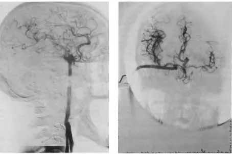
Fig. 3A-Common carotid angiogram with total occlusion of internal carotid artery; superficial temporal artery (STA) has been anastomosed to MCA branch at arrow.
Fig. 3H-Later sequence in angiogram (Fig. 3A). 1
=
site of STA entrance into cranial cavity and 2=
site of anas-tomosis. Note retrograde reflex toward proximal MCA trunk.from its muscular branches for 7-8 cm. The artery remains in situ while the craniotomy flap is turned and the sylvian fissure is explored. A cortical artery of 1.5 mm in diameter or greater is selected as it
Fig. 4A-Vein bypass graft. Reversed vein graft is passed beneath the zygoma and joined to the proximal internal carotid artery.
leaves the sylvian fissure. Using 20-30 magnification, the cortical artery is prepared to receive the trans-plant. Minute penetrating branches are coagulated and incised. The cortical artery is lifted from the sur-face of the brain and a slip of latex is placed under-neath it. The temporal artery is incised obliquely and brought into the field. The adventitia and other tis-sues are stripped from the terminal centimeter of the temporal vessel. The artery is dilated with pressure injection of heparinized saline and bathed in a dilute papaverine solution to reduce spasm. A longitudinal arteriotomy is made in the thin wall of the middle cerebral artery branch by a fragment of sharp razor blade. An incision is made the precise length to ac-commodate the oblique cut of the temporal artery. A polyethylene stint, 1.2 mm in diameter, is placed in the cortical artery after it is irrigated with heparinized saline. Interrupted sutures of 10.0 monofilament nylon are placed at either end of the suture line. The remaining portion is closed with similar interrupted sutures leaving the final three to be placed. The stint is then removed and the final sutures are secured.
The artery is flushed to remove all air and particles of thrombus.
This technique has been employed in 14 cases, there being twelve males and two females with an average age of 50. Twelve have had symptoms of transient ischemia, indicating a lateralized low per-fusion syndrome. Eight cases have had previous sig-nificant cerebral infarctions; two have had symptoms of progressive dementia indicating generalized low perfusion syndromes. Angiography demonstrated that twelve patients had occlusion of the ipsilateral
I.
3.
TEW: RECONSTRUCTIVE INTRACRANIAL VASCULAR SURGERY 143
internal carotid artery; three had bilateral carotid artery occlusion; five had marked stenosis of the contralateral internal carotid artery; and three had unilateral vertebral artery occlusion.
Results. Angiography was performed in all cases, postoperatively, usually one or two weeks following the procedure (Fig. 3). Nine grafts were patent; one, not patent on an earlier angiogram, was opened when the study was repeated two months later. We have recognized that spasm may interfere with early visualization of the graft. Four patients had symp-toms of recurrent cerebral ischemia, one of whom had a patent graft. Two patients who had occluded grafts have not had recurrent attacks of ischemia. Both patients with dementia seem improved. Two patients have had recurrent cerebral infarction; both had had occluded grafts. One patient suffered a post-operative intracerebral hemorrhage presumably due to hemorrhagic infarction. One patient died of myo-cardial infarction. Complications consisted ( one case each) of scalp necrosis, intracranial hemorrhage,
sub-galeal infection, and myocardial infarction; graft oc-clusion occurred in five cases.
The results of analyzing my personal cases and those of my colleagues ( 16) previously mentioned, suggest that the following contraindications to bypass graft procedures be considered: 1) major long-standing neurologic deficit; 2) severe recent cerebral infarction less than six weeks; 3) marked cardiovascu-lar disease; 4) advanced diabetes mellitus; 5) inade-quate donor artery-that is, a superficial temporal artery Jess than one millimeter in diameter. As noted in number five, inadequate donor artery has posed a serious problem in this technique; this inadequacy was the reason that Lougheed ( 18), in 1970, sug-gested the vein bypass procedure. As initially pro-posed by Lougheed, a saphenous vein graft was taken from the leg, the valves were stripped, and the small end was anastomosed to the supraclinoid portion of the internal carotid artery. The graft was brought out over the zygomatic arch. We have subsequently re-vised this procedure somewhat, simply reversing the
graft and suturing the larger end to the internal caro-tid artery (Fig. 4). Flow is restored through this seg-ment as soon as possible, since the hemisphere may be dependent on the collateral supply reaching it through the ophthalmic and posterior communicating arteries, which must, by necessity, be occluded dur-ing the cranial anastomosis. We have not yet devel-oped a satisfactory shunt system to perfuse the hemi-sphere during the 10-15 minutes required for this portion of the procedure. Technically, this is a rela-tively simple procedure; yet the lack of a satisfactory internal bypass prohibits its performance in some of the most worthy candidates. The next major obstacle to bypassing the internal carotid artery is severe plaque formation in the carotid siphon above the an-terior clinoid. Frequently, endarterectomy is required in opening a segment to receive the vein graft. Im-paired runoff may be a problem in spite of careful attention to this maneuver.
The artery is led from the cranium beneath the zygoma into the neck. Prior to completion of the proximal anastomosis to the common carotid artery, a large rubber catheter is passed through the graft to open the valves and fill the vein with heparinized blood. Our experience with this technique includes four cases. Three grafts functioned well; however, one patient, our first, died of a massive hemorrhagic infarction, although it was six weeks following his most recent cerebral infarction. Our second patient suffered delayed thrombosis of the graft, a failure we believe to be related to compression at the zygomatic process. A third graft has functioned well for 14 months and the patient remains free of ischemic com-plications. Figure 5 shows the postoperative angio-grams. A fourth graft was unsuccessful due to severe narrowing of the internal carotid artery. Despite endarterectomy, satisfactory runoff could not be ob-tained.
Conclusion. Although our initial experience with these procedures has certainly been less than spectacular, it does seem to indicate that further pursuit of the project of cerebral revascularization is worthwhile. Careful selection of patients, improve-ment of surgical technique (including the develop-ment of an internal shunt for bypass), and precise tabulation of results remain essential to the continua-tion of this study.
REFERENCES
I. HENSCHEN C: Operative Revascularization des
zirkula-torisch geschadigten Gehirns durch Auflage gestielter
Muskellappen (Encephelo-Myo-Syngangiose).
Langen-becks Arch Klin Chir 264:392, 1950.
2. VINEBERG A, JEWETT BL: Anastomosis between
coro-nary vessels and internal mammary artery. Canad Med
Assoc J 56:609, 1947.
3. DONAGHY, RMP: Patch and bypass in microangiorsal
surgery. In: Microvascular Surgery (ed.) RMP
Don-aghy, MG Yasargil. Thieme, Stuttgart, 1967, pp. 75-86.
4. WELCH K: Excision of occlusive lesions of the middle
cerebral artery. J Neurosurg 13:73, 1956.
5. SHILLITO J: Intracranial arteriotomy in three children,
four adults. In: Microvascular Surgery (ed.) RMP
Donaghy, MG Yasargil, Thieme, Stuttgart, 1967, pp.
138-142.
6. ScHEIBERT CD: Middle cerebral artery surgery for
obstructive lesions. Presented at the meeting of Harvey
Cushing Society, New Orleans, La., May 2, 1962.
7. DRIESEN E: Erfolgreiche Naht der linken. A. Cerebri
media nach Verletzung bei Tumorresektion. Acta
Neurochir 10:462, 1962.
8. CHOU SN: Embolectomy of middle cerebral artery:
Report of a case. J Neurosurg 20:161, 1963.
9. JACOBSON JH, WALLMAN LJ, SCHUMACHER GA,
FLANA-GAN M, SUAREZ FL, DONAGHY RMP: Microsurgery as
an aid to middle cerebral artery endarterectomy.
J Neurosurg 19:108, 1962.
10. LOUGHEED WM, GUNTON RW, BARNETT HJM:
Em-bolectomy of internal carotid, middle, and anterior
cerebral arteries. J Neurosurg 22:607, 1965.
11. SUNDT TM, NOFZINGER JD: Clip-grafts for aneurysm
and small vessel surgery. J Neurosurg 27:477, 1967.
12. YASARGIL MG: Reconstructive operations on the
cere-bral blood vessels. In: Microsurgery Applied to
Neuro-surgery (ed.) MG Yasargil, Thieme, Stuttgart, 1969,
pp. 96-119.
13. MACHLEDER HI, BARKER WF: Stroke on the wrong
side. Arch Surg 105:943, 1972.
14. MARSHALL J: Angiography in the investigation of
TEW: RECONSTRUCTIVE INTRACRANIAL VASCULAR SURGERY 145
15. BLAISDELL WF, HALL AD, THOMAS AM, Ross SJ: Cerebrovascular disease, experience with panarteriogra-phy in 300 consecutive cases. Cal Med 103:321, 1965.
16. REICHMAN H, CHATER N, YASARGIL MG, TEW J: Recent
advances in treatment of cerebrovascular disease.
Ex-hibit presented at American Association of Neuro-logical Surgeons, April 8, 1973.
17. REICHMAN H: Personal communication. December, 1973.