Ali S. Shalash, MD* Thomas W. Rösler, PhD* Stefanie H. Müller, MS* Mohamed Salama, MD Günther Deuschl, MD Ulrich Müller, MD Thomas Opladen, MD Britt-Sabina Petersen,
PhD
Andre Franke, PhD Franziska Hopfner, MD* Gregor Kuhlenbäumer,
MD, PhD* Günter U. Höglinger,
MD*
Correspondence to Prof. Dr. Höglinger: [email protected]
c.207C
.
G mutation in sepiapterin
reductase causes autosomal dominant
dopa-responsive dystonia
ABSTRACT
Objective: To elucidate the genetic cause of an Egyptian family with dopa-responsive dystonia (DRD), a childhood-onset dystonia, responding therapeutically to levodopa, which is caused by mutations in various genes.
Methods:Rare variants in all coding exons ofGCH1were excluded by Sanger sequencing. Exome sequencing was applied for 1 unaffected and 2 affected family members. To investigate the functional consequences of detected genetic variants, urinary sepiapterin concentrations were determined by high-performance liquid chromatography.
Results:A heterozygous rare nonsynonymous variant in exon 1 of sepiapterin reductase (SPR, c.207C.G, p.Asp69Glu) was found in all affected family members. Urinary concentrations of sepiapterin were above the standard of normal controls in mostSPRmutation carriers, suggest-ing functional biochemical consequences of the mutation. Variant filtersuggest-ing of all genes involved in the tetrahydrobiopterin pathway, required for levodopa synthesis, revealed an additional common variant in dihydrofolate reductase (DHFR, rs70991108). The presence of both variants was significantly stronger associated with the biochemical abnormality and the clinical disease state as opposed to 1 variant only.
Conclusions:The rareSPRmutation can cause autosomal dominant DRD with incomplete pene-trance. The commonDHFRvariant might have synergistic effects on production of tetrahydro-biopterin and levodopa, thereby increasing penetrance.Neurol Genet2017;3:e197; doi: 10.1212/ NXG.0000000000000197
GLOSSARY
DHFR5dihydrofolate reductase;DRD5dopa-responsive dystonia;MAF5minor allele frequency;NADPH5nicotinamide adenine dinucleotide phosphate;SPR5sepiapterin reductase.
Dopa-responsive dystonia (DRD) encompasses a group of movement disorders with
heteroge-neous clinical and genetic manifestations, presenting with childhood-onset limb dystonia and
response to levodopa therapy.
1Additional neurologic presentations may include parkinsonian
and pyramidal features.
2,3Variants in the gene of GTP cyclohydrolase I (
GCH1
), a key protein
of the tetrahydrobiopterin (BH
4) pathway, required for the biosynthesis of levodopa (figure, A),
are the most frequent cause of autosomal dominant DRD, also known as DYT5a.
1,4Variants of
sepiapterin reductase (
SPR
), encoding another key protein of the BH
4pathway (figure, A), have
been identified in autosomal recessive DRD.
5At present, only 1 patient with DRD without
family history has been identified with a heterozygous
SPR
variant.
6Here, we describe the first
DRD family carrying a rare
SPR
variant (c.207C.G, p.Asp69Glu, chr2:73114768_C.G,
*These authors contributed equally to the manuscript.
From the Department of Neurology (A.S.S.), Ain Shams University, Cairo, Egypt; German Center for Neurodegenerative Diseases (DZNE) (T.W.R., G.U.H.), Munich, Germany; Department of Neurology (T.W.R., G.U.H.), Technical University of Munich, Germany; Department of Neurology (S.H.M., G.D., F.H., G.K.), University Hospital Schleswig Holstein, Kiel, Germany; Medical Experimental Research Center (MERC) (M.S.), Mansoura University, Egypt; Institute for Human Genetics (U.M.), University of Giessen, Germany; Division of Neuropediatrics and Metabolic Medicine (T.O.), University Children’s Hospital, Heidelberg, Germany; and Institute of Clinical Molecular Biology (B.-S.P., A.F.), Christian-Albrechts-Universität zu Kiel, Germany.
Funding information and disclosures are provided at the end of the article. Go to Neurology.org/ng for full disclosure forms. The Article Processing Charge was funded by Klinikum rechts der Isar der TUM.
undetected in 8,599 alleles annotated in the
Exome Variant Server) with autosomal
domi-nant inheritance and incomplete penetrance.
METHODS Standard protocol approvals, registrations, and patient consents.Affected and nonaffected individuals of a family of Egyptian origin were examined by neurologists special-ized in movement disorders (A.S.S. and G.U.H.) at the Department of Neurology, Ain Shams University, Cairo, Egypt. Ethical approval was obtained at Ain Shams University (RP/42) and Technical University of Munich (203/15s). All participants provided written informed consent. Consanguinity was excluded by interview and genetic analyses.
Sanger and next-generation sequencing. After excluding known rare variants in all 6 coding exons ofGCH1by Sanger sequencing, exome sequencing was applied to 1 unaffected and 2 affected family members. Genomic DNA libraries were captured using the Nextera Rapid Capture Expanded Exome Kit (Illumina, San Diego, CA), and DNA fragments were sequenced on an Illu-mina HiSeq2000 system. Variants were identified by a standard analysis pipeline.
Variant filtering and validation.Variants with a minor allele frequency (MAF) .0.1%, based on .6,500 (The National
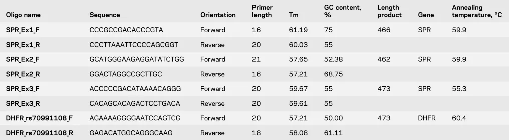
Heart, Lung, and Blood Institute Exome Sequencing Project, version ESP6500siv2), were discarded. Assuming a dominant dis-order, only nonsynonymous variants were retained as candidate alleles, including missense, nonsense, stop-loss, and splice-site mutations, as well as small insertions and deletions present in the heterozygous state and present in both affected sisters (figure B, II.2 and II.4). Hypothesis-based variant filtering was applied to all genes in the SPR pathway, also considering variants with an MAF .1.0%. Candidate variants were validated by Sanger sequencing and cosegregation with disease in the family (table 1).
In silico analyses.The effects of amino acid substitutions on pro-tein function were predicted using MutationTaster (mutationtaster. org), PolyPhen-2 (genetics.bwh.harvard.edu/pph2), and CADD (cadd.gs.washington.edu). Mutagenesis analysis was performed with PyMOL (pymol.org) based on the SPR protein structure 4XWY (PDB file). Functional protein domains were analyzed using Hap-loReg v4.1 (broadinstitute.org/mammals/haploreg/haploreg.php).
Urinary sepiapterin.First-void urine was collected, protected from light, immediately placed on dry ice, and stored at280°C. For sepiapterin quantification, samples were prepared as described7and subjected to high-performance liquid
chromatogra-phy analysis on a C18 reversed-phase column (Spherisorb ODS1, 5 mm, ID 4.6 mm, L250 mm; Waters, Milford, MA) and isocratic elution with 24 mM KH2PO4 (pH 5.0)/CH3OH Figure Sepiapterin reductase and dihydrofolate reductase in the context of dopa-responsive dystonia
(85:15, vol/vol) (flow rate 1.1 mL/min over 15 minutes). A fluo-rescence detector (RF 20A XS; Shimadzu, Kyoto, Japan; excitation l5425 nm; emissionl5530 nm) was used. Sepiapterin eluted with a retention time of 9.9 minutes. Urine samples were masked before analysis. Mean values of 4 technical measurements are presented.
Statistical analyses. Descriptive analyses andx2statistics were
calculated using Statistica 8.0 (StatSoft Inc., Tulsa, OK). Data are presented as mean6SEM.p,0.05 was considered statistically significant.
RESULTS Clinical description. We examined 5 affected and 4 nonaffected family members (figure, B). Symptom onset was between childhood and puberty (table 2). Patient II.4 presented with lower limb dystonia (tiptoe walking) and rigidity since childhood, with progressive course and diurnal variation. He improved markedly with L-DOPA,
however with low-threshold drug-induced dyskine-sia. His current medication comprises fractionated
L-DOPA doses (750 mg/d), anticholinergics, and
amantadine (400 mg/d). The other affected in-dividuals showed diurnal lower limb dystonia, rigid-ity, and mild pyramidal signs. Patients (II.2 and II.5) experienced symptomatic benefit withL-DOPA and
continued on pramipexole. Patients III.2 and III.3 were not treated withL-DOPA because of their young
age, mild symptoms, and fear of their parents of
L-DOPA–induced dyskinesia.
Genetic analysis.We found a nonsynonymous hetero-zygous variant in exon 1 of SPR (NM_003124.4 c.207C.G, p.Asp69Glu, genomic [GRCh37]: chr2:73114768_C.G) in all affected family mem-bers. In SPR, aspartate 69 is involved in binding the cofactor nicotinamide adenine dinucleotide phos-phate (NADPH). In silico mutagenesis analysis pre-dicted that the longer and more flexible side chain of glutamate 69 in mutation carriers leads to an increased distance and loss of hydrogen bonds to NADPH (figure, C).
Structural gene/exon variations ofSPRwere checked in the 3 samples in which whole-exome sequencing was performed using the BreakDancer tool (github.com/ genome/breakdancer).8 We did not detect such an
event nearSPRor elsewhere on chromosome 2. In addition, we found an intronic 19 base-pair insertion (rs70991108) in an active transcription start site of the dihydrofolate reductase (DHFR) gene in all affected family members (table 2). This variant alters 8 regulatory motifs (HaploReg).
Urinary sepiapterin.SPR deficiency increases urinary sepiapterin levels.7 To investigate the consequences
of the variants observed in this study, we measured urinary sepiapterin concentrations in all members (table 2). All clinically affected family members had sepiapterin concentrations slightly above the range measured in normal controls (mean 6 SD: 4.2 6 3.12 mmol/mol creatinine), but below the values in proven SPR-deficiency patients (727 6 230mmol/mol creatinine).7
Genotype-phenotype relationship.The family member without theSPRvariant (II.1) had normal sepiapterin concentrations and was healthy. From the 8SPR var-iant carriers (table 2), 7 had elevated sepiapterin con-centrations; 5 of those were clinically affected.
From 2 members without the DHFR variant (I.2 and III.1), 1 had elevated sepiapterin values, but both were healthy. From 7DHFRvariant carriers (table 2), 6 had elevated sepiapterin concentrations; 5 of those were clinically affected.
Six family members carried bothSPRandDHFR
variants (table 2); all of them had elevated sepiapterin concentrations; 5 of them were clinically affected.
There was a significantly stronger association of both variants simultaneously with the biochemical abnormality (x25.1429,p, 0.05) and the clinical
disease state (x25.625,p,0.05) as opposed to only
1 variant.
Table 1 Primer sequences for theSPRgene encoding sepiapterin reductase (SPR) and for rs70991108 located in theDHFRgene encoding dihydrofolate reductase (DHFR)
Oligo name Sequence Orientation
Primer length Tm
GC content, %
Length
product Gene
Annealing temperature, °C
SPR_Ex1_F CCCGCCGACACCCGTA Forward 16 61.19 75 466 SPR 59.9
SPR_Ex1_R CCCTTAAATTCCCCAGCGGT Reverse 20 60.03 55
SPR_Ex2_F GCATGGGAAGAGGATATCTGG Forward 21 57.65 52.38 462 SPR 59.9
SPR_Ex2_R GGACTAGGCCGCTTGC Reverse 16 57.21 68.75
SPR_Ex3_F ACCCCCGACATAAAACAGGG Forward 20 59.67 55 473 SPR 55.3
SPR_Ex3_R CACAGCACAGACTCCTGACA Reverse 20 59.61 55
DHFR_rs70991108_F AGAAAAGGGGAATCCAGTCG Forward 20 57.21 50.00 473 DHFR 60.4
DISCUSSION We identified a rare heterozygous var-iant of SPR in a family with DRD segregating as autosomal dominant trait (figure, B and table 2).
SPRis essential for levodopa synthesis. In silico anal-ysis suggested pathogenicity of this variant. Consis-tently, we found slightly elevated urine sepiapterin concentrations in 7 variant carriers, 5 of these pre-senting clinical symptoms. Since also 3 nonaffected siblings carried this variant, incomplete penetrance was assumed. OtherSPRvariants are known to cause autosomal recessive DRD. A heterozygousSPR vari-ant in a single patient with DRD without family history had already indicated the possibility of auto-somal dominant causality of SPR variants.6 These
observations suggest that thisSPRvariant has autoso-mal dominant pathogenicity with incomplete penetrance.
In the same family, we also identified a DHFR
variant (rs70991108). In silico analysis suggested a potential effect of this variant on protein expression. Consistently, we found elevated urine sepiapterin concentrations in 6 and clinical symptoms in 5 of the 7 variant carriers. Two nonaffected siblings car-ried the variant as well. Because of its relatively high
allele frequency in the general population,9we do not
assume this variant to be pathogenic per se. Of interest, the presence of the commonDHFR
variant in addition to the SPR variant significantly increased the risk of the presence of the biochemical abnormality (elevated sepiapterin urine concentra-tions) and of clinical symptoms. The 1 sibling carry-ing both variants (SPRandDHFR) without clinical DRD symptoms might be either in a preclinical dis-ease stage or might be protected by unknown resil-ience factors.
While high DHFR expression in the liver may account for a modest biochemical phenotype in the periphery, the very low cerebral DHFR expression might be further reduced by the variant, leading to a clinically significant disturbance ofL-DOPA
synthe-sis.10 This suggests that the presence or absence of
the commonDHFRvariant affects the penetrance of the autosomal dominantSPRvariant. With regard to the sequential metabolic function of the SPR and
DHFRgene products in theL-DOPA synthesis path-way (figure, A), this interpretation appears plausible. The SPR-DHFR pathway metabolizes sepiapterin to generate tetrahydrobiopterin, essential for the Table 2 Clinical, genetic, and biochemical data of the investigated DRD family
Family member Sex
Age at
examination, y Dystonia
Disease onset
Phenomenology (in chronological order)
Response to
L-DOPA
Variant
Sepiapterin (mmol/mol Cr)6SEM
SPR DHFR
I.1 M ND NA NA NA NA NA NA NA
I.2 F 56 2 2 2 ND 1 2 3.7560.63
II.1 M 35 2 2 2 ND 2 1 2.9960.53
II.2 F 32 1 Childhood Dystonic posturing of
both feet; with diurnal fluctuation, mild lower limb bradykinesia and rigidity, and extensor
planter responses
1 1 1 15.4462.07a
II.3 M 29 2 2 2 ND 1 1 8.4760.72a
II.4 F 27 1 Childhood Initially; lower limb dystonia, bradykinesia, and rigidity with diurnal variation progressed to generalized parkinsonism; L-DOPA–induced
dyskinesia and pyramidal signs
1 1 1 28.4364.02a
II.5 M 21 1 Puberty Lower limb dystonia (tiptoe walking) after prolonged walking;
bilateral lower
limb rigidity, postural tremor, and equivocal Babinski sign
1 1 1 7.3960.88a
III.1 M 14 2 2 2 ND 1 2 17.7463.92a
III.2 F 10 1 Childhood Tiptoe walking with activity; fast fatigability with dystonic posturing on the left foot
ND 1 1 22.6764.75a
III.3 M 5 1 Infancy Tiptoe walking; equinovarus deformity
ND 1 1 19.9562.40a
Abbreviations: Cr5creatinine; DHFR5dihydrofolate reductase; DRD5dopa-responsive dystonia; NA5not available; ND5not determined; SPR5 sepiapterin reductase;2 5not present;1 5present; variant DHFR5chr5:79,950,163-/TGGCGCGTCCCGCCCAGGT (19 base-pair insert); variant SPR5 chr2:73,114,768 C/G in exon 1.
formation ofL-DOPA. The enzymes SPR and DHFR
are serially catalyzing this reaction. Homozygous muta-tions affectingSPRon both homologous chromosomes impose a massive bottleneck within this pathway and are known to cause DRD. A heterozygous mutation affectingSPRon only 1 chromosome imposes a minor bottleneck for this pathway. The clinical penetrance of this condition may thus depend on additional cofactors affecting this pathway, such as epigenetic silencing of the healthySPRallele. In our family, this cofactor seems to be the co-occurrence of an additional variant in the downstream enzymeDHFR, causing a“tandem steno-sis”within this pathway.
Taken together, the present study proposes the rareSPRc.207C.G variant as a cause of autosomal dominant DRD with incomplete penetrance and the common DHFR rs70991108 variant as a potential modifier, significantly affecting the penetrance of theSPRvariant.
AUTHOR CONTRIBUTIONS
Ali S. Shalash: study concept and design, acquisition of data, analysis and interpretation of data, and critical revision of the manuscript for intellec-tual content. Thomas W. Rösler: study concept and design, acquisition of data, analysis and interpretation of data, study supervision, and critical revision of the manuscript for intellectual content. Stefanie H. Müller: acquisition of data, analysis and interpretation of data, and critical revi-sion of the manuscript for intellectual content. Mohamed Salama: study concept and design, acquisition of data, analysis and interpretation of data, and critical revision of the manuscript for intellectual content. Günther Deuschl: study concept and design, analysis and interpretation of data, and critical revision of the manuscript for intellectual content. Ulrich Müller and Thomas Opladen: study concept and design, acquisi-tion of data, analysis and interpretaacquisi-tion of data, and critical revision of the manuscript for intellectual content. Britt-Sabina Petersen: acquisition of data, analysis and interpretation of data, and critical revision of the manuscript for intellectual content. Andre Franke: study concept and design, acquisition of data, analysis and interpretation of data, study supervision, and critical revision of the manuscript for intellectual con-tent. Franziska Hopfner: study concept and design, acquisition of data, analysis and interpretation of data, study supervision, and drafting the manuscript for intellectual content. Gregor Kuhlenbäumer: study con-cept and design, acquisition of data, analysis and interpretation of data, study supervision, and critical revision of the manuscript for intellectual content. Günter U. Höglinger: study concept and design, acquisition of data, analysis and interpretation of data, study supervision, and drafting the manuscript for intellectual content.
ACKNOWLEDGMENT
The authors thank Lena Jaschkowitz and Magda Berjas for technical assistance.
STUDY FUNDING
Supported by the German Academic Exchange Service (DAAD, Grants: 56502692, 57070724, 57162390, and 57247022). Günter U. Höglinger was funded by the Deutsche Forschungsgemeinschaft (DFG, HO2402/6-2).
DISCLOSURE
A.S. Shalash reports no disclosures. T.W. Rösler has served on the edi-torial board ofFrontiers in Neurologyand will receive royalty payments for a patented invention (Perk activator for the treatment of neurodegener-ative diseases, WO 2016024010 A1). S.H. Müller and M. Salama report
no disclosures. G. Deuschl has served on the scientific advisory boards of Medtronic, Sapiens, Britannica, and Boston Scientific; has received travel funding/speaker honoraria from Medtronic and Desitin; has served on the editorial boards ofMovement Disorders,Aktuelle Neurologie, and sev-eral other journals (names not indicated); and receives publishing royal-ties from Thieme Publishers. U. Müller has served on the editorial board of Neurogenetics and has received research support from CurePSP. T. Opladen has received speaker honoraria from Swedish Orphan. B.-S. Petersen, A. Franke, and F. Hopfner report no disclosure. G. Kuhlenbäumer has received research support from the International Essential Tremor Foundation. G.U. Höglinger has served on the scien-tific advisory boards of Asceneuron, AbbVie, Bristol-Myers Squibb, Roche, UCB, Novartis, Sanofi, and Biogen; has received travel fund-ing/speaker honoraria from AbbVie, Roche, UCB, and Movement Dis-orders Society; holds a patent for Method for the treatment of neurodegenerative diseases (PCT/EP2015/068734); and has received research support from Sellas Life Sciences Group, Bayerische Forschung-sallianz, German Ministry of Education and Research (BMBF), German Academic Exchange Service (DAAD), German Research Foundation (DFG), German Center for Neurodegenerative Diseases (DZNE), Cur-ePSP, International Parkinson Foundation, and NOMIS Foundation. Go to Neurology.org/ng for full disclosure forms.
Received May 24, 2017. Accepted in final form August 21, 2017.
REFERENCES
1. Klein C. Genetics in dystonia. Parkinsonism Relat Disord 2014;20(suppl 1):S137–S142.
2. Wijemanne S, Jankovic J. Dopa-responsive dystonia— clinical and genetic heterogeneity. Nat Rev Neurol 2015; 11:414–424.
3. Friedman J, Roze E, Abdenur JE, et al. Sepiapterin reduc-tase deficiency: a treatable mimic of cerebral palsy. Ann Neurol 2012;71:520–530.
4. Müller U. The monogenic primary dystonias. Brain 2009; 132:2005–2025.
5. Bonafe L, Thony B, Leimbacher W, Kierat L, Blau N. Diagnosis of dopa-responsive dystonia and other tetrahy-drobiopterin disorders by the study of biopterin metabo-lism in fibroblasts. Clin Chem 2001;47:477–485. 6. Steinberger D, Blau N, Goriuonov D, et al. Heterozygous
mutation in 59-untranslated region of sepiapterin reductase gene (SPR) in a patient with dopa-responsive dystonia. Neurogenetics 2004;5:187–190.
7. Carducci C, Santagata S, Friedman J, et al. Urine sepiap-terin excretion as a new diagnostic marker for sepiapsepiap-terin reductase deficiency. Mol Genet Metab 2015;115: 157–160.
8. Fan X, Abbott TE, Larson D, Chen K. BreakDancer: identification of genomic structural variation from paired-end read mapping. Curr Protoc Bioinformatics 2014;45:15.6.1–15.6.11.
9. Dutta S, Chattopadhayay S, Dasgupta C, Sarkar S. Low molecular weight heparin: a practical approach in deep venous thrombosis in palliative care. Indian J Palliat Care 2011;17:143–145.
10. Blau N, Bonafe L, Thony B. Tetrahydrobiopterin defi-ciencies without hyperphenylalaninemia: diagnosis and genetics of dopa-responsive dystonia and sepiapterin reductase deficiency. Mol Genet Metab 2001;74: 172–185.
DOI 10.1212/NXG.0000000000000197
2017;3;
Neurol Genet
Ali S. Shalash, Thomas W. Rösler, Stefanie H. Müller, et al.
dopa-responsive dystonia
c.207C>G mutation in sepiapterin reductase causes autosomal dominant
This information is current as of November 2, 2017
Services
Updated Information &
http://ng.neurology.org/content/3/6/e197.full.html
including high resolution figures, can be found at:
References
http://ng.neurology.org/content/3/6/e197.full.html##ref-list-1
This article cites 11 articles, 1 of which you can access for free at:
Permissions & Licensing
http://ng.neurology.org/misc/about.xhtml#permissions
its entirety can be found online at:
Information about reproducing this article in parts (figures,tables) or in
Reprints
http://ng.neurology.org/misc/addir.xhtml#reprintsus
Information about ordering reprints can be found online:
reserved. Online ISSN: 2376-7839.
Published by Wolters Kluwer Health, Inc. on behalf of the American Academy of Neurology.. All rights an open-access, online-only, continuous publication journal. Copyright Copyright © 2017 The Author(s).
is an official journal of the American Academy of Neurology. Published since April 2015, it is