Introduction
Recent theories about cluster headache (CH) pathogenesis point out the central role of the trigeminovascular system during an attack. In CH, as well as in migraine, trigeminal antidromic activation may cause the release of nociceptive neuropeptide transmitters in the blood vessel wall with a further excitation of the sensory fibers [1].
Analysis of the blink reflex (BR) may be a reliable method for evaluating the nociceptive reflex circuits con-necting the trigeminal and facial nerves involved in CH attack pathogenesis [1]. BR has been studied in CH with conflicting results as a result of the large methodological variability. Pavesi et al. [2] observed an increased R2 thresh-old on the affected side during the active phase. Formisano et al. [3] showed normal values for latency, duration, ampli-tude of R2 during the non-symptomatic phase, but increased
R2 duration and amplitude on the side of pain with a bilat-eral reduction of R2 habituation during the attacks. Furthermore, a reduced R2 amplitude was found on the affected side when stimulated at the intensity useful to elic-it the maximal R2 response on the unaffected side, in a group of 12 CH patients examined during the active phase [4]. More recently [5], the R2 recovery was evaluated dur-ing the cluster period of CH patients: after paired stimuli delivered at the supraorbital nerve, the R2 recovery was enhanced on the symptomatic side, while after index finger stimulation it was increased on both the symptomatic and non-symptomatic sides. These observations suggest a hyperexcitability of the trigeminal pathways on the side of CH attacks and a hypoactivity in the descending reticular system which bilaterally controls the trigeminofacial reflex-es. These studies are in agreement with the hypothesis of a basic trigeminal dysfunction in CH, but the large variability of methods and patients has prevented the definition of a
The blink reflex in chronic cluster headache:
a comparison with migraine patients suffering
from unilateral pain*
M. de Tommaso ()
First Neurologic Clinic,
Policlinico, Piazza Giulio Cesare 11, I-70124 Bari, Italy
Tel:. +39-080-5478565 Fax: +39-080-5478532
E-mail: ipnobari@cimedoc.uniba.it
* The authors belong to the Interuniversity Center for the Study of Headache and Neuro-transmitter Disorders of the Central Nervous System, with centers in Perugia, Rome, Sassari, Bari, Naples and Florence, Italy.
Marina de Tommaso Marco Guido
Giuseppe Libro Vittorio Sciruicchio Luciana Losito Olimpia Difruscolo Francomichele Puca
Abstract Objective. To evaluate the blink reflex (BR) in chronic cluster headache (CH) patients. Design. The electrophysiological data were col-lected in during the headache-free phase. Setting. Headache patients were recruited from outpatients seen for the first time at the First
Neurologic Clinic of Bari University. Patients and participants. Ten CH patients, 19 migraine without aura patients with strictly unilateral headache (MWoA) and 18 normal controls were selected.
Measurements and results. The BR procedure was applied. In CH, a sig-nificant R2 duration increase was
found on the symptomatic side in comparison with MWoA and con-trols. In both patient groups an early appearance of the R3 component was bilaterally clear. Conclusions. The BR findings confirm the central genesis of CH. The R3 abnormalities suggest a basic dysfunction of the central control on the trigeminal nociceptive circuits. The R2
involvement on the symptomatic side indicates a unilateral facilitation of the trigeminal-facial connections per-sisting after the CH bout.
Key wordsCluster headache • Migraine • Blink reflex Received: 3 January 2000
BR has also been studied in migraine. Bank et al. [6] found a prolonged latency of R2 components in migraine patients; Sand and Zwart [7] did not confirm these results. In a previous examination of migraine without aura patients during headache-free periods, an early appearance and increased amplitude of the R3 component were observed in comparison with controls, probably as a sign of a primary trigeminal system dysfunction [8]. Furthermore, the R3 response was increased on the painful side during the migraine attack [9].
The recognition of CH as a separate clinical entity from migraine is fairly recent: today no doubt exists about the clinical autonomy of the two headache forms, but some common pathophysiological characteristics make the rela-tion between CH and migraine remain an unresolved issue [10]. A central genesis was supposed for the two forms of vascular headache, migraine and CH, although many fea-tures differentiate them. The activation of a specific trigemi-novascular reflex on the side of pain, consisting of an affer-ent in the trigeminal system and an efferaffer-ent in the parasym-pathetic fibers of cranial nerve VII, is clearly observed dur-ing a CH bout but not in a migraine attack [1]. Therefore, study of BR may be a useful tool for investigating the cir-cuits connecting the trigeminal-facial reflexes. The aim of the present study was to evaluate BR in chronic and drug-free CH patients during the asymptomatic phase, and to compare it to that of a select group of migraine without aura patients suffering from a strictly unilateral headache.
Materials and methods
Headache patients were recruited from outpatients seen for the first time at our department. Inclusion criteria were the diagnosis of chronic CH or of migraine without aura with strictly unilateral headache (MWoA). Patients recruited in the MWoA group referred strict unilateral headache in the last 12 months, with a prevalent involvement of the same side in the years before and an average of at least 2 attacks in the last 3 months. They were diag-nosed according to the IHS criteria [11]. All patients were free of pain for at least 72 h. Exclusion criteria were any current or previ-ous general medical, neurological or psychiatric illness as defined by DSM-IV, intake of psychoactive drugs or of headache prophy-lactic, and recent symptomatic treatments (analgesic, ergot or 5HT1B1D drugs taken in the previous 72 h). We also excluded patients who referred attacks in the 48 h following the day of the examination. We selected 10 CH and 19 MWoA outpatients. Three migraine patients, without first-degree inheritance for migraine and with slight neurological signs, were submitted to magnetic res-onance imaging (MRI), which failed to show any abnormality.
The clinical neurophysiologist had no information about the subjects. Subjects were seated on an examination chair with their eyes gently closed. A Micromed EMG apparatus was used with fil-ter settings between 50 and 3000 Hz, 200 ms analysis time, and 8192 Hz sampling rate. Electromyographic activity was recorded
the bridge of the nose, near the inner canthus of the eye; the ground electrode was positioned around the arm. Electrical stimulation was applied by surface electrodes placed longitudinally, 2-cm apart, above the emergence supraorbital nerve. Stimuli were square-wave, negative single pulses of 0.1 ms duration, delivered unilaterally by a constant current isolation unit. To evaluate the thresholds (R1, R2, R3) of the blink reflex components, the sub-jective perceptive threshold (Pth) and pain threshold (Path), elec-trical stimuli were given at unpredicted intervals, with elecelec-trical intensity increasing in 2-mA steps. The length of interstimuli inter-vals was random but always longer than 40 s. During this interval, the examiner verbally interacted with the subjects to keep them awake, without giving them any warning of the subsequent stimu-lus, in order to avoid R2 and R3 habituation and the R3 attenuation due to the attention [12]. All subjects were invited to verbally express the stimulation level at which the subjective perceptive and pain sensations were felt. Signals were amplified, full-wave recti-fied and averaged.
We evaluated the latency, duration and area (mV/s) of the R1, R2 and R3 components elicited by 5x perceptive threshold electrical intensity (5xPth). We further computed the absolute dif-ferences between the direct BR responses elicited by the stimulation of the two sides (right-left). The latency, area and duration of the crossed R2 (CR2) and R3 (CR3) responses were also computed.
Statistical analysis
The electrophysiological and clinical data were analysed by
ANOVA, Student’s ttest for unpaired data and Bonferroni test.
They were correlated with clinical data by Spearman’s correlation test. The values obtained by stimulation of the symptomatic and non-symptomatic sides in patients were considered abnormal when they exceeded the right or left side normal ranges for at least 2 standard deviations (+ 2 SD).
Results
T
able 1
Clinical and electrophysiological features in cluster headache (CH) patients, unilateral migraine (MW
oA) patients and normal co
ntrols (N) Subjects Age Sex Age since T ime since Pain
Perceptive threshold (mA)
Pain threshold (x Pth)
R2 threshold (x Pth)
R3 threshold (x Pt
h) (years) onset (years) onset (days) side SS NS SS NS SS NS D SS NS D CH 1 2 7 M 16 3 R 4 4 4 3 2 2 0 2.5* 2.5* 0 2 2 3 M 15 3 R 4 6 3.5 1.3* 1.5 1.3 0.2 2* 1.6* 0.4 3 2 4 M 15 6 L 4 4 2* 3 1.5 1.5 0 1.5* 1.5* 0 4 6 0 M 35 3 R 4 4 3 3 2 1.5 0.5 3.5 3.5 0 5 2 6 F 22 7 R 4 4 3 3 1.5 1.5 0 1.5* 1.5* 0 6 3 5 F 30 3 L 4 4 3 3 2 1.5 0.5 3.5 3.5 0 7 3 9 F 38 10 L 4 4 3 3 2 1.5 0.5 2.5* 3.5 1 8 32 M 3 0 3 R 2 4 2* 2* 2 0 * 2 4* 2.5* 1.5 9 3 7 M 8 3 L 4 2 0* 2* 1.5 3 1.5 1.5* 2* 0.5 10 42 F 3 0 3 R 6 6 1.6* 1.6* 0* 1.3 1.3 1.3* 1.3* 0 MW oA 1 4 5 F 23 6 L 4 4 0* 0* 2 2 0 2.5* 2.5* 0 2 40 M 1 5 6 R 4 4 4.5 3 2 2 0 2.5* 2.5* 0 3 46 F 3 8 6 R 6 6 1.3* 1.3* 1.3 0* 1.3 1.5* 0* 1.5 4 4 8 F 12 5 R 4 4 3.5 3.5 2 0* 2 3.5 3.5 0 5 3 8 M 11 6 R 4 4 3 3 2 0* 2 2.5* 2.5* 2 6 2 0 M 7 6 R 2 2 6 6 3 3 0 3 * 3 * 0 7 2 1 F 10 3 R 4 4 4 3 2 2 0 2.5* 2.5* 0 8 8 F 23 4 R 4 4 3.5 3.5 1.5 2 0.5 1.5* 2.5* 1 9 2 8 M 12 4 R 4 4 3 3 2 2 0 2.5* 2.5* 0 10 25 F 2 2 6 R 4 4 3.5 3 2 2 0 2.5* 2.5* 0 11 1 0 F 1 0 5 R 2 2 6 6 3 2 0 3 3 0 12 35 M 1 5 7 L 4 4 3 3
2 2.5
0.5 2.5* 1.5* 1 13 52 F 1 6 4 L 4 2 3 6 1.5 4 2.5 1.5* 5 3.5 14 42 F 11 6 L 4 4 3.5 3 0* 1.5 1.5 3.5 4 0.5 15 47 F 1 4 6 R 6 6 2.6 2.6 0* 0* 0 2.3* 0* 2.3 16 32 F 11 6 R 2 4 6 3 2 0* 2 4 2.5* 1.5 17 25 F 2 2 5 R 4 4 3 4.5 1.5 2 0.5 1.5* 2.5 1 18 43 F 3 9 3 L 4 4 3 3 1.5 0* 0.5 NE 3.5 NE 19 47 F 1 8 7 R 4 4 3 3 2 2 0 2.5* 2.5* 0 N (n° 18) 8F R L R L R L R L Mean 27.77 10M 3.75 3.6 3.8 3.6 1.9 1.91 1.03 3.7 3.2 1.5 SD 5.86 1.04 1.1 0.8 0.7 0.81 0.82 0.8 0.4 0.3 1.6
*, value exceeding the normal ranges corresponding to right or left sides ±
2
SD.
SS
, symptomatic side;
NS
, non-symptomatic side;
R
, right side;
L
, left side;
D
, dif
ference between R
and L
(R-L);
NE
, not elicited;
Pth
significantly lower in one CH patient and in one MWoA suf-ferer on both sides; 6 MWoA patients showed an unilater-al R2th reduction on one side and the R2th mean vunilater-alues were not dissimilar between CH and MWoA groups. Also, the asymmetry index was like in the selected groups (mean R2th in CH patients, 1.48 ± 0.44 x Pth on the symptomatic side, 1.51 ± 0.48 x Pth on the asymptomatic side; 0.65 ± 0.21 x Pth mean asymmetry index; mean R2th in MWoA patients, 1.61 ± 0.51 x Pth on the symptomatic side, 1.49 ± 0.46 x Pth on the asymptomatic side; 0.68 ± 0.22 x Pth mean asymmetry index; mean asymmetry index in normal subjects, 0.79 ± 0.34 x Pth). The R3 component was elicit-ed in all patients and controls, except for a migraine patient on the symptomatic side. Six CH and 12 MWoA patients showed a significant R3th reduction on both sides. Two
reduction in comparison with normal controls (Table 1). The R3th mean values were not significantly different between the patients groups and in both CH and MWoA series the R3th asymmetry was like normal controls (CH patients, 2.48 + 0.8 x Pth on the symptomatic side, 2.45 + 0.85 x Pth on the non-symptomatic side; MWoA patients, 2.58 + 0.88 x Pth on the symptomatic side and 2.76 + 0.85 x Pth on the asymptomatic side; mean asymmetry index 0.98 + 0.45 x Pth in CH patients, 1.65 + 0.67 x Pth in MWoA patients, 1.23 + 0.56 in normal subjects).
The R2 duration on the symptomatic side and the rela-tive asymmetry values exceeded the normal ranges in 9 CH patients and in one MWoA patient (Table 2). The mean R2 duration on the painful side was significantly increased in CH in comparison with MWoA patients. The mean R2
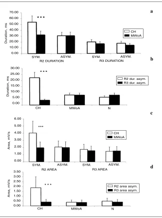
Fig. 1 a, cMean values (SD) of the R2 and
R3 duration (a) and area (c) in cluster headache patients (CH; n= 10) and migraine without aura patients suffering from strictly unilateral pain (MWoA; n= 19) measured on the symptomatic (sym.) and non-symptomatic (asym.) sides. The stimulus was 5 x perceptive threshold.
Results of ttest for unpaired data are
shown (***: p< 0.001). b, d Mean values
(SD) of R2 duration (b) and area (d) inter-side asymmetry (right – left) in CH, MWoA groups and normal subjects (n=18). Results of Bonferroni test are shown. (*** p< 0.001)
70.00
60.00
50.00
40.00
30.00
20.00
10.00
0.00
30.00
25.00
20.00
15.00
10.00
5.00
0.00
6.00
5.00
4.00
3.00
2.00
1.00
0.00
3.50
3.00
2.50
2.00
1.50
1.00
0.50
0.00
CH MWoA N
CH MWoA N
SYM. ASYM.
R3 AREA
SYM. ASYM.
R2 AREA
SYM. ASYM.
R3 DURATION
SYM. ASYM.
R2 DURATION
Duration, ms
Duration, ms
Area, mV/s
Area, mV/s
a
b
c
d
CH MWoA
R2 dur. asym. R3 dur. asym.
CH MWoA
Table 2 Electrophysiological features in cluster headache patients (CH), and unilateral migraine patients (MWoA) and normal controls (N)
R2 duration (ms) R3 duration (ms) R2 area (mV/s) R3 area (mV/s)
Subject
SS NS D SS NS D SS NS D SS NS D
CH
1 47* 43 4 28* 26* 2 4.3* 2.2 1.1 2.3 1.2 1.1
2 47* 26 21* 16 22 6 4.2* 2.8 1.4* 1.2 1.3 0.1
3 59* 30 19* 30* 30* 0 4.9* 2 2.9* 1.7 1 0.7
4 60* 30 30* 30* 28* 2 3.6 2.9 0.7 2.1 1.9 0.2
5 66* 28 38* 14 8 6 4,3* 2.2 2.1* 1.8 1.7 0.1
6 51* 32 19* 12 10 6 4.3* 1.8 2.5* 2.2 1.5 0.7
7 52* 32 20* 12 6 6 3.4 1.5 1.9* 0.8 1.1 0.3
8 51* 28 23* 11 2 8 4.1 1.5 2.6* 2.3 1.9 0.4
9 46 25 21* 20 18 2 2.8 1.4 1.4* 1.3 1.4 0.1
10 60* 32 28* 25* 23 2 4.6* 2.4 2.2* 2 1.7 0.3
MWoA
1 32 32 0 14 8 6 2.2 2.1 0.1 0.9 1.4 0.5
2 30 40 10 8 16 8 1.8 2.5 0.7 2.5 2 0.5
3 42 36 6 9 10 1 2.1 1.3 0.8 1.5 0.7 0.8
4 28 32 4 14 24 10 2.3 2.1 0.2 2.1 1.6 0.5
5 30 19 11 20 20 0 1.7 2 0.3 1.5 1.4 0.1
6 10 20 10 40* 10 30* 1.3 2 0.8 2.3 2 0.3
7 26 26 0 26* 18 8 1.8 2.2 0.4 1.2 1.4 0.2
8 38 44 6 16 16 0 2.4 2.5 0.1 0.9 1.3 0.4
9 48* 34 14 52* 4 48* 2.7 2.3 0.4 1.3 1.3 0
10 8 30 6 26* 14 8 1.9 1.8 0.1 0.8 1.2 0.4
11 38 42 4 12 12 0 2.1 2.4 0.3 2.5 1.9 0.6
12 38 40 2 12 12 0 1.9 2.5 0.6 1.3 1.9 0.6
13 46 32 14 4 16 12 2.1 1.9 0.2 1.7 1.6 0.1
14 38 46* 8 8 12 4 1.7 2.3 0.6 1.5 1.4 0.1
15 20 10 10 10 15 5 1.3 1.8 0.5 2 1.6 0.4
16 20 20 0 6 6 0 1.8 .5 0.3 1.8 1.5 0.3
17 26 34 12 14 8 6 2 1.9 0.1 1.5 1.2 0.3
18 40 34 16 – 28* – 2.2 1.8 0.4 – 1.2 –
19 38 16 22* 14 14 0 2.3 1.9 0.4 1.5 2 0.5
N (n° 18) R L R L R L R L
Mean 34.3 33 6.3 11 9.8 8 2.6 2.8 0.5 1.9 1.8 0.4
SD 6.1 7 6.2 5 7.7 7 0.8 0.7 0.3 1.5 1.4 0.4
*, value exceeding the normal ranges corresponding to right (R) or left (L) sides ± 2 SD; SS, symptomatic side; NS, non-symptomatic side; D, difference between right and left sides
Fig. 2 An example of blink reflex recorded on the non-symptomatic (a) and the symptomatic (b) sides in a chronic cluster headache patient. The electrical intensity was 5x perceptive threshold. A prevalence of the R2 duration was clear on the painful side
a b
10 ms
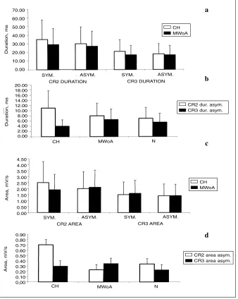
in CH patients in comparison with both MWoA and normal subjects (Fig. 1a,b, Fig. 2a,b). The R2 area was also increased in most CH patients on the painful side, with a sig-nificant asymmetry between the two sides, in comparison with both control subjects and migraine sufferers (Table 2 Fig. 1c,d). The R3 duration and area and the respective asymmetry index were increased only in a few CH and MWoA patients (Table 2). The mean values were like among the groups (Fig. 1). In CH patients, the CR2 response duration and area were slightly but not significantly increased in comparison with migraine sufferers when the painful side was stimulated. The CR2 by the non-symptomatic side stim-ulation were like in the patients group (Fig. 3).
In CH group, the R2 duration on the symptomatic side and the R2 duration asymmetry were not correlated with the interval from the last attack, (R2 duration vs. interval from the
from the last attack, r = –0.3456). In patient groups, the time from the last attack showed no significant correlation with the R3th on both the symptomatic and non-symptomatic sides.
Discussion
The results of the study first showed in chronic CH patients an increase in duration and area of the blink reflex R2 response on the painful side during the pain-free interval. The R2 response is mediated by polysynaptic interneuronal nets of the bulbar lateral reticular formation and corresponds to the objectively observed blink of the lids. Its duration increase in CH could suggest unilateral involvement of the trigeminal reflex circuits, not found in migraine without
70.00
60.00
50.00
40.00
30.00
20.00
10.00
0.00
0.90 0.80 0.70 0.60 0.50 0.40 0.30 0.20 0.10 0,00
Duration, ms
Duration, ms
Area, mV/s
Area, mV/s
20.00 18.00 16.00 14.00 12.00 10.00 8.00 6.00 4.00 2.00 0.00
4.50 4.00 3.50 3.00 2.50 2.00 1.50 1.00 0.50 0.00
SYM. ASYM.
CR3 DURATION
SYM. ASYM.
CR2 DURATION
CH MWoA N
CH MWoA N
a
b
c
d
SYM. ASYM.
CR3 AREA
SYM. ASYM.
CR2 AREA
Fig 3 Mean values (SD) of the crossed R2 (CR2) and R3 (CR3) responses dura-tion (a) and area (c) elicited by the stim-ulation of the symptomatic side (sym) and of the pain free side (asym.) in clus-ter headache patients (CH, n=10), migraine patients (MWoA; n=19) and controls (N; n=18). The crossed R2 by the painful side stimulation was slightly and significantly enhanced in the CH patients. The interside asymmetry of the CR2 and CR3 duration and area were like among groups (b, d). The electrical intensity was 5 x perceptive threshold
CH MWoA
CR2 dur. asym. CR3 dur. asym.
CH MWoA
aura patients suffering from strictly unilateral headache. This type of abnormality was unsteadily described in previ-ous studies, probably for the variability of clinical condi-tions. The R2 amplitude and duration increase found by Formisano et al. [3] on the painful side during the cluster attack restored during the pain-free phase after an undeter-mined time from the last CH bout. Our findings about R2 were recorded in the pain-free intervals of chronic CH and could agree with a possible persistence of abnormalities occurring during the attack. No significant modification was found in our series about the R2th. An increase in R2 thresh-old was previously observed on the symptomatic side in CH patients after an indefinite interval from the last bout and during prophylactic therapy [2, 4], so it could be a persis-tence of an electrophysiological abnormality occurring dur-ing the attack or an effect of drug assumption. In the migraine group no R2 abnormality was detectable during the pain-free periods. In a previous study, the R2 duration and threshold appeared also unmodified during migraine attack in comparison with pain-free interval [9]. In CH patients, the unilateral facilitation of the trigeminal connec-tion to the facial nerve by the activaconnec-tion of the trigemino-vascular reflex, causing the parasympathetic effects during the attacks [1], could be responsible for the unilateral increase of the direct R2 response on the symptomatic side, probably an abnormality persisting in the headache-free phase. The activation of this reflex, evident by the VIP lev-els increase, was shown during the attack only in a minori-ty of migraine patients, suffering from mild symptoms of autonomic activation [13]. According to these findings, in our series only one migraine patient showed an increase of R2 duration on the symptomatic side. In CH patients, the occurrence of a unilateral dysfunction of the interneural trigeminofacial connecting circuits, probably persisting after the last CH bout, could be suggested by the following findings: the prevalence of the direct R2 response on the symptomatic side with a slight and not significant increase of the crossed R2 elicited by the painful side stimulation and normal area and duration of the crossed R2 by the pain-free side stimulation. The reason for unilateral facilitation of the R2 response, which nociceptive quality was denied for the evidence of a selective activation by the A-beta fibers [14], remains to be clarified. In most CH patients, the early appearance of the R3 component was evident on both sides, like in MWoA patients, in comparison with normal subjects. The R3 component of the blink reflex was first described by Penders and Delwaide [15] as a reflex with a latency around
75–90 ms, produced symmetrically in both orbicular oculi muscles and elicited by stimulation anywhere on the face. Rossi et al. [12, 16] suggested the nociceptive quality of the reflex, with a threshold always higher than the R2 one and around the pain sensation, a slow recovery cycle and a strong inhibition by focusing of the attention. This could be interpreted as a defensive reflex reaction to painful stimuli that increases and prolongs the R2 response in order to bet-ter protect the eyes during potentially dangerous events before the onset of the voluntary contraction of the eyelids. Though the anatomic basis of the R3 component is still unknown [14], its appearance after low intensity and poten-tially not dangerous stimuli, could be interpreted as an expression of a possible primary dysfunction of the trigem-inal reflex circuits probably caused by a failure of central control on the brainstem neuronal networks. This abnormal trigeminal reflex behaviour could be a sign of a basic dys-function predisposing to both types of headaches. In his widely discussed pathogenetic theory of migraine, Lance [17] suggested the involvement of raphe dorsalis nucleus, locus coeruleus, raphe magnus nucleus, and the periaque-ductal grey matter brainstem structures in migraine attacks. The latter structures are implicated in the inhibition, under the cortical modulation, of other trigeminal reflex circuits, such as the exteroceptive suppression of temporalis muscu-lar activity, applied by Schoenen [18] in primary headaches; their involvement in the control of the R3 blink reflex response might also not be excluded. The R3 abnormalities observed in our series suggest that cluster headache and migraine might be different clinical manifestations of the same central neuronal circuit dysfunction [19]. Our findings concur with those of a recent study [5], in which R2 elicit-ed after pairelicit-ed supraorbital stimuli recoverelicit-ed more rapidly in CH patients on the symptomatic side, while R2 recovery by index stimulation was bilaterally faster in patients com-pared with controls. Even in that study, CH patients showed two types of BR abnormalities, the former correlated with the side of pain and probably was caused by a unilateral spinal trigeminal nucleus sensitisation; the latter was proba-bly due to a dysfunction of the reticular nuclei.
Taken together, our BR findings seem to confirm the cen-tral genesis of CH, the R3 abnormalities suggesting a basic dysfunction of the central control on the trigeminal nocicep-tive circuits, that could predispose to both migraine and CH, and the specific involvement of the R2 component on the side of pain a selective unilateral facilitation of the trigeminal-facial connections occurring during the CH attack.
References
1. Goadbsy PJ, Edvinsson LH (1994) An in vivo evidence for trigeminovascular activation in cluster headache. Brain 117:427–434
2. Pavesi G, Granella F, Brambilla S, Medici D, Mancia D, Manzoni GC (1987) Blink reflex in cluster
headache: evidence of a trigeminal sys-tem dysfunction. Cephalalgia
7(6):100–102
3. Formisano R, Cerbo R, Ricci M, Agostino R, Cesarino F, Cruccu G, Agnoli A (1987) Blink reflex in cluster headache. Cephalalgia 7(6):353–354 4. Raudino F (1990) The blink reflex in
(1997) Inhibition of the blink reflex R2 component after supraorbital and index finger stimulation is reduced in cluster headache: an indication for both seg-mental and suprasegseg-mental dysfunc-tion? Pain 71:81–88
6. Bank J, Bense E, Kiraly C (1992) The blink reflex in migraine. Cephalalgia 12(5):289–292
7. Sand T, Zwart JA (1994) The blink reflex in chronic tension type headache, migraine, and cervicogenic headache. Cephalalgia 14(6):447–450 8. de Tommaso M, Guido M, Libro G,
Sciruicchio V, Puca F (2000) The three responses of the blink reflex in adult and juvenile migraine. Acta Neurol Belg 100:96–102
9. de Tommaso M, Guido M, Libro G, Sciruicchio V, Puca F (2000) Zolmitriptan reverses blink reflex changes induced during the migraine attack in humans. Neurosci Lett 289:57–60
and clinical aspects of cluster
headache: relation with the migrainous syndrome. Ital J Neurol Sci 20:S4–S6 11. Headache Classification Committee of
the International Headache Society (1988) Classification and diagnostic criteria for headache disorders, cranial neuralgias and facial pain. Cephalalgia 8[Suppl 7]:1–96
12. Rossi B, Vignocchi MG (1993) Methodological considerations of the use of the blink reflex R3 component in the assessment of pain in man. Ital J Neurol Sci 14:217–224
13. Goadbsy PJ, Edvinsson L, Ekman R (1990) Vasoactive peptide release in the extracerebral circulation of humans during migraine headache. Ann Neurol 28:183–187
14. Cruccu G, Ferracuti S, Leardi MG, Fabbri A, Manfredi M (1991) Nociceptive quality of the orbicularis oculi reflexes evaluated by distinct opiate and benzodiazepine induced changes in man. Brain Res 556:209–217
Physiologic approach to the human blink reflex. In: Desmedt JE (ed) New developments in electromyography and clinical neurophysiology, vol. 3. Karger, Basel, pp 649–657
16. Rossi B, Risaliti R, Rossi A (1989) The R3 component of the blink reflex in man: a reflex response induced by acti-vation of high threshold cutaneous afferents. Electroencephalogr Clin Neurophysiol 73:334–340
17. Lance JW (1993) Current concepts of migraine pathogenesis. Neurology 43(3):S11–S15
18. Schoenen J (1993) Exteroceptive sup-pression of temporalis muscle activity in patients with chronic headache and in normal volunteers: methodology, clinical and pathophysiological rele-vance. Headache 33:3–17