Evaluation of the performance of parallel-hole collimator for high
resolution small animal SPECT: A Monte Carlo study
Alireza Sadremomtaz and Zeinab Telikani
Department of Physics, Faculty of Science, University of Guilan, Rasht, Iran
(Received 24 April 2016, Revised 20 May 2016, Accepted 25 May 2016)
ABSTRACT
Introduction: Image quality and accuracy of in vivo activity quantification in SPECT are affected by collimator penetration and scatter components, especially in high energy imaging. These phenomena highly depend on the collimator characteristic and photon energy. The presence of penetrated and scattered photons from collimator in SPECT images degrades spatial resolution, contrast and image quality. Knowledge of penetration and scatter distribution is essential for optimization of collimator design and development of reconstruction algorithms.The aim of this study to survey the collimator performance of the newly developed HiReSPECT dual head gamma camera with pixelated array CsI(Na).
Methods:We modeled the HiReSPECT, by using SIMIND Monte Carlo simulation code. The contribution of geometric, scatter and penetration components were quantitatively calculated for the different energy sources. Then we compared these results with simulation results of another small animal SPECT with compact pixelated array CsI(Tl) detector.
Results: The simulated System spatial resolution and energy resolution of the HiReSPECT at 140keV respectively are 1.9mm and 29.72 keV (21.23%) FWHM at 2.5cm distance from detector surface also Geometric, penetration, and scatter at 140keV for the HiReSPECT collimator are 96.42%, 2.22%, 1.30%, respectively. Similarly, geometric, penetration, and scatter at 159keV and 245keV for this system collimator are (95.24%, 3.08%, 1.68%) and (87.21%, 8.10%, 4.69%), respectively.
Conclusion: The results verified that the magnitude of these components depend on collimator geometric structure and photons energy. The measured performances indicated that the HiReSPECT scanner is well suited for preclinical molecular imaging research and provide high resolution for small animal imaging.
Key words:SPECT; Compact pixelated gamma camera; Septal penetration; Scatter; Monte Carlo simulation
Iran J Nucl Med 2016;24(2):136-143 Published: July, 2016
http://irjnm.tums.ac.ir
Corresponding author:Dr.Alireza Sadremomtaz, Department of Physics, Faculty of Science, University of Guilan, Rasht, Iran. E-mail: [email protected]
O
ri
g
in
a
l
A
rti
c
Ir
a
n
J
N
u
c
l
M
e
d
2
0
1
6
,
V
o
l
2
4
,
N
o
2
(
S
e
ri
a
l
N
o
4
6
)
h
tt
p
:/
/i
rj
n
m
.t
u
m
s
.a
c
.i
r
J
u
ly
,
2
0
1
6
137 INTRODUCTION
The collimator is an important component in determining reconstructed SPECT image quality [1]. Photons can be classified after leaving the patient, according to their history in the collimator, as belonging to one of several components of an image: the geometric component (passed through a collimator hole), the penetration component ( passed through septa without attenuation), or the scatter component ( scattered at least once in the septa). In addition, photons that are absorbed in the septa collimator can produce a characteristic X-ray component as a result of photoelectric interaction [2]. Only first event provides correct positional information. Considerable effort was expended in the design of gamma cameras to reduce or eliminate the detection of the others events [3-5]. The magnitude of penetrated and scattered photons strongly depends on the energy of photons, object under study and collimator design parameters. Knowledge of penetration and scatter distribution is essential for optimization of collimator design, selection of imaging protocols and development of optimum correction algorithms. Image quality and accuracy of in vivo activity quantification in SPECT are affected by penetration and scatter components of the collimator, particularly in high energy imaging [6]. The presence of high levels of penetration and scatter components in the projection data complicate the collimator performance and potentially degrade quantitative accuracy. In addition to, image quality depends on sensitivity and resolution of the collimator-detector system. The presence of penetrated and scattered photons from collimator body in SPECT images degrades spatial resolution, contrast and quantification. As well as, image quality and quantification accuracy are affected by these factors. Absolute quantification depends on information of the sensitivity of the camera collimator system in counts per second per MBq. Sensitivity is usually assumed to be independent of distance from the collimator [7]. While the geometric efficiency of a parallel-hole collimator is relatively easy to calculate analytically and is independent of the distance from the collimator but penetration and scatter contributions are difficult to calculate analytically and depend on the source distance. Thus the investigation of the penetration and scatter components of the collimator response function is very important for the evaluation and interpretation of SPECT images [8].
It is possible to track and record the life history of the individual photon originating from the source that ultimately deposits its complete energy inside the crystal using Monte Carlo Simulation. Therefore, with the help of Monte Carlo Simulation technique,
accurate assessment of the geometric, penetration and scatter contribution inside the photopeak window can be made [9]. In this study, we estimate the geometric, penetration, and scatter components for parallel-hole collimator of animal SPECT using Simulation of Imaging Nuclear Detectors (SIMIND) Monte Carlo Simulation code.
Animal models of human diseases are widely used in the field of drug development and investigating potential therapies and gene research in the preclinical phase [10]. Over the recent years, animal SPECT systems have been developed in order to achieve high-resolution and high-sensitivity systems. Several small-animal SPECT systems with compact high resolution gamma camera consisting of a pixelated crystal array coupled to position- sensitive photomultiplier tubes (PSPMTs) have been developed in different groups and are available commercially [11-13].
In this paper, we intend to survey the collimator performance of the newly developed the HiReSPECT scanner. So we model the HiReSPECT, using Monte Carlo code and evaluate the geometric, scattered and septal penetration components. In order to investigate effective factors on these components, we compare these results with simulation results of another small animal SPECT with different geometric configuration.
METHODS
Detection system description
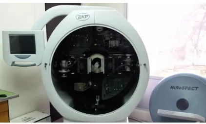
The HiReSPECT is a preclinical dedicated SPECT scanner with dual head has been developed in Institute for Advanced Medical Technologies of Tehran University of Medical Sciences, Iran. This imaging system was installed in Guilan University, Iran, as illustrated in Figure 1, was used in this study and also for the Monte Carlo simulations.
Ir
a
n
J
N
u
c
l
M
e
d
2
0
1
6
,
V
o
l
2
4
,
N
o
2
(
S
e
ri
a
l
N
o
4
6
)
h
tt
p
:/
/i
rj
n
m
.t
u
m
s
.a
c
.i
r
J
u
ly
,
2
0
1
6
138 Two heads of this scanner positioned at 180 degrees
angular distance from each other. Each head consists of 50×100 mm2 pixelated scintillator array of 1×1×5 mm3 CsI(Na) crystal tightly attached to two H8500 (Hamamatsu photonic Co., Hamamatsu City, Japan) position sensitive PMTs (PSPMT). The crystal enclosed in 50 µm-thick aluminum and also a 3mm thick glass window. Each head is equipped with a high resolution hexagonal parallel-hole collimator with an active collimating area of 108×56 mm2 [14-17].
Other imaging system is a compact gamma camera that developed by Loudos et al consisting of a pixelated CsI(Tl) scintillator with 3mm thickness that coupled to a PSPMT Hamamatsu R2486. The crystal array is 46 mm in diameter and is made of square pixels, covering the whole circular section of the crystal. Each pixel is separated from the others by a 250 µm-thick diffusive white layer (epoxy). The maximum number of pixels along the diameter of the crystal array is 41. The gamma camera is equipped with a removable low energy high resolution collimator with a 1.12 mm flat-to-flat distance of the hexagonal parallel holes. The whole detection head is surrounded with a 5 mm thick lead shielding [18, 19].
Monte Carlo simulation
The SIMIND dedicated Monte Carlo simulation code has been developed by Professor Michael Ljungberg, Medical Radiation Physics Department of Clinical Sciences, and Lund University, Sweden. The code has been extensively tested by different research groups as a tool for modeling of gamma camera and collimator design [20-23]. The SIMIND system has two main programs, named CHANGE and SIMIND. The CHANGE program provides a way of defining the system parameters to be simulated and writing data to external data files. The actual MC simulation is made by the program SIMIND that reads input files created by CHANGE and outputs results to screen or to different data files. The CHANGE program enables the user to easily define the desired imaging system. Also CHANGE contains a series of menus that prompt the user to input specific parameters to the description of the system. These parameters are then written to a data file used in SIMIND [24]. This code can also accurately simulate all interaction of photons in collimator. In addition SIMIND be able to separate the geometric, penetration and scatter components of detected photons in the interest energy window.
In this study, we modeled the HiReSPECT dual head gamma camera and the Loudos’s scanner with their dedicated LEHR collimator accurately by using the SIMIND Monte Carlo code. (1×10⁹) Photons with energy window setting at 20% were considered. The energy spectra create by SIMIND contains a
spectrum of 512 channels. The pixel size in the simulated planar source images is 0.3 cm, and they were stored in 128 × 128 matrix. The SIMIND code created a binary matrix image having float values (real *4). The simulations were set up in such a way that during each simulation gamma photons must impinge on whole camera surface, and at the end of simulation SIMIND writes the value of each component (geometric, penetration, and scatter) and images in separate files. The energy spectrum was displayed by using Origin8.5.1 program. The gamma camera characteristics are shown in Table 1.
Table 1:The design parameters of collimator and gamma camera characteristic.
HiReSPECT Loudos scanner
Hole length (mm) 34 27.5
Septal thickness (mm) 0.2 0.4625
Hole diameter (mm) 1.2 1.12
Crystal Scintillator CsI(Na) CsI(Tl)
Crystal thickness (mm) 5 3
Energy resolution 20% 15.6%
Simulation to characterize geometric, scatter and penetration components
Point Source in air- we simulated a point source with
37MBq activity in air, 2.5cm away from the detector surface. In order to survey the collimator response as function of photons energy, we do simulations with different sources with different energy.
Line source in air- a 99mTc line source with 1.12mm
inner diameter and 75mm length with 37MBq activity in air, 2.5cm away from the detector surface was used for system spatial resolution simulation. (1×10⁹) photons with energy window setting at 20% are considered. We did simulations with different energy sources.
The impact of different components in projection data were investigated using analysis of these sources planar image.
RESULTS
Ir
a
n
J
N
u
c
l
M
e
d
2
0
1
6
,
V
o
l
2
4
,
N
o
2
(
S
e
ri
a
l
N
o
4
6
)
h
tt
p
:/
/i
rj
n
m
.t
u
m
s
.a
c
.i
r
J
u
ly
,
2
0
1
6
139
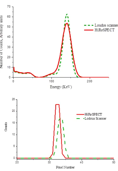
Fig 2. (a) Comparison between simulated Energy spectra of a
99m
Tc point source in air placed at 10cm away from the detector surface for HiReSPECT and Loudos Scanner (above). (b) Comparison betweensimulated line spread function of a 99m
Tc point source in air placed at 10cm from detector surface for HiReSPECT and Loudos Scanner (below).
Figure 2b shows the Line Spread Function of these two imaging systems. System spatial resolution of the HiReSPECT and the Loudos’s scanner at 2.5cm distance from detector surface are 1.9mm and 2.4mm respectively. These simulation results of HiReSPECT are good agreement with experimental result [15]. In order to survey the collimator response as function of the photons energy, we simulate different radionuclide point sources in air. Variation process of geometric, scatter and penetration components as function of energy for the HiReSPECT collimator is shown in Figure 3.
As shown in Figure 4, simulated energy spectra of different point source in air in 20% of photopeak window placed at 2.5cm away from detector surface. As shown in Figure 5 by increasing the energy, system spatial resolution decreases so image quality and quantification accuracy are affected by these changes.
The Contribution of the geometric, septal penetration and scattering component in parallel-hole collimator (LEHR) of two imaging system for radioactive sources using Monte Carlo Simulation are given in Table 2.
Fig 3. Variation of geometric, penetration and scatter components in collimator as function of energy for HiReSPECT system.
Fig 4. Comparison between simulated Energy spectra of different point sources in air placed at 10cm away from the detector surface in HiReSPECT.
Ir
a
n
J
N
u
c
l
M
e
d
2
0
1
6
,
V
o
l
2
4
,
N
o
2
(
S
e
ri
a
l
N
o
4
6
)
h
tt
p
:/
/i
rj
n
m
.t
u
m
s
.a
c
.i
r
J
u
ly
,
2
0
1
6
140
Table 2: Comparison of the simulation results in the two different imaging systems for the different radionuclides.
Imaging system Detector hits (absolute number of photons)
Geometric Collimation
(%)
Penetration (%)
Scatter in collimator
(%)
Sensitivity (cps/MBq)
99m
Tc (140 KeV) HiReSPECT 1720365 96.42 2.22 1.30 33.47
Loudos scanner 1289252 95.75 2.92 1.33 32.59
123
I ( 159 KeV) HiReSPECT 1755350 95.24 3.08 1.68 27.52
Loudos scanner 1321033 94.49 3.96 1.55 26.57
111In (245 KeV ) HiReSPECT 2076382 87.21 8.10 4.69 16.80
Loudos scanner 1559963 86.59 9.91 3.49 13.78
As shown in Table 2, the value of geometric component and sensitivity for 99mTc point source in the HiReSPECT are 96.42% and 33.47 CPS/MBq and for the Loudos’s system are 95.75% and 32.59 CPS/MBq, respectively.
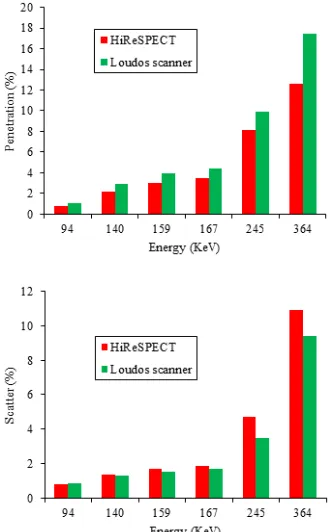
In this article we calculate and compare Contribution value of penetrated and scattered components as function of energy for a point source in the HiReSPECT and the Loudos’s scanner with different septa thickness and length collimator. The results simulations are seen in Figure 6.
Fig 6. (a) Comparison of penetrated photons as a function of energy for a point source in air for HiReSPECT and Loudos scanner with different collimator thickness (above). (b) Comparison of scatter photons as a function of energy for a point source in air for HiReSPECT and Loudos scanner with different septa thickness (below).
DISCUSSION
We evaluated the performance of parallel-hole collimator (LEHR) of two imaging systems for radioactive sources by using Monte Carlo Simulation. A most useful property of a CsI( doped with Tl) is its variable decay time for various exciting particles but CsI( doped with Na) has relatively slow decay time so this is disadvantages of CsI( doped with Na) [25]. As shown in Figure 2a when we compare energy spectra of the HiReSPECT and the Loudos’s scanner with each other, we observe that due to the lack of uniformity of the count rate, height of photopeak in these two imaging systems is not identical. According to the contribution of penetration and scatter components in projection data, the shape of Compton edge and Compton region are different in two imaging systems and as it can be seen the contribution of septal penetration and scatter in the HiReSPECT less than the Loudos’s scanner so Compton region in the Loudos’s scanner deeper than in the HiReSPECT.
The general effect of scattering is the addition of events in the lower energy region of spectrum. This is a result of the registration of scatter events. These events lead to a loss of contrast and resolution in the images. On the other hand these phenomena cause to decrease the count rate. If the scattered events are included in the image, the image will be a false representation of the radionuclide source within the patient. Scattering correction is most often made either in the energy domain where scatter in the photopeak energy window is modeled by collecting data in additional energy windows or by using analytical methods that model the scatter on photopeak data directly [26].
Ir
a
n
J
N
u
c
l
M
e
d
2
0
1
6
,
V
o
l
2
4
,
N
o
2
(
S
e
ri
a
l
N
o
4
6
)
h
tt
p
:/
/i
rj
n
m
.t
u
m
s
.a
c
.i
r
J
u
ly
,
2
0
1
6
141 As seen in Figure 3 and 4, it is clear that by
increasing the energy of photons the contribution of scattered and penetrated photons increase while the contribution of geometric photons decrease. These phenomena affect the pulse height distribution of different radionuclide sources. In the radionuclide sources emit photons with high energy, because of increasing of septal penetration and scatter component, the photopeak become broadened and scattering peaks are more obvious.
For example, due to high level of scattering in 131I with 364 keV energy, broadening response function is very much. Broadening energy response function causes to loss of energy resolution so using radionuclides with high energy is not suitable in low energy collimator particular in preclinical research. It should be emphasized that although the geometric component in projections is independent from the energy of photons but decreasing the geometric components in Figure 3 is due to the decreasing the interaction with crystal with increasing the energy of photons [25].
The two main factors that determine the spatial resolution of a system are (i) the collimator design and (ii) the accuracy with which scintillation events can be localized, which is limited by statistical variation in the number of scintillation photons produced. This gives rise to variations in the PM-tube positioning signals. The effects of energy and source location on gamma camera intrinsic and extrinsic spatial resolution have been studied experimentally, SIMIND and GATE Monte Carlo simulations [27]. In this article we studied the effect of energy on spatial resolution of the HiReSPECT by using SIMIND code.
Also by increasing in the energy of gamma photons the value of geometric component has decreased as result by reduction of the passed photons number from collimator holes, the efficiency is decreased so when we select a collimator, it is necessary to consider the energy of gamma photons. It is advised not to use LEHR collimator for high energy radioisotopes imaging [9]. On the other hand simulated components are different for these two imaging systems, because geometric characteristic of collimators and crystals in these two imaging systems are different.
The collimator septa prevent photons from penetrating from one hole to another; this depends on the relationship between photon energy and the thickness of the metal septa separating the holes. Relatively thin septa are adequate for low energy photons. The advantage of thin septa is that holes can be located in a given area and this result in a higher sensitivity [28]. The influence of septa thickness, hole size and hole length of collimator on image quality have been studied by MCNP code [29].
Septa thickness of these two imaging systems is different. As shown in Figure 6a, the simulated values of penetrated components in these systems are not identical. The HiReSPECT with 0.2mm septa thickness has thinner septa than the Loudos’s scanner with 0.46mm septa thickness so more number of photons must pass through septa without attenuation but here this not happen, because collimator length of the HiReSPECT is taller than one so the simulated penetrated component of the HiReSPECT is less than the Loudos’s system. With increasing photons energy, the amounts of this value are increased. In Figure 6b can be observed the impact of different energy of photons on the scattering component in the HiReSPECT and the Loudos’s Scanner with different thickness of collimator.
The collimator length of the HiReSPECT camera is 34mm and collimator thickness of the Loudos’s camera is 27.5mm.The amounts of scattering in longer collimator are more than shorter one while increasing energy causes to increase this component. The radionuclides that emit photons with high energy have a higher probability of scattering or penetrating the lead septa of collimator than being absorbed [30]. The presence of scatter photons in SPECT images degrades contrast, spatial resolution and quantification. In addition, image quality and quantification accuracy are affected by septal penetration [31].The results in this study are in good agreement with the pervious publications [6, 15, 17, 21, 32].
According to the obtained results from this article, the HiReSPECT is suitable for preclinical molecular imaging research because the HiReSPECT has better spatial resolution than Loudos’s scanner so it can provide high system spatial resolution for millimeter structure of small animal imaging.
CONCLUSION
Ir
a
n
J
N
u
c
l
M
e
d
2
0
1
6
,
V
o
l
2
4
,
N
o
2
(
S
e
ri
a
l
N
o
4
6
)
h
tt
p
:/
/i
rj
n
m
.t
u
m
s
.a
c
.i
r
J
u
ly
,
2
0
1
6
142 System spatial resolution and energy resolution of the
HiReSPECT at 140keV respectively are 1.9mm and 29.72 keV (21.23%) FWHM at 2.5cm distance away from detector surface. Validation of this simulation results was verified by experimental result. In order to survey the performance of the newly developed HiReSPECT camera, the geometric, penetration and scatter components for the HiReSPECT collimator at different energy of photons were modeled by using the SIMIND Monte Carlo code. The results of this study showed that by increasing the energy of photons, the penetration and scatter components increase while the geometric components decrease so a LEHR collimator do not well work when a high- energy SPECT scan is desired. Also for investigating of the effective factor on these components, we do this simulation for other gamma camera. Comparing of simulation results of two imaging systems verify that these components depend on the collimator geometric structure and the energy of photons.
REFERENCES
1. Lu Y, Chen L, Gindi G. Collimator performance evaluation for In-111 SPECT using a detection/localization task. Phys Med Biol. 2014;59(3):679-96.
2. De Vries DJ, Moore S. Approximation of Approximation of hexagonal holes by square holes in Monte Carlo simulation of gamma-camera collimation. IEEE Trans Nucl Sci. 2002;49(5):2186-95.
3. Polo IO. Evaluation of the scattered radiation components produced in a gamma camera using Monte Carlo method. Braz J Biom Eng. 2014;30(2):179-188.
4. Ljungberg M, Larsson A, Johansson L. A New Collimator simulation in SIMIND Based on the delta-scattering technique. IEEE Trans Nucl Sci. 2005; 52(5):1370-75.
5. Sundin K, Ljungberg M. SIMIND based pinhole imaging: development and validation IEEE Nucl Sci Symp Conf Rec. 2007;5:3998-4005.
6. Shafaei M, Ay MR, Sardari D, Dehestani N, Zaidi H. Monte Carlo assessment of geometric, scatter and septal penetration components in DST-XLi HEGP collimator. IFMBE Proceedings. 2008; 22:2479–82.
7. Fleming JS, Alaamer AS. Influence of collimator characteristics on quantification in SPECT. J Nucl Med. 1996 Nov;37(11):1832-6.
8. He X, Frey E, Links J, Song X, Tusi B. Comparison of penetration and scatter effects on defect contrast for GE and Siemens LEHR collimators in myocardial perfusion SPECT—A simulation study. IEEE Trans Nucl Sci. 2005;52(5):1359- 64.
9. Pandey AK, Sharma SK, Karunanithi S, Kumar P, Bal C, Kumar R. Characterization of parallel-hole collimator using Monte Carlo Simulation. Indian J Nucl Med. 2015 Apr-Jun;30(2):128-34.
10. Green MV, Seidel J, Vaquero JJ, Jagoda E, Lee I, Eckelman WC. High resolution PET, SPECT and projection imaging in small animals. Comput Med Imaging Graph. 2001 Mar-Apr;25(2):79-86.
11. Xi W, Seidel J, Kakareka JW, Pohida TJ, Milenic DE, Proffitt J, Majewski S, Weisenberger AG, Green MV, Choyke PL. MONICA: a compact, portable dual gamma camera system for mouse whole-body imaging. Nucl Med Biol. 2010 Apr;37(3):245-53.
12. Magota K, Kubo N, Kuge Y, Nishijima K, Zhao S, Tamaki N. Performance characterization of the Inveon preclinical small-animal PET/SPECT/CT system for multimodality imaging. Eur J Nucl Med Mol Imaging. 2011 Apr;38(4):742-52.
13. Weisenberger A, Bradley E, Majewski S, Saha M. Development of a novel radiation imaging detector system for in vivo gene imaging in small animal studies. IEEE Trans Nucl Sci. 1998;45(3):1743–49.
14. Pashazadeh AM, Tanha K, Jafarian-Dehkordi F, Assadi M, Moji V, Zeraatkar N, Ay MR. Experimental evaluation of the performance of HiReSPECT scanner: A high-resolution SPECT system for small animal imaging. Front Biomed technol. 2014;1(3):222-27.
15. Moji V, Zeratkar N, Farahani MH, Aghamiri MR, Sajedi S, Teimourian B, Ghafarian P, Sarkar S, Ay MR. Performance evaluation of a newly developed high-resolution, dual-head animal SPECT system based on the NEMA NU1-2007 standard. J Appl Clin Med Phys. 2014;15(6):4936.
16. Sajedi S, Zeraatkar N, Moji V, Farahani MH, Sarkar S, Arabi H, Teymoorian B, Ghafarian P, Rahmim A, Ay MR. Design and development of a high resolution animal SPECT scanner dedicated for rat and mouse imaging. Nucl Instrum Methods Physic Res. 2014;741:169–76.
17. Zeraatkar N, Sajedi S, Farahani MH, Arabi H, Sarkar S, Ghafarian P, Rahmim A, Ay MR. Resolution-recovery-embedded image reconstruction for a high-resolution animal SPECT system. Phys Med. 2014 Nov;30(7):774-81.
18. Lazaro D, Buvat I, Loudos G, Strul D, Santin G, Giokaris N, Donnarieix D, Maigne L, Spanoudaki V, Styliaris S, Staelens S, Breton V. Validation of the GATE Monte Carlo simulation platform for modelling a CsI(Tl) scintillation camera dedicated to small-animal imaging. Phys Med Biol. 2004 Jan 21;49(2):271-85.
19. Giokaris N, Loudos G, Maintas D, Karabarbounis A, Spanoudaki V, Stiliaris E, Boukis S, Gektin A, Boyarintsev A, Pedash V, Gayshan V. Crystal and collimator optimization studies of a high-resolution γ-camera based on a position sensitive photomultiplier. Nucl Instrum Methods Phys Res, Sect A. 2004;527(1):134–9.
20. Khosravi HR, Sarkar S, Takavar A , Saghari M, Shahriari M. Planar and SPECT Monte Carlo acceleration using a variance reduction technique in I-131 imaging. Int J radiat Res. 2007;4(4):175-82.
21. Zeniya T, Watabe H, Aoi T, Kim KM, Teramoto N, Takeno T, Ohta Y, Hayashi T, Mashino H, Ota T, Yamamoto S, Iida H. Use of a compact pixellated gamma camera for small animal pinhole SPECT imaging. Ann Nucl Med. 2006 Jul;20(6):409-16.
22. Pirayesh Islamian J, Bahreyni Toossi MT, Momennezhad M, Zakavi R, Sadeghi R . Monte Carlo study of the effect of backscatter material thickness on 99mTc source response in single photon emission computed tomography. Iran J Med Phys. 2013;10(1):69-77.
Ir
a
n
J
N
u
c
l
M
e
d
2
0
1
6
,
V
o
l
2
4
,
N
o
2
(
S
e
ri
a
l
N
o
4
6
)
h
tt
p
:/
/i
rj
n
m
.t
u
m
s
.a
c
.i
r
J
u
ly
,
2
0
1
6
143
adding the phantom as an accessory to the program. Iran J Med Phys. 2012;9(2):135-40.
24. Ljungberg M. The SIMIND Monte Carlo program Home Page. http://www2.msf.lu.se/simind.
25. Knoll GF. Radiation detection and measurement. 4nd ed. New York: John Wiley & Sons, Inc; September 2010.
26. Khalil M. Basic sciences of nuclear medicine. New York: Springer-Verlag; 2011.
27. Holstensson M, Partridge M, Buckley SE, Flux GD. The effect of energy and source location on gamma camera intrinsic and extrinsic spatial resolution: an experimental and Monte Carlo study. Phys Med Biol. 2010 Mar 21;55(6):1735-51.
28. Sprawls P. Physical principles in medical imaging. 2nd ed. Madison: Medical Physics Publishing; 2000.
29. Rajaee A, Shahriari M, Kamali Asl A, Hosseini SH. Simulation study of the influence of collimator material on image quality improvement for high energy photons in nuclear medicine using MCNP code. J Theor Appl Phys. 2011;4(4):13-18.
30. Autret D, Bitar A, Ferrer L, Lisbona A, Bardiès M. Monte Carlo modeling of gamma cameras for I-131 imaging in targeted radiotherapy. Cancer Biother Radiopharm. 2005 Feb;20(1):77-84.
31. Dewaraja YK, Ljungberg M, Koral KF. Characterization of scatter and penetration using Monte Carlo simulation in 131I imaging. J Nucl Med. 2000 Jan;41(1):123-30.
32. Pirayesh Islamian J, Bahreyni Toossi M.T, Momennezhad M, Naseri Sh, Ljungberg M. Monte Carlo study of the effect of collimator thickness on Tc-99m source response in single photon emission computed tomography. World J Nucl Med. 2012;11(2):70-74.