Thioperamide-induced Antinociception is Mediated through Endogenous
Opioid System in the Dentate Gyrus of Adult Rats
Emad Khalilzadeh1 Esmaeal Tamaddonfard1*
Amir-Abbas Farshid2 Amir Erfanparast1
1
Department of Basic Sciences, Faculty of Veterinary Medicine, Urmia University, Urmia, Iran
2
Department of Pathobiology, Faculty of Veterinary Medicine, Urmia University, Urmia, Iran
Received: 30 October 2010, Accepted: 29 November 2010
Abstract
The present study investigated the effects of intra-dentate gyrus microinjection of naloxone (an opioid antagonist) and thioperamide (an antagonist of histamine H3 receptors) in the
formalin test in rats. Subcutaneous injection of formalin (50 µl, 2.5 %) in the ventral surface of right hind paw produced a biphasic pattern (first phase: 0-5 min and second phase: 15 - 60 min) of licking/biting and shaking of the injected paw. Intra-dentate gyrus microinjections of thioperamide (2 and 4 µg) significantly (P < 0.05) suppressed the pain responses. Microinjections of naloxone (1, 2 and 4 µg) alone into the dentate gyrus non-significantly increased the intensity of pain. Pretreatment with naloxone (4 µg) significantly (P < 0.05) reversed the antinociceptive effect of thioperamide (4 µg). The results indicated that at the level of the dentate gyrus, blockade of histamine H3 receptors with thioperamide produced an
analgesic effect. This thioperamide-induced antinociception may be mediated through the endogenous opioid system.
Key words: Dentate gyrus, Formalin-induced pain, Naloxone, Thioperamide, Rats
*
Corresponding author:
Esmaeal Tamaddonfard, DVM, DVSc
Department of Basic Sciences, Faculty of Veterinary Medicine, Urmia University, Urmia, Iran E-mail address: etamaddonfard@yahoo.com, e_tamaddonfard@urmia.ac.ir
Veterinary Research Forum
Introduction
Some studies suggest that the dentate gyrus may involve in modulation of pain. Microinjections of acetylcholine and pilocarpine into the dentate gyrus have decreased the discharge frequency of pain-excited neurons, and increased the discharge frequency of pain-inhibited neurons in the sciatic nerve electrical stimulation model of nociception in rats.1 Besides, lidocaine, a local anesthetic, produced analgesia in the formalin test of rats when microinjected into the dentate gyrus.2
The histamine H3 receptors are widely
distributed in the limbic system areas such as the hippocampus, the dentate gyrus and the amygdale.3 In the dentate gyrus, histamine H3 receptors play important
roles in modulation of excitatory synaptic transmission, information flow and memory consolidation.4-6 On the other hand, opioid receptors are expressed in the hippocampal formation (i.e., the hippocampus and the dentate gyrus), and are involved in mediation of hippocampal functions including adult neurogenesis, the action of gonadal hormones, development of neonatal transmitter system and pain.7-9
By intracerebroventricular route of administration of thioperamide, some researchers have suggested an important role of histamine H3 receptors in
modulation of pain.10,11 At the level of the brain, the opioidergic system, through mu (µ), delta (δ), and kappa (κ) receptors, exerts a major role in modulation of pain.12-14
The present study was aimed to investigate the implication of histamine H3
receptor in pain perception by microinjection of a histamine H3 receptor
antagonist, thioperamide, into the dentate gyrus using formalin test in rats. In addition, to identify the mechanism that possibly mediating the effect of thioperamide on pain, we assessed the contribution of the endogenous analgesic opioid system using microinjection of
naloxone prior to thioperamide. Formalin test has been used frequently to study pain mechanisms in laboratory animals and according to these studies a biphasic pattern of pain-related behaviors was produced by subcutaneous injection of small amounts (20–100 µl) of dilute solutions (0.1–10 %) of formalin into the various parts of the body.15,16 The first phase in turn may be attributed to a direct algogenic effect of formalin on the nociceptors and the second phase to release of local inflammatory mediators responsible for sensitization of primary and spinal sensory neurons and subsequent signal transduction into the brain.15-17
Materials and Methods
Animals. Healthy adult male Wistar rats, weighing 300–350 g were used in this study. Rats were maintained in polyethylene cages with food and water available ad libitum in a laboratory with controlled ambient temperature (22 ± 0.5 °C) and under a 12 h light-dark cycle (lights on from 07:00 a.m.). Six rats were used in each experiment. Experiments were performed between 12:00 and 15:00 o’clock. All research and animal care procedures were approved by the Veterinary Ethics Committee of the Faculty of Veterinary Medicine of Urmia University and were performed in accordance with the National Institutes of Health Guide for Care and Use of Laboratory Animals.
Drugs. Drugs used in the present study included thioperamide maleate (Sigma-Aldrich) and naloxone dihydrochloride (Sigma–Aldrich). All drugs were dissolved in sterile normal saline 30 min before intra-dentate gyrus microinjection.
Surgical procedure. To deliver the
bregma, 2 mm left and right sides of the midline and 3.6 mm below the top of the skull.18 The cannulas were then fixed to the skull using three screws and dental acrylic (Acropars, Tehran, Iran). At least 14 days were allowed for recovery from the surgery.
Intra-dentate gyrus microinjection.
Intra-dentate gyrus microinjections of normal saline (control), thioperamide (1, 2 and 4 µg) and naloxone (1, 2 and 4 µg) were performed using a 5 µl Hamilton syringe. The volume of 0.5 µl of each solution was injected slowly into each dentate gyrus over a period of 1 min. Intra-dentate gyrus microinjections of thioperamide and naloxone were performed 10 and 5 min before intra-plantar injection of formalin, respectively.
Formalin test. The formalin test was applied as follows: Fifty microlitres of 2 . 5 % f o r m a l i n w a s i n j e c t e d subcutaneously into the ventral surface of right hind paw using a 29-gauge injection needle.19-22 Nociceptive behaviors including licking/biting and shaking of the injected paw were observed every 5 min for 1 h. In the present study, data collected between 0-5 min after formalin injection represented the first (early) phase and data collected between 15-60 min after injection of formalin represented the second (late) phase of pain.15, 20, 23
Cannula verification. At the end of each experiment, 0.25 µl methylene blue was injected into the each dentate gyrus. The animals were killed with the high dose ether, and perfused intracardially with physiological saline followed by 10 % formalin solution. Brains were removed and placed in the formalin (10%) solution. At least 3 days later, the brains were sectioned coronally (50-100 µm), and viewed under a loupe to localize the injection site (Fig. 1).18
Statistical analysis. To evaluate
significance differences among intra-dentate gyrus treated groups, one-way analysis of variance (ANOVA) and Duncanۥ s test were applied. In figures, all
values are expressed as the mean ± SEM. A value of P < 0.05 was considered statistically significant.
Results
The placements of the tip of the cannulas in the dentate gyrus of rats are shown in Fig. 1. The rat brain section was modified from the atlas of Paxinos and Watson18 (Fig. 1A).
The location of the cannula tip placements in the dentate gyrus was confirmed with intra-dentate gyrus injection of methylene blue (Fig. 1B).
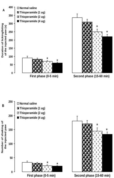
Fig. 2 shows the effects of intra-dentate gyrus microinjection of thioperamide on the formalin-induced nociceptive behaviors. Intra-dentate gyrus microinjection of thioperamide at dose of 2 and 4 µg, but not at a dose of 1 µg, significantly decreased the duration of licking/biting of the formalin-injected paw in the first (F(3,20) = 3.319, P < 0.05 ) and
second (F(3,20) = 9.223, P < 0.05) phases
(Fig. 2A).
The number of shakes of the formalin-injected paw was significantly decreased in the first (F(3,20) = 2.942, P < 0.05) and
second (F(3,20) = 6.483, P < 0.05) phases
when 2 and 4 µg, but not 1 µg, of thioperamide were microinjected into the dentate gyrus (Fig. 2B).
Fig. 3 shows the effects of intra-dentate gyrus microinjection of naloxone on the formalin-induced nociceptive behaviors. Microinjections of naloxone (1, 2 and 4 µg) non-significantly increased the first (F(3,20) = 0.882, P > 0.05) and second
(F(3,20) = 0.642, P > 0.05) phases of
formalin-induced licking/biting of the injected paw (Fig. 3A).
Naloxone at doses of 1, 2 and 4 µg also non-significantly increased the number of shakes in the first (F(3,20) = 0.882, P >
0.05) and second (F(3,20) = 0.642, P > 0.05)
Fig 1. Verified section was taken from the atlas of Paxinos and Watson18 (A). The black circles represent the cannulas tip placements. Location of the injection cannula tips in the dentate gyrus of all rats included in the data analysis (B). DG: dentate gyrus.18
0 50 100 150 200 250 300 350 400
Duration of lic
king/biting
of the
inje
cted pa
w (s)
Normal saline Thioperamide (1 ug) Thioperamide (2 ug) Thioperamide (4 ug)
* *
* *
First phase (0-5 min) Second phase (15-60 min) A
0 50 100 150 200 250
Number of s
haking of
the injecte
d paw
Normal saline Thioperamide (1 ug) Thioperamide (2 ug) Thioperamide (4 ug)
B
* *
* *
First phase (0-5 min) Second phase (15-60 min)
Fig 2. Effects of intra-dentate gyrus microinjection of thioperamide on the formalin-induced licking/biting (A) and shaking (B) of the hind paw in rats. *P < 0.05 as compared with normal saline (control) group, n = 6 rats in each group.
0 50 100 150 200 250 300 350 400
Duration of lic
king/biting
of the
inje
cted pa
w (s)
Normal saline Naloxone (1 ug) Naloxone (2 ug) Naloxone (4 ug)
A
First phase (0-5 min) Second phase (15-60 min)
0 50 100 150 200 250
N
u
m
b
er
o
f sh
akes o
f
th
e i
n
ject
ed
p
aw
Normal saline Naloxone (1 ug) Naloxone (2 ug) Naloxone (4 ug) B
First phase (0-5 min) Second phase (15-60 min)
Fig 3. Effects of intra-dentate gyrus microinjection of naloxone on the formalin-induced licking/biting
(A) and shaking (B) of the hind paw in rats. n = 6 rats in each group.
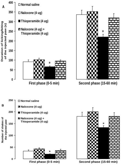
Fig. 4 shows the effects of intra-dentate gyrus microinjection of naloxone on the thioperamide-induced antinociception in the formalin test. Microinjection of naloxone (4 µg) prior to thioperamide (4 µg) significantly reversed the suppressive effect of thioperamide on the licking/biting of the formalin-injected paw in the first (F(3,20) = 4.522, P < 0.05) and second
(F(3,20) = 7.461, P < 0.05) phases (Fig. 4A).
The suppressive effects of intra-dentate gyrus microinjected thioperamide (4 µg) on shaking behavior induced by formalin were significantly inhibited by prior microinjection of naloxone (4 µg) in the first (F(3,20) = 3.396, P < 0.05) and second
0 50 100 150 200 250 300 350 400
Duration of lic
king/biting
of the
inje
cted pa
w (s)
Normal saline
Naloxone (4 ug)
Thioperamide (4 ug)
Naloxone (4 ug) + Thioperamide (4 ug)
*
*
First phase (0-5 min) Second phase (15-60 min) A
0 50 100 150 200 250
Number of s
hake
s of
the injecte
d paw
Normal saline Naloxone (4 ug) Thioperamide (4 ug) Naloxone (4 ug) + Thioperamide (4 ug)
*
*
First phase (0-5 min) Second phase (15-60 min) B
Fig 4. Effects of intra-dentate gyrus microinjection pretreatment with naloxone on the thioperamide-induced antinociception in the formalin-thioperamide-induced licking/biting (A) and shaking (B) of the hind paw in rats. *P < 0.05 as compared with other groups, n = 6 rats in each group.
Discussion
In this study, intra-dentate gyrus microinjection of thioperamide produced an antinociceptive effect in the formalin-induced pain. Histamine H3 receptors act
as pre-synaptic auto-receptors as well as post-synaptic hetero-receptors.24,25 Activation of histamine H3 auto-receptors
by R-α-methylhistamine, immepip and imetit (histamine H3 receptor agonists)
results in the inhibition of histamine synthesis and release from histaminergic neurons.26,27 On the other hand, blockade of histamine H3 auto-receptors with
histamine H3 receptor antagonists
including clobenpropit, ciproxifan and thioperamide can increase the release of histamine from histaminergic endings.26,27 Although the majority of histamine H3
receptors are located in brain,3 histamine H3 receptor mRNA is also found in various
non-brain tissues including skin, stomach,
intestines, brown adipose tissue, dorsal root ganglion and spinal cord.28,29 The evidences taken from acute and chronic pain tests have suggested peripheral, spinal and supraspinal roles for histamine H3
receptor in modulation of pain. Local activation of histamine H3 receptor with
sub-plantar injection of R- α-methylhistamine potentiated the suppressive effect of fentanyl in thermal hyperalgesia induced by sub-plantar injection of Complete Freund’s Adjuvant in mice.30 In addition, administration of immepip and thioperamide to the cholestatic rats increased and decreased tail-flick latencies, respectively.31 It has been reported that activation of histamine H3 receptors by immepip on peripheral and
spinal sites of pain pathways attenuates formalin-induced swelling and flinching.32 Using histamine H3 receptor gene
knockout mice, Mobarakeh et al. (2009)33 reported an inhibitory effect of histamine through its H3 receptors on the
morphine-induced antinociception in hot-plate, tail-flick, paw-withdrawal and formalin tests of nociception at the spinal level. At the supraspinal level, intracerebroventricular injection of thioperamide increased the nociceptive threshold in a rat model of neuropathic pain.11 In contrast, intracerebroventricular injection of thioperamide did not exert any analgesic activities in the tail flick and hot plate tests of nociception in rats.34 However, Malmberg-Aiello et al. (1994).10 reported analgesic and hyperalgesic effects after intracerebroventricular injection of thioperamide and R-α-methylhistamine, respectively, in rats and mice.
frequently used to explore the role of endogenous opioid analgesic system in pain modulation.9, 36-38 Several interactions exist between histamine H3 and opioid
receptors in modulation of pain. Local activation of histamine H3 receptor with
sub-plantar injection of R- α-methylhistamine potentiated the suppressive effect of fentanyl in thermal hyperalgesia induced by sub-plantar injection of Complete Freund’s Adjuvant in mice.30 Subcutaneous injection of naloxone attenuated immepip-induced antinociception in cholestatic rats.31 Using the histamine H3 receptors gene knockout
mice, Mobarakeh et al. (2009)33 reported that histamine, through its H3 receptors,
exerted inhibitory effects on the morphine-induced antinociception at the spinal level. In addition, microinjection of naloxone into periaqueductal gray reversed the antinociceptive effect induced by microinjection of histamine into the same site.39
In the present study, intra-dentate gyrus microinjection of thioperamide, without any significant effect on interphase (data not shown) suppressed the first and the second phases of formalin-induced licking/biting and shaking responses. The first phase of formalin-induced pain may be attributed to a direct algogenic effect of formalin on the nociceptors and the second phase to release of local inflammatory mediators responsible for sensitization of primary and spinal sensory neurons and subsequent signal transduction into the brain.15-17 The interphase of formalin test is under active inhibition of spinal cord mechanisms.40 The pain-related behaviors can be associated with distinct brain structures including spinal, brainstem and cerebrally mediated responses to nociceptive stimulation.41 Regarding the formalin-induced nociceptive behaviors including licking/biting and shaking of the injected paw, it was found that these behaviors are mediated by supraspinal structures.42
In conclusion, the results of the present study indicated that blockade of brain histamine H3 receptor by thioperamide in
the dentate gyrus of the brain could produce an antinociceptive effect. The endogenous opioid analgesic system may be involved in thioperamide-induced antinociception.
Acknowledgments
This work was financially supported by Education Vice Chancellor of Urmia University.
References
1. Jiao R, Yang C, Zhang Y, et al. Cholinergic mechanism involved in the nociceptive modulation of dentate gyrus. Biochem Biophys Res Commun 2009; 379: 975-979.
2. McKenna JE, Melzack R. Analgesia induced by lidocaine microinjection into the dentate gyrus. Pain 1992; 49: 105-112.
3. Pillot C, Heron A, Cochois V, et al. A detailed mapping of the histamine H3
receptor and its gene transcripts in rat brain. Neuroscience 2002; 114: 173-193.
4. Chang M, Saito H, Abe K. Histamine H3-receptor mediated inhibition of
excitatory synaptic transmission in the rat dentate gyrus in vivo. Jpn J Pharmacol 1998; 77: 251-255.
5. Manhan-Vaughan D, Reymann KG, Brown RE. In vivo electrophysiological investigations into the role of histamine in the dentate gyrus of the rat. Neuroscience 1998; 84: 783-790.
6. Foley AG, Prendergats A, Barry C, et al.
H3 receptor antagonism enhances
7. Ding YQ, Kaneko T, Nomura S, et al. Immunohistochemical localization of mu-opioid receptors in the central nervous system of the rat. J comp Neurol 1996; 367: 375-402.
8. Drake CT, Chavkin C, Milner TA. Opioid system in the dentate gyrus. Prog Brain Res 2007; 163: 245-263. 9. Erfanparast A, Tamaddonfard E,
Farshid AA, et al. Antinociceptive effect of morphine microinjections into the dorsal hippocampus in the formalin-induced orofacial pain in rats. Vet Res Forum 2010; 2: 83-89.
10. Malmberg-Aiello P, Lamberti C, Ghelardini C, et al. Role of histamine in rodent antinociception. Br J Pharmacol 1994; 111: 1269-1279.
11. Huang L, Adachi N, Nagaro T, et al. Histaminergic involvement in neuropathic pain produced by partial ligation of the sciatic nerve in rats. Reg Anesth Pain Med 2007; 32: 124-129. 12. Xiong W, Yu LC. Involvement of mu-
and kappa-opioid receptors in morphine-induced antinociception in the nucleus accumbens of rats. Neurosci Lett 2006; 399: 167-70.
13. Duale C, Sierralta F, Dallel R. Analgesia induced by morphine microinjected into the nucleus raphe magnus: effect on tonic pain. Curr Drug Deliv 2007; 4: 181-184.
14. Feng J, Jia N, Han LN, et al. Microinjection of morphine into thalamic nucleus submedicus depresses bee venom-induced inflammatory pain in the rat. J Pharm Pharmacol 2008; 60: 1355-1363.
15. Tjolsen A, Berge OG, Hunskaar S, et al. The formalin test: an evaluation of the method. Pain 1992; 51: 5-17.
16. Raboisson P, Dallel R. The orofacial formalin test. Neurosci Biobehav Rev 2004; 28: 219-226.
17. Porro CA, Cavazzuti M. Spatial and temporal aspects of spinal cord and brainstem activation in the formalin pain model. Prog Neurobiol 1993; 41: 565-607.
18. Paxinos G, Watson C. The rat brain in stereotaxic coordinates. Compact Third Edition, Academic Press, San Diego, USA, 1997.
19. Guidon J, Desroches J, Beaulieu P. The antinociceptive effects of intraplantar injections of 2-arachidonoyl glycerol are mediated by cannabinoid CB2 receptors. Br J
Pharmacol 2007; 150: 693-701.
20. Heughan CE, Sawynok J. The interaction between gabapentin and amitriptyline in the rat formalin test after systemic administration. Anesth Analg 2002; 94: 975-980.
21. Lee OI, Jeong YS. Effects of different concentrations of formalin on paw edema and behaviors in rats. J Korean Med Sci 2002; 17: 81-85.
22. Marcil J, Walczak JS, Guindon J, et al. Antinociceptive effects of tetrodotoxin (TTX) in rodents. Br J Anesth 2006; 96: 761-768.
23. Capone F, Aloisi AM. Refinement of pain evaluation techniques. Ann Ist Super Sanita 2004; 40: 223-229.
24. Arrang JM, Garbarg M, Lancelot JC, et al. Highly potent and selective ligands for histamine H3-receptors. Nature
1987; 327: 117-123.
25. Pollard H, Moreau J, Arrange JM, et al. A detailed authoradiographic mapping of histamine H3 receptors in rat brain areas. Neuroscience 1993; 52: 169-189.
26. Brown RE, Stevens DR, Haas HL. The physiology of brain histamine. Prog Neurobiol 2001; 63: 637-672.
27. Haas HL, Sergeeva OA, Selbach O. Histamine in the nervous system. Physiol Rev 2008; 88: 1183-1241. 28. Cannon KE, Chazot PL, Hann V, et al.
Immunohistichemical localization of histamine H3 receptors in rodent skin,
dorsal root ganglion, superior cervical ganglion and spinal cord: potential antinociceptive targets. Pain 2007a; 129: 76-92.
receptor in the developing CNS and brown fat suggests novel roles for histamine. Mol Cell Neurosci 2003; 24: 614-623.
30. Fernandez-Duenas V, Ciruela F, Gandia J, et al. Histamine H3 receptor activation potentiates peripheral opioid-mediated antinociception: substance P role in peripheral inflammation in mice. Eur J Pharmacol 2010; 638: 72-77.
31. Hasanein P. Histamine H3 receptor modulates nociception in a rat model of cholestasis. Pharmacol Biochem Behav 2010; 96: 312-316.
32. Cannon KE, Leurs R, Hough LB. Activation of peripheral and spinal histamine H3 receptors inhibits
formalin-induced inflammation and nociception, respectively. Pharmacol Biochem Behav 2007b; 88: 122-129. 33. Mobarakeh JI, Takahashi K, Yanai K.
Enhanced morphine-induced antinociception in histamine H3
receptor gene knockout mice. Neuropharmacology 2009; 57: 409-414.
34. Hough LB, Nalwalk JW, Li YB. et al. Novel qualitative structure-activity relationships for the antinociceptive actions of H2 antagonists, H3
antagonists and derivatives. J Pharmacol Exp Ther 1997; 283: 1534-1543.
35. Trescot AM, Datta S, Lee M, et al. Opioid Pharmacology. Pain Physician 2008; 11: 133-153.
36. Tamaddonfard E, Hamzeh-Gooshchi N. Effect of crocin on the morphine-induced antinociception in the formalin test in rats. Phytother Res 2010a; 24: 410-413.
37. Tamaddonfard E, Hamzeh-Gooshchi N. Effects of intraperitoneal and intracerebroventricular injection of crocin on acute corneal pain in rats. Phytother Res 2010b; 24: 1463-1467. 38. Tamaddonfard E, Erfanparast A,
Farshid AA, et al., Interaction between histamine and morphine at the level of
the hippocampus in the formalin-induced orofacial pain in rats. Pharmacol Rep 2011; 63: (Accepted for publication).
39. Thoburn KK, Hough LB, Nalwalk JW, et al. Histamine-induced modulation of nociceptive responses. Pain 1994; 58: 29-37.
40. Henry JL, Yashpal K, Pithcher GM, et al. Physiological evidence that the 'interphase' in formalin test is due to active inhibition. Pain 1999; 82: 57-63. 41. Millan MJ. The induction of pain: an
integrative review. Prog Neurobiol 1999; 57: 1-154.