C A S E R E P O R T
Open Access
A novel
PAX5
rearrangement in
TCF3-PBX1
acute lymphoblastic leukemia: a case report
Thayana Conceição Barbosa
1,2, Bruno Almeida Lopes
1, Caroline Barbieri Blunck
1, Marcela Braga Mansur
1,
Adriana Vanessa Santini Deyl
3, Mariana Emerenciano
1†and Maria S. Pombo-de-Oliveira
2*†Abstract
Background:Chromosome translocations are a hallmark of B-cell precursor acute lymphoblastic leukemia (BCP-ALL). Additional genomic aberrations are also crucial in both BCP-ALL leukemogenesis and treatment management. Herein, we report the phenotypic and molecular cytogenetic characterization of an extremely rare case of BCP-ALL harboring two concomitant leukemia-associated chromosome translocations: t(1;19)(q23;q13.3) and t(9;17)(p13;q11. 2). Of note, we described a new rearrangement between exon 6 ofPAX5and a 17q11.2 region, where intron 3 of SPECC1is located. This rearrangement seems to disruptPAX5similarly to aPAX5deletion. Furthermore, a distinct karyotype between diagnosis and relapse samples was observed, disclosing a complex clonal evolution during leukemia progression.
Case presentation:A 16-year-old boy was admitted febrile with abdominal and joint pain. At clinical investigation, he presented with anemia, splenomegaly, low white blood cell count and 92% lymphoblast. He was diagnosed with pre-B ALL and treated according to high risk GBTLI-ALL2009. Twelve months after complete remission, he developed a relapse in consequence of a high central nervous system and bone marrow infiltration, and unfortunately died.
Conclusions:To our knowledge, this is the first report of a rearrangement betweenPAX5andSPECC1. The presence ofTCF3-PBX1andPAX5-rearrangement at diagnosis and relapse indicates that both might have participated in the malignant transformation disease maintenance and dismal outcome.
Keywords:der(9)t(9;17)(p13;q11.2) translocation,PAX5-SPECC1, Near-triploidy karyotype,TCF3-PBX1
Background
B-cell precursor acute lymphoblastic leukemia (BCP-ALL) is characterized by recurrent chromosomal alterations in-cluding translocations, which result in aberrant fusion genes.TCF3-PBX1is one of these fusions and is found in 3–6% of BCP-ALL patients; moreover, the acquisition of
secondary genomic aberrations, such as loss ofCDKN2A/
B,PAX5 orRB1, is common and crucial to
leukemogen-esis in those cases [1,2].
PAX5, a master regulator of B-cell differentiation, is frequently disrupted by deletions, point mutations and amplifications in BCP-ALL. These disruptions might
result in potentially oncogenic proteins [3, 4]. For
example, PAX5 amplifications (PAX5AMP) were
identi-fied in diagnostic-relapse matched samples of patients who lacked common cytogenetic abnormalities at
re-lapse, indicating that PAX5AMP may be an important
driver of leukemogenesis [5]. Notably,PAX5 can be la-beled a“promiscuous” gene since over 16 partnergenes have been identified in leukemia-associated rearrange-ments. These rearrangements result in fusion genes encoding chimeric proteins that modify PAX5 function and occur in 2–3% of pediatric BCP-ALL patients.
Here, we report an extremely rare case of BCP-ALL harboring two concomitant leukemia-associated
alter-ations: TCF3-PBX1 and PAX5 rearrangement (PAX5-r).
Additionally, to the best of our knowledge, this is the
first report of PAX5 disruption caused by a
rearrange-ment with a 17q11.2 region, where SPECC1 gene is
located. * Correspondence:mpombo@inca.gov.br
†Mariana Emerenciano and Maria S. Pombo-de-Oliveira contributed equally
to this work.
2Pediatric Hematology-Oncology Program, Research Center, Instituto Nacional de Câncer, Rio de Janeiro, RJ, Brazil
Full list of author information is available at the end of the article
Case presentation
Clinical course
A 16-year-old boy was admitted to the Hospital das Clíni-cas de Porto Alegre, Porto Alegre, Brazil, febrile with abdominal and joint pain. At clinical investigation, he presented with anemia, splenomegaly and leukocytosis (white blood cells count 19.6 × 10⁹/L) with 72% lympho-blast. Bone marrow (BM) aspiration disclosed lymphoblast cells infiltration (92%). The central nervous system (CNS) was not infiltrated by blast cells. The immunophenotyping was characterized by nTdT, cCD10, CD20, CD22, CD38
and CD45(low) and cCD9, CD19, cCD79, and CD58
(in-term)
-positive cells in 45% of blast cells. Myeloid and T-cell markers were negative. The patient was treated according to the GBTLI-ALL2009 at high-risk arm. He was a pred-nisone poor responder (at day 8; > 1000 circulating lym-phoblasts), minimal residual disease at day 35 was ne gative, and he was considered in complete remission (CR). Twelve months after CR, he was hospitalized with a CNS infiltration and BM highly infiltrated with lymphoblasts. The laboratorial investigations demonstrated a similar immunophenotype profile and distinct karyotype. Despite undergoing the relapse treatment-rescue, the patient died due to complications from an opportunistic infection.
Molecular analysis
The diagnosis and characterization of leukemia were established by morphology, immunophenotyping, and molecular-cytogenetic analysis according to the World Health Organization classification [6].
Cytogenetic analysis of leukemic BM was performed using GTG-banding standard procedures, and the karyo-type was described according to the International System
for Human Cytogenetic Nomenclature (ISCN) of 2013 [7,
8]. The karyotype of the diagnostic sample showed
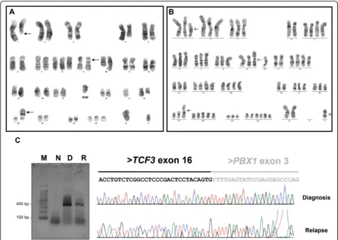
evi-dence of two concomitant chromosomal translocations (48,XY,t(1;19)(q23;q13.3),del(4)(q27q35),der(9)t(9;17)(p13; q11.2),del(10)(q24q26),del(18)(q21q3,+ 8,+ 22,+marc[20]) (Fig.1a). In addition to the two rearrangements observed at diagnosis, the karyotype of the BM at relapse also sho wed near-triploidy: 73,XX,t(1;19)(q23;p13),+ 1,+ 2,+ 3,+ 5,+ 5,+ 6,+ 7,+ 8,i(9q),+ 9,10,+ 12,+ 12,+ 13,+ 14,+ 14,+ 15,+ 15,+ 17,+ 18,+ 19,+ 20,+ 20,+ 20,+ 21,+ 22,+ 22,+ 22,+ 22,+ mar [2] (Fig.1b). The presence ofTCF3-PBX1 was con-firmed in both diagnostic and relapse samples by reverse transcription PCR (RT-PCR) followed by sequencing (Fig. 1c, d).
Additional copy number alterations (CNA) were identi-fied by multiplex ligation-dependent probe amplification (MLPA) using the SALSA MLPA P335-A4/B1 and P202-B1 kits (MRC-Holland, Amsterdam, the Nether lands) as previously described [9]. MLPA data were ana-lyzed using Coffalyser.Net. The relative copy number was obtained after the normalization of peaks against controls.
Values between 0.70 and 1.3 were considered to be within the normal range. Values below 0.70 or above 1.3 indi-cated deletion or gain, respectively. Values below 0.25 in-dicated homozygous deletion. Submicroscopic deletions
were only identified at relapse moment affectingCDKN2A
(exons 2 and 5),CDKN2B(exon 2),IKZF2(exons 2 and 5)
JAK2(exon 23), and miR31 (exon 1), as well as a partial gain inRB1(exons 14, 19, 24 and 26) (Fig.2a, b).
To investigate the presence of a putative gene fusion derived from the PAX5-r we performed a 3’Rapid
amplifi-cation of cDNA ends (3’RACE)-PCR based on
Scotto-Lavino’s protocol [10]. After cDNA synthesis, including an oligonucleotide (dT)-tailed primer (QT), two rounds of
PCR were performed to enrich the reaction for fragments containing the 3’end ofPAX5transcripts. The first round of amplification used the forward primer PAX5.E4.F1
(5’-AACCAACCAGTCCCAGCTTC-3′), while the
sec-ond was performed using the PAX5.E5.F1 primer (5’-TAC
TCCATCAGCGGCATCC-3′). The results showed a head
-to-head fusion between exon 6 ofPAX5and a 17q11.2 re-gion, where intron 3 ofSPECC1is located (Fig.2c).
Subsequently, we used SuperScript™III One-Step
RT-PCR System (Invitrogen, Carlsbad, EUA) and sequencing on ABI®3500 Genetic Analyzer (Applied Biosystems, Foster City, EUA) to confirm thatPAX5-r was present at both time points diagnosis and relapse (Fig.2d). Further-more, the predicted truncated PAX5 protein consisted of 266 amino acid residues, preserving the paired domain, the octapeptide motif and the homeodomain of PAX5, and a tail of 6 amino acids coded by the contiguous
in-tron 3 sequence ofSPECC1, which does not correspond
to any predictive functional domain (Fig.2e, f ). Discussion and conclusions
In this case report, we describe the identification of a der(9)t(9;17)(p13;q11.2) translocation, resulting inPAX5-r,
in a 16-year-old boy withTCF3-PBX1 positive BCP-ALL.
To date, this is the first report of a rearrangement between PAX5-exon 6 andSPECC1-intron 3. SixteenPAX5-partner genes had been previously described, composing a hetero-geneous group of genes encoding proteins that play dis-tinct roles in signaling pathways, transcription regulation, chromatin remodeling and cell structure [3,4]. However, in this case report, due to the absence of amino acid
resi-dues from SPECC1in the truncated protein formed, the
effect of the new protein identified is likely to be similar to aPAX5recurrent deletion [11–13].
The SPECC1 gene, also known as HCMOGT-1, has
three different splicing variants and encodes the NSP5a3a protein, which belongs to the cytospin-A family. NSP5a3a is highly expressed in head and neck squamous cell car-cinoma and in testis cell lines, but not in other normal cells [14]. Furthermore, NSP5a3a interacts with the nucle-olar phosphoprotein B23, which plays multiple roles in
cell growth and proliferation [15]. To the best of our
knowledge, the only report of SPECC1 altered in the
leukemic context was a case of juvenile myelomonocytic
leukemia withPDGFRB-SPECC1[16].
With regard to other CNAs and in concordance with previous findings inPAX5-rearranged leukemias, few dele-tions and/or gains are observed [3,4]. Here, we detected a
heterozygous deletions affecting CDKN2A/B, JAK2 and
miR31, confirming the presence of an unbalanced t(9;17)
with 9p13.2-pter deletion.CDKN2A/Bdeletions are
com-mon in ALL and frequently observed in concomitance
with other PAX5 gene fusions, such as PAX5-C20orf112
and PAX5-ETV6[4]. RegardingTCF3-PBX1 patients, the additional CNAs observed in this case report are in fact the most frequent alterations found in this cytogenetic -subtype. Furthermore, they are known to have a cumula-tive effect on the leukemic development [17]. Our analysis of CNAs at diagnosis and relapse revealed a high degree of clonal heterogeneity and a complex evolution between diagnosis and relapse samples, suggesting a clonal selec-tion pattern during leukemia progression, which could
have implications for the treatment efficiency. Moreover, based on the FMC characterization, we assume that the
major clone harboring a TCF3-PBX1 fusion. Although
there is no description of BCP-ALL subsets associated withPAX5- r, the presence of a second clone is not dis-charged and would be explored.
In conclusion, although the occurrence of
near-trip-loidy has been found in BCP-ALLTCF3-PBX1 patients,
the concomitant presence ofPAX5-r should be revisited.
The translocationPAX5-SPECC1is a new report that
re-quires further investigations. We also showed that both
TCF3-PBX1 and PAX5-r were present at diagnosis and
relapse, indicating that both might have participated in the malignant transformation disease maintenance and dismal outcome.
Abbreviations
3’RACE:3’Rapid amplification of cDNA ends; BCP-ALL: B-cell precursor acute lymphoblastic leukemia; BM: Bone marrow; CNA: Copy number alterations; CNS: Central nervous system; CR: Complete remission; ISCN: International System for Human Cytogenetic Nomenclature; MLPA: Multiplex ligation-dependent probe amplification;PAX5AMP:PAX5amplifications; RT-PCR: Reverse transcription PCR
Fig. 1Karyotype andTCF3-PBX1confirmation.aRepresentative GTG-banded metaphase of the leukemic clone at diagnosis andbat relapse.c
RT-PCR toTCF3-PBX1at diagnosis and relapse, respectively. M, marker (100pb); N, negative control; D, diagnosis sample; R, relapse sample.d
Acknowledgments
We are grateful to Dr. Archana Ganji for the commentaries on karyotype interpretation.
Funding
This study was supported by Brazilian National Counsel of Technological and Scientific Development-CNPq (CNPQ#301594/2015–5 and
CNPQ-Fig. 2Characterization of additional abnormalities identified.a, bMLPA profile of matched sample of case described using the P335 ALL SALSA
MLPA kit at diagnosis and relapse sample, respectively.c3’RACE-PCR PAX5 and sequencing of truncated PAX5 after 3’RACE-PCR with diagnosis sample. M, marker (1 kb); N, negative control; D, diagnosis sample; R, relapse sample.dRT-PCR toPAX5-SPECC1and sequencing ofPAX5transcript at diagnosis and relapse.eExpected protein sequence of the truncated PAX5 transcript derived from thePAX5-SPECC1head-to-head fusion. The translated protein refers to the PAX5–201 (NM_016734), showing its alternating exons (black and green) with splice acceptor amino acid residue (red), and amino acid residues coded from the fusion site on SPECC1 (green).fSchematic representation of the PAX5 wild-type protein and the fusion transcript detected
2017#305529/2017–0) and Fundação Carlos Chagas Filho de Amparo à Pes-quisa do Estado do Rio de Janeiro-FAPERJ (E_26/201.539/2014, E_26/200.388/ 2016 and E_26/203.214/2017).
Availability of data and materials
The data sets generated and/or analyzed during the current study are available from the corresponding author upon reasonable request.
Authors’contributions
TCB, ME and MSPO conceived the study; TCB, BAL, CBB and MBM performed cytogenetic-molecular experiments, analyzed and interpreted data; TCB. and ME wrote the manuscript; ASVD provided leukemia samples and clinical data used in this study; and all authors contributed with writings and approved the final manuscript.
Ethics approval and consent to participate
Informed consent was obtained from the patient’s parent in accordance with the Declaration of Helsinki and ethics was approved by both participating center. The research ethics committee from INCA approved the sample and data collection (n° CAAE 33243214.7.0000.5274).
Consent for publication
Written informed consent was obtained from the patient’s parents for publication of patient’s clinical and molecular data.
Competing interests
The authors declare that they have no competing interests.
Publisher’s Note
Springer Nature remains neutral with regard to jurisdictional claims in published maps and institutional affiliations.
Author details
1Division of Clinical Research, Research Center, Instituto Nacional de Câncer, Rio de Janeiro, RJ, Brazil.2Pediatric Hematology-Oncology Program, Research Center, Instituto Nacional de Câncer, Rio de Janeiro, RJ, Brazil.3Hospital de Clínicas de Porto Alegre, Porto Alegre, RS, Brazil.
Received: 25 July 2018 Accepted: 6 December 2018
References
1. Barbosa TC, Mansur MB, Blunck CB, Emerenciano M, Pombo-de-Oliveira MS. Characterization of RB1 in pediatric TCF3-PBX1+ acute lymphoblastic leukemia. Blood. 2017;130:3976.
2. Duque-Afonso J, Feng J, Scherer F, Lin CH, Wong SH, Wang Z, Iwasaki M, Cleary ML. Comparative genomics reveals multistep pathogenesis of E2A-PBX1 acute lymphoblastic leukemia. J Clin Invest. 2015;125(9):3667–80. 3. Nebral K, Denk D, Attarbaschi A, König M, Mann G, Haas OA, Strehl S.
Incidence and diversity of PAX5 fusion genes in childhood acute lymphoblastic leukemia. Leukemia. 2009;23(1):134–43.
4. Coyaud E, Struski S, Prade N, Familiades J, Eichner R, Quelen C, et al. Wide diversity of PAX5 alterations in B-ALL: a Groupe francophone de Cytogenetique Hematologique study. Blood. 2010;115(15):3089–97. 5. Schwab C, Nebral K, Chilton L, Leschi C, Waanders E, Boer JM, et al.
Intragenic amplification of PAX5: a novel subgroup in B-cell precursor acute lymphoblastic leukemia? Blood Adv. 2017;1(19):1473–7.
6. Arber DA, Orazi A, Hasserjian R, Thiele J, Borowitz MJ, Le Beau MM, et al. The 2016 revision to the World Health Organization classification of myeloid neoplasms and acute leukemia. Blood. 2016;127:2391–405.
7. Yunis JJ. Comparative analysis of high-resolution chromosome techniques for leukemic bone marrows. Cancer Genet Cytogenet. 1982;7(1):43–50. 8. Shaffer LG, McGowan-Jordan J, Schmid M. An international system for human
cytogenetic nomenclature. In: Recommendations of the international standing committee on human cytogenetic nomenclature; 2013.
9. Barbosa TC, Terra-Granado E, Quezado Magalhães IM, Neves GR, Gadelha A, GuedesFilho GE, et al. Frequency of copy number abnormalities in common genes associated with B-cell precursor acute lymphoblastic leukemia cytogenetic subtypes in Brazilian children. Cancer Genet. 2015;208(10):492–501. 10. Scotto-Lavino E, Du G, Frohman MA. 3’end cDNA amplification using
classic RACE. Nat Protoc. 2006;1(6):2742–5.
11. Artimo P, Jonnalagedda M, Arnold K, Baratin D, Csardi G, de Castro E, Duvaud S, et al. ExPASy: SIB bioinformatics resource portal. Nucleic Acids Res. 2012;40(W1):W597–603.
12. Fortschegger K, Anderl S, Denk D, Strehl S. Functional heterogeneity of PAX5 chimeras reveals insight for leukemia development. Mol Cancer Res. 2014;12(4):595–606.
13. Kawamata N, Pennella MA, Woo JL, Berk AJ, Koeffler HP. Dominant-negative mechanism of leukemogenic PAX5 fusions. Oncogene. 2012;31(8):966–77. 14. Sang N, Fath DM, Giordano A. A gene highly expressed in tumor cells
encodes novel structure proteins. Oncogene. 2004;23(58):9438–46. 15. D'Agostino L, Giordano A. Possible functional role of NSPs in cancer. Cell
Cycle. 2008;7(12):1810–27.
16. Morerio C, Acquila M, Rosanda C, Rapella A, Dufour C, Locatelli F, et al. HCMOGT-1 is a novel fusion partner to PDGFRB in juvenile myelomonocytic leukemia with t(5;17)(q33;p11.2). Cancer Res. 2004;64(8):2649–51.