RESEARCH
Rapid conjugative mobilization of a
100 kb segment of
Bacillus subtilis
chromosomal
DNA is mediated by a helper plasmid with no
ability for self-transfer
Megumi Miyano
1, Kosei Tanaka
2, Shu Ishikawa
1, Shinji Takenaka
3, Andrés Miguel‑Arribas
4, Wilfried J. J. Meijer
4*and Ken‑ichi Yoshida
1*Abstract
Background: The conjugative plasmid, pLS20, isolated from Bacillus subtilis natto, has an outstanding capacity for rapid self‑transfer. In addition, it can function as a helper plasmid, mediating the mobilization of an independently replicating co‑resident plasmid.
Results: In this study, the oriT sequence of pLS20cat (oriTLS20) was eliminated to obtain the plasmid, pLS20catΔoriT. This resulted in the complete loss of the conjugative transfer of the plasmid but still allowed it to mobilize a co‑ resident mobilizable plasmid. Moreover, pLS20catΔoriT was able to mobilize longer DNA segments, up to 113 kb of chromosomal DNA containing oriTLS20, after mixing the liquid cultures of the donor and recipient for only 15 min. Conclusions: The chromosomal DNA mobilization mediated by pLS20catΔoriT will allow us to develop a novel genetic tool for the rapid, easy, and repetitive mobilization of longer DNA segments into a recipient chromosome.
Keywords: Bacillus subtilis, Conjugation, Gram positive, Plasmid transfer
© The Author(s) 2018. This article is distributed under the terms of the Creative Commons Attribution 4.0 International License (http://creativecommons.org/licenses/by/4.0/), which permits unrestricted use, distribution, and reproduction in any medium, provided you give appropriate credit to the original author(s) and the source, provide a link to the Creative Commons license, and indicate if changes were made. The Creative Commons Public Domain Dedication waiver (http://creativecommons.org/ publicdomain/zero/1.0/) applies to the data made available in this article, unless otherwise stated.
Background
Bacterial cell division is asexual, and the genetic traits of mother and daughter cells with normal development are not changed fundamentally. However, horizontal gene transfer (HGT) can provide genetic plasticity for
bac-teria, even among different species [1]. HGT can lead
sometimes to problematic effects for the recipient bac-teria that accept the transferred genes. Those receiving undesirable genetic traits are excluded generally from the bacterial population, and it becomes less likely that harmful genes are inherited by the next generation. How-ever, mobile genetic elements (MGE) capable of self-transfer and self-replication often confer genetic traits
that are advantageous to the recipient bacterial cells, and that can be propagated rapidly within the population
[2]. HGT includes at least three types of mechanisms,
including transformation, transduction, and conjugation [1]. In the event of transformation, recipient cells uptake foreign DNAs depending on their own genetic compe-tence, and the foreign DNAs do not necessarily have a MGE function. In transduction, foreign DNAs are trans-ferred proactively together by bacteriophage infection, but the infection itself is often harmful for the recipient
cell [3]. Conjugation refers to the transfer of DNA to a
recipient bacterium from a donor bacterium through a mating event. During conjugation, the DNA trans-fers through dedicated conjugation machinery, encoded by genes on the conjugative DNA element. Conjugative DNA elements may be either plasmids or conjugative transposons, which are now more commonly referred to as integrative and conjugative elements or ICEs. HGT is environmentally important, as it increases the diversity
Open Access
of bacterial communities; however, it also serves as a useful tool for engineering desired bacterial strains via recombinant DNA technology for research and industrial purposes.
The typical mechanism for conjugative plasmid transfer between two bacterial strains proceeds as follows [4–8]. First, in the donor, a specific enzyme, relaxase, cleaves a phosphodiester bond of the plasmid DNA to produce a nick at a specific site on its “origin of transfer,” or oriT.
As a result, relaxase is covalently linked to the 5′-end
of the single-stranded plasmid DNA at the nick, and a DNA–protein complex, called relaxosome, is formed. Next, another specific protein, the type IV coupling pro-tein (T4CP), interacts with the relaxosome and targets it for transfer through a type IV secretion system from the donor to the recipient. When one unit of the plasmid DNA is transferred completely into the recipient cell, the single-stranded DNA is circularized and begins to repli-cate as maturation of the double-stranded circular plas-mid DNA occurs in the transconjugant.
Generally speaking, plasmids are classified into two types: conjugative and non-conjugative ones. The for-mer can transfer itself to the recipient using self-coding specific enzymes and a secretion system that is neces-sary for the transfer to occur; the latter cannot transfer itself because it lacks the required set of genes to do so.
However, non-conjugative plasmids containing the oriT
sequence, or mobilizable plasmids, can be mobilized by a co-resident conjugative plasmid within the same cell, in a phenomenon known as mobilization. Mobilizable
plas-mids are believed to need the oriT and its cognate mob
gene, the gene encoding relaxase [9, 10], but recently it was discovered that relaxase is not necessary for plasmid
mobilization if a plasmid carries a “mimic” of the oriT
region found in conjugative plasmids [11].
pLS20 is a 65-kbp-conjugative plasmid isolated from a strain of Bacillus subtilis natto [12]. It can transfer itself
between various B. subtilis-related Gram positive
bacte-ria, including Bacillus anthracis, Bacillus cereus, Bacil-lus licheniformis, Bacillus megaterium, Bacillus pumilus, and Bacillus thuringiensis [13]. pLS20cat, a derivative of pLS20 carrying a chloramphenicol resistance gene [14–17], possesses the incredible quality of being able to very rapidly transfer itself between cells within 15 min by simply mixing the liquid cultures containing donor and recipient cells [18]. In addition, pLS20cat can func-tion as a helper plasmid, with the ability to mobilize an independently replicating and co-resident mobilizable
plasmid containing a short oriT sequence from pLS20cat
(oriTLS20) that is unaccompanied by its cognate mob gene [19].
In this study, we inactivated the oriTLS20 region of
pLS-20cat to obtain pLSpLS-20catΔoriT, rendering it completely
immobile, but it was still able to facilitate
mobiliza-tion of the plasmid containing the oriTLS20. Moreover,
pLS20catΔoriT was able to mobilize longer chromosomal
DNA segments containing oriTLS20 independently of
the natural competence of the recipient cell. The larger DNA mobilization was achieved after mixing the liquid cultures of the donor and recipient for only 15 min. This
pLS20catΔoriT-mediated chromosomal DNA
mobiliza-tion will allow us to develop a novel genetic tool for rapid and repetitive accumulation of longer DNA segments into the recipient chromosome.
Methods
Bacterial strains and growth conditions
The bacterial strains and plasmids used in this study are listed in Table 1. Synthetic oligonucleotides used as
PCR primers are shown in Table 2. Bacterial strains were
grown on Lysogeny Broth (LB) medium (Difco) at 37 °C. When necessary, the medium was supplemented with antibiotics: 5 μg ml−1 chloramphenicol, 1 μg ml−1 eryth-romycin, 100 μg ml−1 spectinomycin, and 10 μg ml−1 kanamycin.
Construction of the recipient strain
The comK gene of strain 168 was inactivated by
replace-ment with a spectinomycin resistance gene, as follows. Two DNA fragments, each of which corresponded to
upstream and downstream regions of comK, were
ampli-fied by PCR using 168 DNA as a template with prim-ers comK-uF/comK-uR for the upstream fragment and comK-dF/comK-dR for the downstream fragment
(Table 2). Another DNA fragment containing the
spec-tinomycin resistance gene of strain TMO310 (Table 1)
was amplified using primers spc-F/spc-R (Table 2). The
three fragments were ligated together by recombinant PCR using primers comK-uF/comK-dR to sandwich the spectinomycin resistance gene between the upstream
and downstream regions of comK. The recombinant PCR
fragment was transformed into strain 168 conferring spectinomycin resistance and yielding the new strain, YNB001 (comK::spc), which was used as the recipient for the conjugative DNA transfer in this study.
Construction of pLS20catΔoriT
The oriTLS20 region of pLS20cat was inactivated by
marker-free deletion, as previously described [20]. Two
DNA fragments corresponding to the upstream
(frag-ment 1) and downstream (frag(frag-ment 2) regions of oriTLS20
responsible for the later deletion of the oriTLS20 region by intramolecular recombination. Another DNA fragment
of the mazF kan cassette (fragment 3) was amplified from
TMO311 DNA (Table 1) using primers mazF-F/mazF-R
(Table 2). The three PCR fragments were designed to be
connected in the order 1–3–2 by recombinant PCR using primers oriT-uF/oriT-dR. The recombinant PCR
frag-ment was transformed then into strain PKS11 (Table 1)
to confer kanamycin resistance, obtaining the new strain YNB022, in which pLS20cat was altered by integrat-ing the PCR fragment through a double crossover event
at the oriTLS20 region. YNB022 was grown overnight at
37 °C in LB liquid medium containing kanamycin. An ali-quot of the culture was transferred into a fresh LB liquid medium containing 1 mM isopropyl-thiogalactopyrano-side (IPTG) and the cells were allowed to grow for 2 h at 37 °C. Then, an aliquot of the culture was spread on an LB plate containing 1 mM IPTG and incubated at 37 °C
overnight. In the presence of IPTG, mazF was expressed,
producing a suicidal toxin so that only the cells that
could pop-out the mazF kan cassette through
intramo-lecular recombination could survive. Of those colonies appearing on the plate, kanamycin-sensitive colonies were sequenced to confirm the correct deletion of the
oriTLS20 region. The resulting plasmid was designated as
pLS20catΔoriT.
Construction of the donor strains
The donor strain YNB060 was constructed as follows
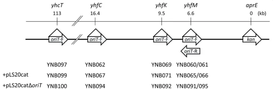
(Fig. 1). Two fragments corresponding to upstream
(frag-ment 1) and downstream (frag(frag-ment 4) regions of yhfM
were amplified from 168 DNA using primers yhfM-uF/yhfM-uR1 (for upstream) and yhfM-dF/yhfM-dR
(for downstream) (Table 2). Fragment 2 containing the
oriTLS20 was amplified using pLS20cat as a template with
primers oriT-F/oriT-R (Table 2). In addition, fragment
Table 1 Strains and plasmids used in this study
Strains and plasmids Relevant genotype or description Source or references
Strains
B. subtilis
PKS11 trpC2 pLS20cat [15]
GR138 trpC2 pLS20cat pGR16B [19]
TMO310 trpC2 aprE::(spc lacI Pspac‑mazF) [20]
TMO311 trpC2 aprE::(kan lacI Pspac‑mazF) [20]
YNB001 trpC2 comK::spc This study
YNB022 trpC2 pLS20cat (kan lacI Pspac‑mazF) This study
YNB026 trpC2 pLS20catΔoriT This study
YNB031 trpC2 pLS20catΔoriT pGR16B This study
YNB060 trpc2 aprE::kan yhfM::(oriTLS20‑F erm) This study YNB061 trpc2 aprE::kan yhfM::(oriTLS20‑R erm) This study YNB069 trpc2 aprE::kan yhfK::(oriTLS20‑F erm) This study YNB062 trpc2 aprE::kan yhfC::(oriTLS20‑F erm) This study YNB097 trpc2 aprE::kan yhcT::(oriTLS20‑F erm) This study YNB065 trpc2 aprE::kan yhfM::(oriTLS20‑F erm) pLS20cat This study YNB066 trpc2 aprE::kan yhfM::(oriTLS20‑R erm) pLS20cat This study YNB071 trpc2 aprE::kan yhfK::(oriTLS20‑F erm) pLS20cat This study YNB067 trpc2 aprE::kan yhfC::(oriTLS20‑F erm) pLS20cat This study YNB099 trpc2 aprE::kan yhcT::(oriTLS20‑F erm) pLS20cat This study YNB091 trpc2 aprE::kan yhfM::(oriTLS20‑F erm) pLS20catΔoriT This study YNB095 trpc2 aprE::kan yhfM::(oriTLS20‑R erm) pLS20catΔoriT This study YNB092 trpc2 aprE::kan yhfK::(oriTLS20‑F erm) pLS20catΔoriT This study YNB094 trpc2 aprE::kan yhfC::(oriTLS20‑F erm) pLS20catΔoriT This study YNB100 trpc2 aprE::kan yhcT::(oriTLS20‑F erm) pLS20catΔoriT This study Plasmids
pLS20cat Conjugative plasmid pLS20 with a chloramphenicol resistance gene inserted in the unique
Sall site [18]
pLS20catΔoriT pLS20cat without oriTLS20 This study
3 carrying the erythromycin resistance gene was ampli-fied using plasmid pMutin2 as the template with
prim-ers erm-F1/erm-R (Table 2). Fragments 1–4 were ligated
in the order 1–2–3–4 by recombinant PCR using
prim-ers yhfM-uF/yhfM-dR. Strain TMO311 (aprE::kan) was
transformed with the recombinant PCR fragment to select colonies resistant both to erythromycin and kan-amycin. The resulting strain was designated as YNB060
(Table 1), which had the erythromycin marker with
oriTLS20 and kanamycin marker at both the yhfM and
aprE loci, located 6.6 kb apart from each other on the
same chromosome. In addition, the direction of
repli-cation of oriTLS20 was oriented toward the kanamycin
marker located 6.6 kb downstream.
Another strain, YNB061, was constructed similarly
as described above. Two fragments of yhfM,
represent-ing upstream and downstream regions, were amplified using primers yhfM-uF/yhfM-uR2 and yhfM-dF/yhfM-dR (Table 2), respectively. The oriTLS20 fragment (fragment 2) and the erythromycin resistance fragment (fragment 3) were amplified using primers oriT-F/oriT-R and erm-F2/erm-R (Table 2), respectively. The four fragments were ligated by recombinant PCR using primers yhfM-uF/yhfM-dR. Strain TMO311 was transformed with the recombinant PCR frag-ment selecting erythromycin-resistant colonies as YNB061 (Table 1). In contrast to YNB060, in YNB061 the direction of replication of oriTLS20 on the yhfM locus was oriented oppositely to the kanamycin marker on the aprE locus.
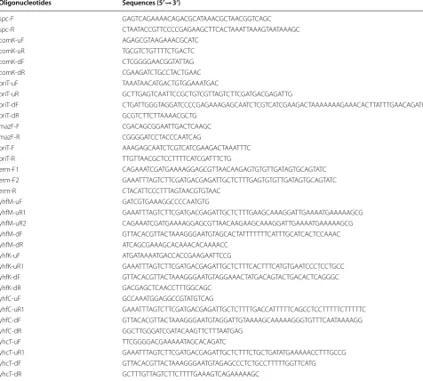
Table 2 Oligonucleotides used in this study
Oligonucleotides Sequences (5′→3′)
spc‑F GAGTCAGAAAACAGACGCATAAACGCTAACGGTCAGC spc‑R CTAATACCGTTCCCCGAGAAGCTTCACTAAATTAAAGTAATAAAGC
comK‑uF AGAGCGTAAGAAACGCATC
comK‑uR TGCGTCTGTTTTCTGACTC
comK‑dF CTCGGGGAACGGTATTAG
comK‑dR CGAAGATCTGCCTACTGAAC
oriT‑uF TAAATAACATGACTGTGGAAATGAC
oriT‑uR GCTTGAGTCAATTCCGCTGTCGTTAGTCTTCGATGACGAGATTG
oriT‑dF CTGATTGGGTAGGATCCCCGAGAAAGAGCAATCTCGTCATCGAAGACTAAAAAAAGAAACACTTATTTGAACAGATC
oriT‑dR GCGTCTTCTTAAAACGCTG
mazF‑F CGACAGCGGAATTGACTCAAGC
mazF‑R CGGGGATCCTACCCAATCAG
oriT‑F AAAGAGCAATCTCGTCATCGAAGACTAAATTTC oriT‑R TTGTTAACGCTCCTTTTCATCGATTTCTG
erm‑F1 CAGAAATCGATGAAAAGGAGCGTTAACAAGAGTGTGTTGATAGTGCAGTATC erm‑F2 GAAATTTAGTCTTCGATGACGAGATTGCTCTTTGAGTGTGTTGATAGTGCAGTATC
erm‑R CTACATTCCCTTTAGTAACGTGTAAC
yhfM‑uF GATCGTGAAAGGCCCCAATGTG
yhfM‑uR1 GAAATTTAGTCTTCGATGACGAGATTGCTCTTTGAAGCAAAGGATTGAAAATGAAAAAGCG yhfM‑uR2 CAGAAATCGATGAAAAGGAGCGTTAACAAGAAGCAAAGGATTGAAAATGAAAAAGCG yhfM‑dF GTTACACGTTACTAAAGGGAATGTAGCACTATTTTTTTCATTTGCATCACTCCAAAC
yhfM‑dR ATCAGCGAAAGCACAAACACAAAACC
yhfK‑uF ATGATAAAATGACCACCGAAGAATTCCG
yhfK‑uR1 GAAATTTAGTCTTCGATGACGAGATTGCTCTTTCACTTTCATGTGAATCCCTCCTGCC yhfK‑dF GTTACACGTTACTAAAGGGAATGTAGGAAACTATGACAGTACTGACACTCAGGGC
yhfK‑dR GACGAGCTCAACCTTTGGCAGC
yhfC‑uF GCCAAATGGAGGCCGTATGTCAG
yhfC‑uR1 GAAATTTAGTCTTCGATGACGAGATTGCTCTTTTGACCATTTTTCAGCCTCCTTTTTCTTTTTC yhfC‑dF GTTACACGTTACTAAAGGGAATGTAGGATTGTAAAAGCAAAAAGGGTGTTTCAATAAAAGG yhfC‑dR GGCTTGGGATCGATACAAGTTCTTTAATGAG
yhcT‑uF TTCGGGGACGAAAAATAGCACAGATC
yhcT‑uR1 GAAATTTAGTCTTCGATGACGAGATTGCTCTTTCTGCTGATATGAAAAACCTTTGCCG yhcT‑dF GTTACACGTTACTAAAGGGAATGTAGAGCCCTCTGCCTTTTTGGTTCATG
The other additional strains, YNB069, YNB062, and YNB097, were constructed similarly to those described
above (Fig. 1). For YNB069, two fragments of upstream
(fragment 1) and downstream (fragment 4) regions of
yhfK were amplified from 168 DNA using primers
yhfK-uF/yhfK-uR1 and yhfK-dF/yhfK-dR (Table 2),
respec-tively. For YNB062, two fragments of upstream and
downstream regions of yhfC were amplified using
prim-ers yhfC-uF/yhfC-uR1 and yhfC-dF/yhfC-dR (Table 2),
respectively. For YNB097, two fragments of upstream and
downstream regions of yhcT were amplified using
prim-ers yhcT-uF/yhcT-uR1 and yhcT-dF/yhcT-dR (Table 2),
respectively. The oriTLS20 fragment (fragment 2) and the erythromycin resistance fragment (fragment 3) were the same as those used above for YNB060 construction. For each case, the respective four fragments were ligated by recombinant PCR using primers yhfK-uF/yhfK-dR for YNB069, primers yhfC-uF/yhfC-dR for YNB062, and
primers yhcT-uF/yhcT-dR for YNB097 (Table 2). Each of
the recombinant PCR fragments was used to transform TMO311 (aprE::kan) to select colonies resistant to both erythromycin and kanamycin. The resulting strains were
designated as YNB069, YNB062, and YNB097 (Table 1),
which all had the erythromycin marker with oriTLS20 at
the yhfK, yhfC, and yhcT loci, and the kanamycin marker at the aprE locus set apart from each other by 9.5, 16.4, and 113 kb within the chromosome, respectively. In addi-tion, in all these strains, the direction of replication of
oriTLS20 was forward-oriented to the kanamycin marker.
Conjugative DNA mobilization
Conjugative DNA mobilization was performed in the
liquid medium, as previously described [18]. Donor and
recipient strains were cultured independently overnight in 5 ml of LB liquid medium containing the appropriate
antibiotics at 37 °C with shaking at 180 rpm. Each of the cultures was diluted to an optical density for the cell
of 0.05 at 600 nm (OD600) in 5 ml of fresh LB medium
without antibiotics and incubated at 37 °C with
shak-ing at 180 rpm. When OD600 reached 0.5–0.7, 500 μl of
the donor and recipient cultures were mixed in a 1.5 ml microtube to stand at 37 °C for 15 min. The mixture was serially diluted and spread on LB plates containing vari-ous combinations of antibiotics to grow colonies over-night. On their respective plates, colonies were counted as colony forming units (CFU) of transformed recipi-ents produced by conjugative transfer and mobilization (transconjugants) to calculate mobilization efficiencies
[CFU of transconjugants/CFU of total recipients × 106
(ppm)].
Results
pLS20catΔoriT cannot transfer itself but can help to mobilize a co‑resident plasmid carrying oriTLS20
As previously described [14–16], pLS20cat has the
complete set of genes required for its own conjuga-tive transfer. In fact, pLS20cat transfers itself from the
donor PKS11 (Table 1, 168 with pLS20cat) or GR138
(strain 168 with pLS20cat and pGR16B) to the recipient
YNB001 (comK::spc) within only 15 min after mixing the
two parental liquid cultures, resulting in a large number (more than 2500 ppm) of recipient cells with acquired chloramphenicol resistance appearing as transconjugants (Fig. 2). It is also known that pLS20cat is capable of
mobi-lizing a co-resident plasmid, pGR16B, carrying oriTLS20
and erythromycin resistance gene [19], as we observed
the donor, GR138, confer erythromycin resistance on nearly 1000 ppm of recipient cells (Fig. 2). These results imply that the helper pLS20cat could be nearly twice more efficient at transforming recipient cells than the
Fig. 1 Schematic representation of the integration of the loci of oriTLS20 and the kanamycin resistance gene of the donor strains. The gene loci
where oriTLS20 was integrated and the distances from the aprE loci where the kanamycin resistance gene (kan) was integrated are shown (top). The
mobilizable plasmid pGR16B. In addition, about 100 ppm of the recipients obtained resistance to both
erythromy-cin and chloramphenicol (Fig. 2), suggesting that about
10% of the transconjugants that accepted pGR16B also may have acquired pLS20cat.
Since the bacterial cells carrying pLS20cat do not
accept the pLS20cat-mediated genetic transfer [14–16],
the transconjugants that have accepted pLS20cat could not be transformed again using the same conjugative transfer system. On the other hand, the cells carrying pLS20cat could transfer not only pLS20cat itself but also mobilize co-resident pGR16B to other strains further. If these transconjugants were released into the environ-ment, the antibiotic resistance genes would be spread to other bacterial cells, causing the undesirable emergence
of new antibiotic-resistant bacteria [21, 22]. To avoid
the self-transfer of pLS20cat, we aimed at
knocking-out oriTLS20 in pLS20cat to construct pLS20catΔoriT.
As expected, the donor YNB026 did not transfer
pLS20catΔoriT at all, whereas YNB031 mobilized the
co-resident pGR16B to confer erythromycin resistance on the recipients (Fig. 2). Furthermore, the mobilization effi-ciency of pGR16B was nearly the same whether pLS20cat
or pLS20catΔoriT served as the helper plasmid. These
results indicate that the pLS20cat-dependent mobiliza-tion of pGR16B did not require self-mobility of the helper plasmid, pLS20cat. In addition, knocking-out oriTLS20 in pLS20cat did not affect the mobilization efficiency of the co-resident, pGR16B.
pLS20catΔoriT can mobilize chromosomal DNA containing oriTLS20
As shown above and in previous studies [19], pLS20cat
can efficiently mobilize the co-resident mobilizable
plas-mid with oriTLS20 but without its cognate mob gene.
Recently, conjugative transfer was shown to mobilize a large DNA fragment, representing the entire
chromo-some of Mycoplasma [23]. Thus, we conceived the idea
that pLS20cat may be able to mobilize chromosomal DNA, depending on the status of the oriTLS20 region.
In the donor chromosome, oriTLS20 was introduced at the
yhfM locus, 6.6 kb upstream of the kanamycin resistance
gene at the aprE locus; in strains YNB060 and YNB061
(Table 1), the direction of replication of oriTLS20 was for-ward- and reverse-oriented to the kanamycin resistance
gene, respectively. pLS20cat or pLS20catΔoriT was
intro-duced into the donor as the helper plasmid, yielding these new strains: (1) YNB065 (YNB060 with pLS20cat), (2)
Fig. 2 Mobilization efficiencies of the mobilizable plasmid, pGR16B, and the helper plasmids, pLS20cat and pLS20catΔoriT. Liquid cultures of the
YNB066 (YNB061 with pLS20cat), (3) YNB091 (YNB060
with pLS20catΔoriT), and (4) YNB095 (YNB061 with
pLS20catΔoriT). On the other hand, in the recipient strain
YNB001, comK encoding the key transcription factor for
natural competence was inactivated so that the strain com-pletely lost its natural competence (data not shown).
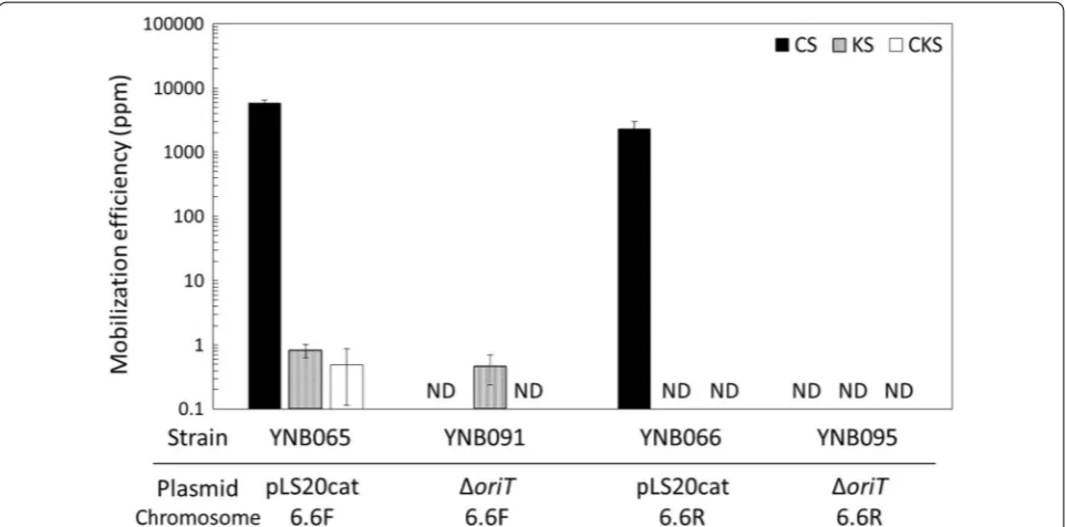
Strains YNB065 and YNB066, both carrying pLS20cat, conferred chloramphenicol resistance on more than
2300 ppm of the recipients (Fig. 3), but YNB065 was able
to confer kanamycin resistance on only 1 ppm of the recipient cells; YNB066 did not confer kanamycin resist-ance at all (Fig. 3). These results indicate that pLS20cat could transfer the kanamycin resistance gene located 6.6 kb downstream of oriTLS20, if the direction of oriTLS20 replication was forward-oriented to the kanamycin resistance gene. In addition, since the recipients had no natural competence, the acquisition of kanamycin resist-ance depended solely on the conjugative transfer. On the other hand, YNB065 conferred not only kanamycin resistance but also chloramphenicol resistance, on nearly 1 ppm of the recipients (Fig. 3). These results suggest that a large majority of the kanamycin-resistant recipients that accepted the chromosomal DNA could additionally have acquired the helper plasmid, pLS20cat.
When pLS20catΔoriT was introduced as the helper
plasmid into YNB060 (YNB091) and YNB061 (YNB095), it appeared that no recipient acquired chlorampheni-col resistance (Fig. 3), confirming the loss of
self-mobil-ity in pLS20catΔoriT. On the other hand, and more
importantly, when YNB091 was used as the donor,
pLS20catΔoriT transferred kanamycin resistance to
recipients with a similar efficiency and oriTLS20
-orien-tation dependency to pLS20cat (Fig. 3). These results
clearly indicate that pLS20catΔoriT can exert its helper
activity as efficiently as the original helper plasmid, pLS-20cat, not only in the case of mobilizing pGR16B but also when mobilizing the chromosomal DNA. YNB095 did not confer kanamycin resistance on the recipients, con-firming that the DNA mobilization depended on the for-ward-oriented oriTLS20.
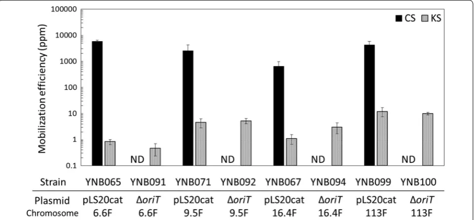
The distance between oriTLS20 and the
kanamy-cin marker was extended by 9.5 kb, 16.4 kb, and 113 kb in a stepwise fashion in strains YNB069, YNB062, and YNB097, respectively (Fig. 1). In all of these strains, the direction of replication of oriTLS20 was forward-oriented to the kanamycin resistance gene. A helper plasmid, either
pLS20cat or pLS20catΔoriT, was introduced into each
donor to create new donor strains: YNB071 (YNB069 with
Fig. 3 Mobilization efficiencies of the kanamycin resistance gene at the aprE locus and the helper plasmids, pLS20cat and pLS20catΔoriT. YNB060
pLS20cat), YNB067 (YNB062 with pLS20cat), YNB099 (YNB097 with pLS20cat), YNB092 (YNB069 with
pLS20catΔoriT), YNB094 (YNB062 with pLS20catΔoriT),
and YNB100 (YNB097 with pLS20catΔoriT). All donors
with pLS20cat conferred chloramphenicol resistance on more than 600 ppm of recipient cells, whereas the other
donors, with pLS20catΔoriT, did not confer
chloram-phenicol resistance at all (Fig. 4). However, and more
importantly, all of the strains with pLS20catΔoriT were
able to confer kanamycin resistance on 0.5–10.0 ppm of the recipient cells. Efficiencies were nearly equivalent to those achieved with YNB091 as the donor (Fig. 4), indicat-ing that the length of mobilized DNA could be extended at least to 113 kb. In addition, pLS20catΔoriT exhibited similar efficiencies to pLS20cat when mobilizing longer
segments of chromosomal DNA (Fig. 4). These results
also indicate that self-mobility of pLS20cat was not neces-sary for its helper function for mobilizing longer segments of chromosomal DNA.
Discussion
Strains of B. subtilis 168 derivatives have an advantage
over other bacteria because their natural competence and high recombination efficiency allow for plasticity of their
genome. A number of artificial introductions and compi-lations of various sizes and kinds of DNA segments have
been performed successfully into the B. subtilis genome
[24]. Therefore, B. subtilis has been regarded, generally, as one of the most promising platforms for designing, assembling, and modifying synthetic DNA, or even on larger scales with an entire synthetic genome. Accord-ingly, there is increasing demand of novel genetic tools for mobilizing longer DNA segments, which will push forward the research and development in synthetic
genome approaches [25].
Here we demonstrated that pLS20cat conjugative transfer was capable of mobilization of not only a
mobi-lizable plasmid carrying oriTLS20 but also chromosomal
DNA. In this study, however, both the donors and recipients were derived from the same parental strain,
B. subtilis 168, and recombination events between the chromosome and the mobilized DNA could occur at any homologous locations; therefore, we are not able to state that the entire length of mobilized DNA accurately replaced the respective part of the chromosome. Nev-ertheless, we can assume, at least, that the mobilization of chromosomal DNA initiated at the integration point
of oriTLS20 and continued until the kanamycin marker
Fig. 4 Mobilization efficiencies of the kanamycin resistance gene at the aprE locus and the helper plasmids, pLS20cat and pLS20catΔoriT.
because mobilization was seen only when the direction of replication of oriTLS20 was forward-oriented to the
kana-mycin resistance gene (Fig. 3). Furthermore, the present
results indicate that DNA segments up to 113 kb could
be mobilized (Fig. 4), which may be one of the longest
segments of DNA mobilized artificially from one cell to another within such a short period of only 15 min. For further applications, it would be worthwhile to test vari-ous conditions in order to extend the length of mobiliz-able DNA.
As mentioned above, the mobilization of chromosomal DNA depended upon the forward-orientation of the
oriTLS20 replication region, implying that the replication origin may function unidirectionally; however, a previous study suggested the possibility that oriTLS20 may be able to replicate bidirectionally [19]. If this is true, it is likely that
oriTLS20 replication in the reverse direction would be too weak to enable the mobilization of longer DNA segments.
On the other hand, the reverse-oriented oriTLS20 could
lead the counterclockwise replication of the other side of chromosome, which requires synthesis of much longer DNA to encounter the kanamycin marker. As we failed to detect any kanamycin-resistant transformant using the reverse-oriented oriTLS20, there could be a certain limit in the length of mobilizable DNA, which is yet to be defined.
We inactivated the oriTLS20 of pLS20cat to make
pLS20catΔoriT, which never transferred itself between
cells but was able to mobilize longer segments of chro-mosomal DNA with nearly the same efficiency as the self-transfer of pLS20cat. To our knowledge, this is the first demonstration that the oriT function of pLS20cat is not required for performing its helper function in
mobi-lizing DNA fragments containing oriTLS20. The DNA
mobilization by the pLS20cat conjugative system only
occurs to a recipient not harboring pLS20cat [14–16].
As described above, when donors with pLS20cat were used, more than 2000 ppm of the recipient cells became chloramphenicol resistant by accepting pLS20cat. On the other hand, the transfer of kanamycin resistance was seen for only 1–10 ppm of recipients. These results imply that nearly all the recipients that acquired the chromo-somal DNA also could have accepted pLS20cat. There-fore, recipients that previously acquired chromosomal DNA with the help of pLS20cat could no longer accept a conjugative DNA mobilization based on the pLS20 sys-tem. On the other hand, recipients that acquired
chro-mosomal DNA with the help of pLS20catΔoriT did not
have pLS20catΔoriT and could accept new rounds of
DNA mobilization. This acquired knowledge will be ben-eficial for developing a novel genetic tool for repetitive accumulation of longer DNA segments into the
recipi-ent chromosome. Furthermore, the pLS20catΔoriT also
would be useful for transforming other B. subtilis-related
Gram positive bacteria, including B. anthracis, B. cereus,
B. licheniformis, B. megaterium, B. pumilus, and B. thur-ingiensis [13].
Conclusions
In this study, the oriTLS20 region of pLS20cat was
elimi-nated to obtain pLS20catΔoriT, which resulted in
com-pletely eliminating the plasmid’s own mobility, while maintaining an ability to efficiently mediate the conju-gative mobilization of a neighboring mobilizable
plas-mid. Moreover, pLS20catΔoriT was able to mobilize
longer DNA segments, up to 113 kb of chromosomal DNA, containing the oriTLS20 region after mixing the liq-uid cultures of the donor and recipient for only 15 min. Understanding this chromosomal DNA mobilization by
pLS20catΔoriT will allow us to develop a novel genetic
tool for the rapid, easy, and repetitive accumulation of longer DNA segments into a recipient chromosome.
Abbreviations
CFU: colony forming units; HGT: horizontal gene transfer; IPTG: isopropyl‑thi‑ ogalactopyranoside; LB: Lysogeny Broth; MGE: mobile genetic elements; T4CP: type IV coupling protein; PCR: polymerase chain reaction.
Authors’ contributions
KY and WM conceived the idea for the project and initiated the study in CSIC‑ UAM with the MEXT grant of Open Partnership Joint Research Projects/Semi‑ nars. MM conducted most of the experiments and analyzed the results, under the supervision and with indispensable assistance of KT in strain construction, SI in data analysis, and ST in conjugation experiments in Kobe, and with indis‑ pensable assistance of AA in plasmid preparation in Madrid. KY wrote the final manuscript with MM. All authors read and approved the final manuscript.
Author details
1 Department of Science, Technology and Innovation, Kobe University, 1‑1 Rokkodai Nada, Kobe 657 8501, Japan. 2 Organization of Advanced Science and Technology, Kobe University, 1‑1 Rokkodai Nada, Kobe 657 8501, Japan. 3 Department of Agrobioscience, Kobe University, 1‑1 Rokkodai Nada, Kobe 657 8501, Japan. 4 Centro de Biología Molecular ‘Severo Ochoa’ (CSIC‑ UAM), Instituto de Biología Molecular ‘Eladio Viñuela’ (CSIC), Universidad Autónoma, Canto Blanco, 28049 Madrid, Spain.
Acknowledgements
The authors thank Mr. Jorge Val Calvo for his help in experiments.
Competing interests
The authors declare that they have no competing interests.
Availability of data and materials
The datasets and materials obtained and analyzed during the current study are available from the corresponding author on reasonable request.
Consent for publication
Not applicable.
Ethics approval and consent to participate
Not applicable.
Funding
• We accept pre-submission inquiries
• Our selector tool helps you to find the most relevant journal
• We provide round the clock customer support
• Convenient online submission
• Thorough peer review
• Inclusion in PubMed and all major indexing services • Maximum visibility for your research
Submit your manuscript at www.biomedcentral.com/submit
Submit your next manuscript to BioMed Central
and we will help you at every step:
Research Projects/Seminars. Work in the Meijer lab was funded by grants Bio2013‑41489‑P and BIO2016‑77883‑C2‑1‑P of the Ministry of Economy and Competitiveness of the Spanish Government to WM.
Publisher’s Note
Springer Nature remains neutral with regard to jurisdictional claims in pub‑ lished maps and institutional affiliations.
Received: 17 October 2017 Accepted: 20 December 2017
References
1. Frost LS, Leplae R, Summers AO, Toussaint A. Mobile genetic elements: the agents of open source evolution. Nat Rev Microbiol. 2005;3:722–32. 2. Thomas CM, Nielsen KM. Mechanisms of, and barriers to, horizontal gene
transfer between bacteria. Nat Rev Microbiol. 2005;3:711–21. 3. Davison J. Genetic exchange between bacteria in the environment.
Plasmid. 1999;42:73–91.
4. Abajy MY, Kopeć J, Schiwon K, Burzynski M, Döring M, Bohn C, et al. A type IV‑secretion‑like system is required for conjugative DNA transport of broad‑host‑range plasmid pIP501 in Gram‑positive bacteria. J Bacteriol. 2007;189:2487–96.
5. Alvarez‑Martinez CE, Christie PJ. Biological diversity of prokaryotic type IV secretion systems. Microbiol Mol Biol Rev. 2009;73:775–808.
6. Grohmann E, Muth G, Espinosa M. Conjugative plasmid transfer in Gram‑ positive bacteria. Microbiol Mol Biol Rev. 2003;67:277–301.
7. Wallden K, Rivera‑Calzada A, Waksman G. Type IV secretion systems: versatility and diversity in function. Cell Microbiol. 2010;12:1203–12. 8. Zechner EL, Lang S, Schildbach JF. Assembly and mechanisms of bacterial
type IV secretion machines. Philos Trans R Soc B. 2012;367:1073–87. 9. Francia MV, Varsaki A, Garcillán‑Barcia MP, Latorre A, Drainas C, De La Cruz
F. A classification scheme for mobilization regions of bacterial plasmids. FEMS Microbiol Rev. 2004;28:79–100.
10. Smillie C, Garcillan‑Barcia MP, Francia MV, Rocha EPC, de la Cruz F. Mobility of plasmids. Microbiol Mol Biol Rev. 2010;74:434–52.
11. Ramsay JP, Firth N. Diverse mobilization strategies facilitate transfer of non‑conjugative mobile genetic elements. Curr Opin Microbiol. 2017;38:1–9.
12. Tanaka T, Kuroda M, Sakaguchi K. Isolation and characterization of four plasmids from Bacillus subtilis. J Bacteriol. 1977;129:1487–94. 13. Koehler TM, Thorne CB. Bacillus subtilis (natto) plasmid pLS20 mediates
interspecies plasmid transfer. J Bacteriol. 1987;169:5271–8.
14. Meijer WJJ, Boer AJ, Tongeren SV, Venema G, Bron S. Characterization of the replication region of the Bacillus subtilis plasmid pLS20: a novel type of replicon. Nucleic Acids Res. 1995;23:3214–23.
15. Singh PK, Ramachandran G, Durán‑Alcalde L, Alonso C, Wu LJ, Meijer WJJ. Inhibition of Bacillus subtilis natural competence by a native, conjugative plasmid‑encoded comK repressor protein. Environ Microbiol. 2012;14:2812–25.
16. Singh PK, Ramachandran G, Ramos‑Ruiz R, Peiro‑Pastor R, Abia D, Wu LJ, et al. Mobility of the native Bacillus subtilis conjugative plasmid pLS20 is regulated by intercellular signaling. PLoS Genet. 2013;9:10.
17. Ramachandran G, Singh PK, Luque‑Ortega JR, Yuste L, Alfonso C, Rojo F, Wu LJ, Meijer WJJ. A complex genetic switch involving overlapping diver‑ gent promoters and DNA looping regulates expression of conjugation genes of a Gram‑positive plasmid. PLoS Genet. 2014;10:10.
18. Itaya M, Sakaya N, Matsunaga S, Fujita K, Kaneko S. Conjugational transfer kinetics of pLS20 between Bacillus subtilis in liquid medium. Biosci Bio‑ technol Biochem. 2006;70:740–2.
19. Ramachandran G, Miguel‑Arribas A, Abia D, Singh PK, Crespo I, Gago‑ Córdoba C, et al. Discovery of a new family of relaxases in Firmicutes bacteria. PLoS Genet. 2017;13:2.
20. Morimoto T, Ara K, Ozaki K, Ogasawara N. A new simple method to intro‑ duce marker‑free deletions in the Bacillus subtilis genome. Genes Genet Syst. 2009;84:315–8.
21. Davies J, Davies D. Origins and evolution of antibiotic resistance. Micro‑ biol Mol Biol Rev. 2010;74:417–33.
22. Berglund B. Environmental dissemination of antibiotic resistance genes and correlation to anthropogenic contamination with antibiotics. Infect Ecol Epidemiol. 2015;5:28564.
23. Dordet‑Frisoni E, Sagné E, Baranowski E, Breton M, Nouvel LX, Blanchard A, et al. Chromosomal transfers in mycoplasmas: when minimal genomes go mobile. MBio. 2014;5:6.
24. Itaya M, Fujita K, Kuroki A, Tsuge K. Bottom‑up genome assembly using the Bacillus subtilis genome vector. Nat Methods. 2008;5:41–3.