R E S E A R C H
Open Access
Natural IgM antibodies that bind
neoepitopes exposed as a result of spinal
cord injury , drive secondary injury by
activating complement
Aarti Narang
1, Fei Qiao
1, Carl Atkinson
1, Hong Zhu
1, Xiaofeng Yang
1, Liudmila Kulik
2, V. Michael Holers
2and Stephen Tomlinson
1,3*Abstract
Background:Natural IgM antibodies (Abs) function as innate immune sensors of injury via recognition of neoepitopes expressed on damaged cells, although how this recognition systems function following spinal cord injury (SCI) exposes various neoepitopes and their precise nature remains largely unknown. Here, we investigated the role of two natural IgM monoclonal Abs (mAbs), B4 and C2, that recognize post-ischemic neoepitopes following ischemia and reperfusion in other tissues.
Methods:Identification of post-SCI expressed neoepitopes was examined using previously characterized
monoclonal Abs (B4 and C2 mAbs). The role of post-SCI neoepitopes and their recognition by natural IgM Abs in propagating secondary injury was examined in Ab-deficient Rag1−/−or wild type C57BL/6 mice using Ab reconstitution experiments and neoepitope-targeted therapeutic studies, respectively.
Results:Administration of B4 or C2 mAb following murine SCI increased lesion size and worsened functional outcome in otherwise protected Ab-deficient Rag1−/−mice. Injury correlated with colocalized deposition of IgM and C3d in injured spinal cords from both mAb reconstituted Rag1−/−mice and untreated wild-type mice. Depletion of peritoneal B1 B cells, a source of natural Abs, reduced circulating levels of IgM with B4 (annexin-IV) and C2 (subset of phospholipids) reactivity, reduced IgM and complement deposition in the spinal cord, and protected against SCI. We therefore investigated whether the B4 neoepitope represents a therapeutic target for complement inhibition. B4-Crry, a fusion protein consisting of a single-chain Ab derived from B4 mAb, linked to the complement inhibitor Crry, significantly protected against SCI. B4-Crry exhibited a dual function in that it inhibited both the binding of pathogenic IgM and blocked complement activation in the spinal cord.
Conclusions:This study identifies important neoepitopes expressed within the spinal cord after injury. These neoepitopes are recognized by clonally specific natural IgM Abs that activate complement and drive pathology. We demonstrate that these neoepitopes represent novel targets for the therapeutic delivery of a complement inhibitor, and possibly other payload, to the injured spinal cord.
Keywords:Complement, Spinal cord injury, Natural antibodies, IgM, Neoepitope, Therapy
* Correspondence:[email protected]
1Department of Microbiology and Immunology, Medical University of South
Carolina, 173 Ashley Ave, CRI 213, Charleston, SC 29425, USA 3Ralph H Johnson VA Medical Center, Charleston, SC, USA
Full list of author information is available at the end of the article
Background
Traumatic spinal cord injury (SCI) results in severe debilitation, often with complete paralysis and loss of sensory function below the injury site. Following a primary mechanical insult, a secondary wave of cell death ensues, that is accompanied by ischemia, vascu-lar damage, glutamate excitotoxicity, ionic dysregula-tion, and inflammation. The area of secondary injury emanating from the site of trauma represents a thera-peutic target.
There is strong evidence that complement plays an important role in secondary SCI, and both the clas-sical [1] and alternative [1, 2] pathways of comple-ment activation have been implicated in propagating injury (reviewed in [3]). The alternative pathway func-tions as an amplification loop for the classical and lectin pathway of activation, although the role of the lectin pathway has not been investigated in SCI. Also, complement fragment levels have been found to be elevated in sera of spinal cord injury patients [4]. In an experimental model, it has been shown that com-plement activation and the propagation of SCI can be caused by pathogenic autoantibodies that arise follow-ing SCI-mediated activation of B cells [5, 6]. However, the role of naturally occurring pre-existing self-reactive antibodies (nAbs) produced by innate B1 B cells, and how complement is activated acutely after SCI, has not been investigated. Since SCI shares some pathophysiological characteristics with ischemia reper-fusion injury (IRI), and since IRI is driven by natural IgM Ab-mediated activation of complement in some organs [7–10], we investigated a role for natural IgM Abs in propagating SCI.
Tissue ischemia exposes neoepitopes known as damage-associated molecular patterns (DAMPs) on the cell surface, and these neoepitopes are recognized by circulating natural IgM Abs that subsequently acti-vate complement [11]. Some of these post-ischemic neoepitopes have been identified in the context of IRI and include non-muscle myosin [8], annexin IV [9], and various phospholipids [10, 12]. However, the na-ture of the neoepitopes exposed as a result of trauma on the injured spinal cord has not been investigated. Natural IgM Abs differ from adaptive immune Abs in that they are germline encoded, arise without exogen-ous antigen stimulation, and are produced by the B1 subset of B cells. B1 cells differ from conventional B2 cells by their phenotype, self-renewing capacity, and production of natural Abs. Although most natural IgM Abs are polyreactive, the subsets that initiate IRI are unique in that they recognize a specific set of epi-topes or patterns and, importantly, are specific for neoepitopes on stressed or injured cells. Here, we in-vestigate a pathogenic role for natural IgM Abs in
promoting spinal cord injury, but it is important to note that natural Abs also have important physio-logical roles, such as protection from pathogens and the removal of cellular and molecular waste [13, 14]. Indeed, a monoclonal IgM Ab corresponding to a natural Ab specificity is currently undergoing clinical evaluation as a potential therapeutic for multiple sclerosis [15, 16].
We previously isolated two natural IgM mAbs from unmanipulated mice, B4 mAb that recognizes annexin IV and C2 mAb that recognizes a subset of phospho-lipids, and demonstrated that these mAbs specifically bind to the intestine [9], brain [10], and heart [17] tissue following ischemia and reperfusion. In this study, we make use of these mAbs to investigate neoepitope expression post-SCI.
Methods
IgM mAbs and annexin IV purification
Hybridomas expressing B4 and C2 IgM mAbs were isolated after fusion of splenocytes from unmanipu-lated C57BL/6 mice with SP2/0-AG14 myeloma cells, with screening of hybridomas for specificity to apop-totic cells [9]. B4 mAb has been shown to recognize annexin IV [9] and C2 mAb to recognize a subset of phospholipids [10]. F632 and D5 IgM mAbs recognize 4-hydroxy-3-nitrophenylacetyl [17] and cytokeratin-19 [9], respectively, and were used as control mAbs. The D5 mAb was isolated from the same panel of hybrid-omas from which B4 and C2 mAbs were isolated, and does not bind to apoptotic cells. The IgM mAbs were purified from culture supernatant by affinity chroma-tography using an agarose bead column conjugated with goat anti-mouse IgM (Sigma-Aldrich, St Louis, MO) as described [17]. Recombinant annexin IV, the B4 ligand, was expressed in 293 F cells and purified from cul-ture supernatant as described [9].
Injury model and treatments
monitored, and bladders were voided twice daily until the bladder function was restored. For IgM mAb re-constitution experiments, Rag1−/− mice were ran-domized into different mAb treatment groups and a control group. Rag1−/− mice were injected i.v. with 100 μg C2, B4 or control mAbs 30 min after surgery. For therapeutic studies, animals were randomized into vehicle and B4Crry treatment groups and a single dose of 0.2 mg of protein was administered i.v. 30 min after injury. All procedures were approved by the Institutional Animal Care and Use Committee (IACUC) at the Medical University of South Carolina.
B1 B cell depletion
Peritoneal B1 cell depletion was achieved by a modifi-cation of the method described by Peterson et al. [19]. Briefly, starting at 4 weeks of age, mice were ad-ministered progressively increasing amounts of dis-tilled water, injected i.p. once per week over a 6-week period (2 injections each of 0.2, 0.5, and 1 ml). One group of mice received PBS injections as control. One day after the final injection, mice were used for pro-cedures. For analysis of B1 B cell depletion, peritoneal cells were obtained by injecting 5 ml of ice cold PBS into the peritoneal cavity, gently massaging the cavity, and collecting lavage fluid containing peritoneal cells using a 22-gauge needle. Single-cell suspensions of these cells were incubated with CD16/32 for 5 min at 4 °C to block Fc receptor binding [19] and were then
analyzed by flow cytometry using
flourochrome-conjugated Abs: anti-mouse CD19 APC and CD45R (B220) FITC (eBioscience, San Diego, CA). Gate was placed on live (7-Aminoactinomycin D viable stain) peritoneal cells, and CD19+ IgMhi B220lo populations were quantified.
Natural IgM levels in mouse serum
Blood was collected immediately following harvest of peritoneal cells, and serum prepared. Levels of anti-annexin IV and anti-phosphatidylcholine IgM Abs in mouse serum were quantified by ELISA using purified recombinant annexin IV (see above) and PC-BSA (Sigma) as capture reagents as previously described [10]. An ELISA kit was used to measure bound IgM in mouse serum according to the manufacturer’s in-structions (Bethyl Laboratories, Montgomery, TX).
Behavioral tests
Open-field assessment of locomotor function was done using the BMS scoring system with a scale of 0–9. The BMS scale is based on operational defini-tions of hind limb movement. Briefly, individual mice were simultaneously observed by two blinded investi-gators for a 4-min testing period, during which hind
limb movements, trunk/tail stability, and
forelimb-hind limb coordination were assessed and then
graded according to published methods [20]. All mice subjected to contusion injury exhibited a BMS score of 0 immediately after injury. The grip walk test (also referred to as the foot fault task), modified from the grid walk/ladder rung walking task [21], assesses defi-cits in descending motor control and is a test of motor coordination [22]. The apparatus consists of a grid floor with the grid raised above the surface. Mice were placed at one end of the grid and monitored from the side as they walked across the grid. The number of hind limb grips instead of placement er-rors as the animal traverses the grid was scored. Each time the paw gripped the bar was counted and com-pared between different treatment groups. All analyses were performed by investigators blinded to the treat-ment groups.
Tissue histology
Spinal cord tissue obtained from mice at 72 h, 7 days, or 21 days after injury was fixed in 10% formalin and embedded in paraffin. Seven-micrometer transverse sections were cut using a microtome and stained with standard hematoxylin and eosin stains for stereologi-cal analysis. Photographic documentation was made using Axio Vision Software (Carl Zeiss) at the reso-lution of 1024 × 1024 pixels. The hemorrhaged area and lesional tissue area was outlined using the meas-ure tool from axiovision (refer to Additional file 1: Figure S2), and the extent of hemorrhage and lesion expressed as percentage of the total area per section. This analysis was done using 100-μm step sections across the length of 1 mm starting at 100 μm from the epicenter on each side [22].
Immunohistochemistry and immunoflourescence
ImageJ and Fiji plugin (NIH) were used for analysis of images for fluorescence intensity. The intensity was measured and averaged from 100-μm step sections across a length of 1 mm, centered at the epicenter and starting at 100 μm from the epicenter on each side.
Statistical analysis
Statistical differences between parametric data were assessed using one-way analysis of variance (ANOVA) test with Bonferroni’s multi-group comparison, and non-parametric data were compared with the Kruskal-Wallis test with Dunn’s comparison (Prism 5.0,
b
a
Rag-/-Rag
-/-+ B4
Rag
-/-+ F632
Rag
-/-+ D5
0 10 20 30
* *
n.s.
***
% area lesioned
Rag-/-
Rag-/- +F632Rag-/- + D5Rag-/- + C2Rag-/-+B4
WT Sham 0
5 10 15
**
* *
d
e
g
a
hr
r
o
m
e
h
a
er
a
%
Rag
-/-+ C2
0 3 6 9 12 15 18 21
0 1 2 3 4 5 6 7 8 9
Rag-/-
WT Rag-/- + B4 Rag-/- + F632
Rag-/- + C2 Sham
Days post injury (d.p.i)
BMS score
c
d
WT
Sham
Coronal Sagittal
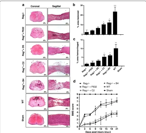
Fig. 1Specific IgM mAbs restore injury in otherwise protected Rag1−/−mice.aRepresentative H&E stained coronal and sagittal sections of WT
GraphPad). Differences between data were considered statistically significant when p< 0.05.
Results
Specific IgM mAbs restore SCI in otherwise protected Rag1−/−mice
Recognition of ischemic tissue by natural self-reactive IgM promotes injury in various organs subsequent to
reperfusion. To better understand the pathophysiology of inflammatory secondary damage following SCI, we in-vestigated whether a similar IgM recognition system is involved in promoting secondary injury to the spinal cord after trauma. Antibody deficient Rag1−/− mice were protected from SCI, but injury outcomes worsened when Rag1−/−mice were injected with either B4 or C2 IgM mAb within 30 min of injury. We found
a
Rag-/- +D5
Rag-/- + C2
Rag-/- +B4
WT
Sham
Rag-/-+D5Rag-/- +C2Rag-/-+B4 WT
Sham 0
10 20 30 40 50
** **
*
C3d flourescence
Rag-/-+D5Rag-/- +C2Rag-/-+B4 WT
Sham 0
10 20 30 40 50
**
**
*
IgM flourescence
b
c
WT
60X
IgM C3d Merged
Hoechst
Fig. 2IgM binding and C3 deposition in injured spinal cords of wt and B4 mAb reconstituted Rag1−/−mice. Coronal step sections were
significantly reduced lesion size and hemorrhaging at 3 days post-injury in Rag−/−mice after injury (Fig. 1a– c). Injection of specific natural Abs caused greater extent of injury in otherwise protected Rag−/−mice. Reconsti-tution of Rag1−/− mice with control F632 mAb or D5 mAb (Fig. 1a–c) did not alter outcome measures, dem-onstrating that the enhanced injury phenotype is neoepi-tope specific. We utilized one of these two specificities as controls for further studies. Compared to wild-type (wt) mice, Rag1−/− mice had significantly improved locomotor activity over a 21-day period post-injury (Fig. 1d). BMS measures in Rag1−/− mice were sig-nificantly worse upon reconstitution with B4 or C2
mAb, while injection of F632 mAb did not cause any significant changes.
Analysis of IgM binding and C3 deposition following SCI Complement has been shown to play a key role in modu-lating the progression of SCI, and we investigated the spe-cificity of B4 mAb binding to the injured spinal cord together with analysis of complement activation. Spinal cord sections obtained from wt mice at the lesion site 3 days post injury demonstrated co-localization of IgM and the complement activation fragment C3d (Fig. 2). Co-localization of IgM and C3d was similarly demonstrated in Rag1−/−mice reconstituted with B4 mAb, but not in
Untreated PBS (i.p.) Water (i.p.)
0 500 1000 1500
***
Anti-Annexin IV
(B4) IgM level (ng/ml)
PBS (i.p.) Water (i.p.)
0 20 40 60 80
**
Percentage of B1 cells (CD19+ IgMhi B220lo)
Untreated PBS (i.p) Water (i.p.) 0
200 400 600
***
Anti-Phosphatidylcholine
(C2) IgM level (
g/ml)
a
b
c
d
PBS (i.p) Water (i.p)
Sham Untreated
UntreatedPBS (i.p.)Water (i.p.) Sham 0
5 10 15
20 *
percent hemorrhag
e
UntreatedPBS (i.p.)Water (i.p.) Sham
0 10 20
30 *
percent lesion
e
Water (i.p.) PBS (i.p.) Untreated
0 100 200 300
T
otal IgM (ug/ml)
f
g
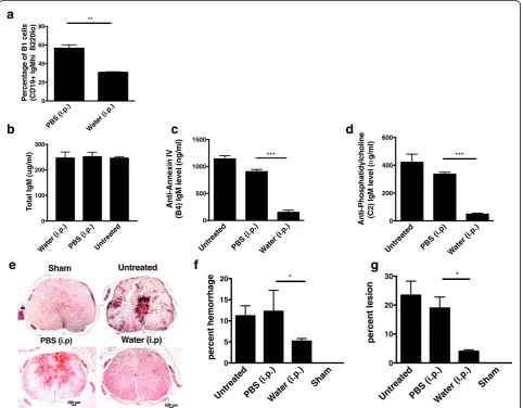
Fig. 3B1 B cell depletion reduces circulating levels of neoepitope-specific natural IgM Abs and protects against SCI.aQuantification of B1a B cell
depletion by flow cytometry analysis of peritoneal cells following a 6-week hypotonic shock procedure, demonstrating depletion of peritoneal B1 B cells (B220loCD5+). Mean ± SE, *p< 0.05,n= 3, and data is representative of three separate experiments.b–dAnalysis of serum levels of total
IgM, specific natural IgM antibodies with B4 mAb reactivity (annexin-IV) and C2 reactivity (phosphatidylcholine), determined by ELISA. Serum from mice treated with peritoneal injections of either PBS or water, as well as serum from WT mice, was analyzed. Mean ± SE, *p< 0.01,p< 0.001 respectively, n= 3–5.eFollowing B1 cell depletion or a control procedure, WT mice were subjected to SCI, and spinal cords isolated 72 h later for analysis.
Rag1−/−mice reconstituted with control D5 mAb, correl-ating with the injury profiles shown above. Together with previous data showing an important role for complement in SCI, these results indicate specific IgM-mediated activa-tion of complement is propagating injury.
Peritoneal B1 B cell depletion reduces levels of circulating pathogenic natural IgM antibodies and provides
protection in the acute phase of injury
Natural IgM Abs are produced by innate like B1 B cells that are capable of self-renewal and are present in high numbers in the peritoneum. To confirm that the patho-genic effect in this model is mediated by natural Abs, we investigated the effect of peritoneal B1 B cell depletion on SCI in wt mice. B1 B cells were depleted using a hypotonic shock method involving intraperitoneal injection of water, and depletion was confirmed by flow cytometric analysis that demonstrated water treatment resulted in a signifi-cant reduction in CD19+ B220loperitoneal cells (Fig. 3a) (see also additional file Figure S2). As previously shown [19], peritoneal B cell depletion did not result in a
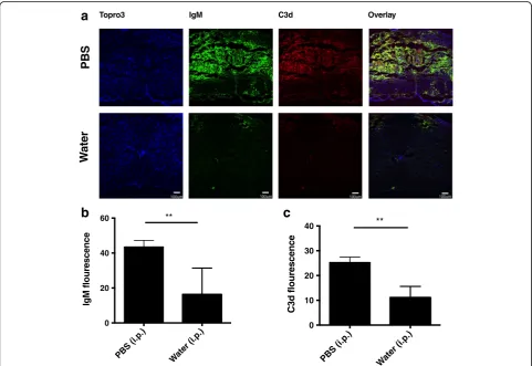
reduction of total circulating IgM (Fig. 3b). However, peritoneal B1 B cell depletion did result in a signifi-cant decrease in circulating Abs of B4 and C2 speci-ficities, further indicating the natural Ab status of anti-annexin IV and anti-phospholipid IgM mAbs (Fig. 3c, d). Analysis of spinal cord sections 3 days post injury revealed significantly less hemorrhaging in spinal cords from B1 B cell-depleted mice compared to controls, and this correlated with significantly less IgM and C3d deposition (Figs. 3f, g, Fig. 4). Taken to-gether, the data demonstrate that SCI is propagated in large part by peritoneal-derived B cell natural IgM and its recognition of annexin IV and phospholipid neoepitopes in the injured spinal cord that subse-quently activate complement. Further, the recognition of tissue and enhanced injury is neoepitope specific.
Targeting complement inhibition to IgM recognition sites in the injured spinal cord
On the basis of the above data and previous data show-ing both efficacy and safety benefits of site targeted
a
b
Wa
te
r
PBS
Topro3 IgM C3d Overlay
c
PBS (i.p.) Water (i.p.)
0 20 40
60 **
IgM flou
rescence
PBS (i.p.) Water (i.p.)
0 10 20 30
40 **
C3d flourescence
Fig. 4B1 B cell depletion reduced IgM deposition and complement activation in the injured spinal cord.aImmunofluorescence images of IgM
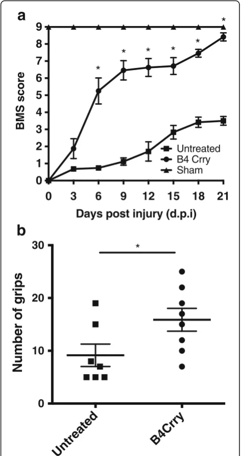
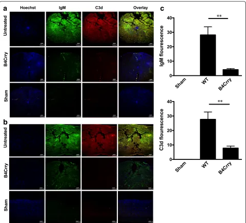
complement inhibition [17, 23], we investigated whether the B4 neoepitope is a therapeutic target for comple-ment inhibition post-SCI. We recently described the construction and functional characterization of B4-Crry, a fusion protein consisting of a scFv derived from B4 mAb linked to the mouse complement inhibitor Crry [17]. Administration of B4-Crry to WT mice 30 min after SCI significantly improved locomotor activity com-pared to vehicle-treated mice, and locomotor functional activity was restored by day 21 in B4-Crry-treated mice (Fig. 5a). B4-Crry treatment also significantly improved performance on a grip walk test, indicating improved restoration of sensorimotor function (Fig. 5b). The per-centage lesion area per section was averaged over step sections obtained across 1 mm around the epicenter. We found significant reduction in lesion size and hemorrhaged area with B4-Crry treatment (Fig. 6).
Effect of B4-Crry on IgM deposition and complement activation in the injured spinal cord
Immunoflourescence microscopy of spinal cord sections demonstrated that B4-Crry reduced IgM binding and abolished C3d deposition at the injury epicenter (Fig. 7a), as well as rostrocaudally as indicated by analysis of lon-gitudinal sections (Fig. 7b). Quantification of both IgM and C3d fluorescence signal suggested that the animals treated with B4Crry had significant decrease in both these signals 3 days post injury (Fig. 7c). These data indi-cate a dual functionality of B4-Crry in providing protec-tion from SCI, since it not only blocks complement activation, but also inhibits the binding of endogenous pathogenic self-reactive IgM in the context of a full nat-ural Ab repertoire.
Discussion
Natural Abs are present from birth and are germline encoded without a requirement for exogenous antigen exposure, and it is increasingly recognized that subsets of natural Abs have varying and distinct roles. Natural IgM Abs can have homeostatic roles, for example by recognizing DAMPs expressed on stressed or dying cells and facilitating their clearance, and they can serve pro-tective roles by a variety of cellular mechanisms [13]. However, they can also have pathogenic roles as has been shown in models of ischemia and reperfusion in-jury where pathogenic levels of IgM bind to ischemic tis-sue and activate complement [11].
It has been shown previously that following murine SCI, B cells become activated and produce pathogenic autoantibodies that propagate secondary injury in the spinal cord and impair recovery. Precisely, how trau-matic injury to the spinal cord results in B cell dysfunc-tion and autoantibody producdysfunc-tion is not clear, but pathology is caused, at least in part, via complement
activation and Fc receptor ligation [6]. However, while there is accumulating evidence that B cell activation and autoantibody production play an important role in propagating SCI (reviewed in [24]), the mechanisms by which secondary spinal injury is initiated and therein the functional significance of self-reactive natural IgM Abs (as opposed to autoreactive Abs resulting from a patho-genic adaptive immune response) in this process have
0
3
6
9
12
15
18
21
0
1
2
3
4
5
6
7
8
9
Untreated B4 Crry Sham
*
*
*
*
*
*
Days post injury (d.p.i)
BMS score
Untreated
B4Crry
0
10
20
30
*
Number of grips
a
b
Fig. 5Neoepitope-targeted complement inhibition with B4-Crry
not been investigated. Here, we show a role for a specific subset of natural IgM Abs in activating complement and propagating secondary injury after trauma to the spinal cord and show that SCI mimics the pathological para-digm of IgM-mediated IRI in other tissues. Thus, in addition to a role for an adaptive autoantibody response in contributing to post-SCI pathology, we demonstrate a role for pre-formed self-reactive IgM Abs in an innate immune response to SCI.
Identification of the human counterpart of murine B1 B cells has been controversial, partly because CD5 is an activation marker on human B cells. However, studies have identified CD20+CD27+CD43+ B cells as the human B1 cell equivalent [25, 26]. Although B1 cells had been thought to be located predominantly in the peritoneal and pleural cavities of mice, recent evidence indicates that the bone marrow and spleen may be an important source [27]. Nevertheless, the current study indicates that peritoneal B1 cell-derived natural IgM plays an important role in propagating SCI, since peritoneal depletion of B1 B cells both significantly reduced levels of circulating pathogenic IgM of B4 and C2 specificity and significantly improved recovery. Both our data and previous studies have shown that these B1 deple-tion procedures do not alter total IgM levels [28]. To-gether, these data suggest a role for distinct subsets of naturally occurring IgM Abs and raise the possibility of
different Ab repertoires originating from different B1 cell locations.
The B4 and C2 IgM mAbs that we utilize in this study have been previously described: B4 recognizes a post-translational modification of annexin IV [9] and C2 recognizes a subset of phospholipids [10].The nat-ural Ab status of these mAbs is indicated by the fact that they were derived from unmanipulated mice, they are universally present in C57BL/6 mice, and the circu-latory levels of IgM Abs of B4 and C2 specificity are re-duced following peritoneal B1 cell depletion. Also, natural IgM Ab recognition of neoepitopes is the ac-cepted paradigm for complement activation following ischemia and reperfusion of other tissues. We show here that the B4 and C2 antigens represent DAMPs expressed in the injured spinal cord. Injury was contin-gent upon specific recognition of the injured spinal cord, since unlike B4 and C2 mAbs, control IgM mAbs, including one that was isolated from the same panel containing the B4 and C2 mAbs, neither bound within the injured spinal cord nor reconstituted injury in otherwise protected Ab-deficient Rag1−/− mice. In an earlier report, Rag2−/− mice were shown to have im-proved recovery following spinal cord contusion com-pared to wt mice, with improved locomotor function and increased axonal regrowth [29]. Improved recovery in Rag2−/− mice was associated with decreased
Untreated
B4Crry
b
Untreated B4Crry
Sham 0
5 10
15 ***
Hemorrhaged ara(%)
Untreated
B4Crry Sham
0 10 20
30 **
% age area lesioned
c
Sham
Coronal Sagittal
a
Fig. 6Histopathology of spinal cords from B4-Crry treated showed reduced spread of lesion 72 h after SCI.aH&E stained coronal and sagittal
microglia/macrophage infiltration, which the authors attributed to the absence of T cell-derived cytokines. Certainly, this could be a contributing mechanism of injury, besides the critical role for natural Abs in acti-vating complement and initiating injury.
It is interesting that specific natural IgM Abs are also implicated in CNS repair mechanisms, beyond their role in identifying injured and dying cells for removal, and some IgM mAbs have shown potential for therapeutic application following CNS injury (reviewed in [16]). These protective Abs appear to
function by providing intracellular signals that pro-mote neuron or glial cell survival. The mechanistic relationship between pathogenic and protective nat-ural Abs is not clear. There may be quantitative and/or qualitative, as well as temporally acting or context-dependent differences between Abs that are involved in injury vs. repair. It is also noteworthy that complement activation products can have both neurodegenerative and neuroregenerative effects fol-lowing CNS injury [30, 31]. While complement-mediated protective mechanisms likely become more
a
b
c
Fig. 7Crry treatment reduces both IgM binding and C3d deposition after SCI. Analysis of of IgM and C3d deposition in vehicle treated,
important in the subacute phase after CNS injury [32], this balance between complement-mediated in-jury and repair is an area in need of further investigation.
Based on the identification of neoepitopes exposed in the injured spinal cord and the pathogenic and complement-activating properties of mAbs recognizing these neoepitopes, we investigated a site-targeted thera-peutic strategy for the local delivery of a complement in-hibitor. Site-targeted complement inhibition obviates the need for systemic inhibition, increases bioavailability, and markedly increases efficacy (over 10-fold) [23]. We have also shown previously that unlike systemic inhibi-tors, therapeutic doses of site-targeted complement in-hibitors do not adversely affect important physiological functions such as host defense [17, 23]. This latter point is an important consideration for the treatment of SCI patients, since CNS injury is immunosupressive and there is a high incidence of morbidity and mortality caused by infection in neurotrauma and SCI patients.
In our therapeutic paradigm, we used our recently de-scribed fusion protein consisting of a single chain (scFv) derived from the B4 hybridoma linked to Crry, a murine orthologue of human sCR1 [17]. In our previous report, we also demonstrated that B4-Crry does not increase sus-ceptibility to infection [23, 33]. Here, the protective effect of B4-Crry correlated not only with significantly reduced complement activation, but also reduced IgM deposition at the injury site. This indicates that in addition to comple-ment inhibition, B4-Crry is inhibiting the binding of IgM Abs to the injured spinal cord in the context of a full nat-ural Ab repertoire. This could be due to B4 scFv competing for IgM binding sites, but also due to decreased complement-mediated injury with a reduction in the asso-ciated exposure of neoepitopes. While it can be expected that there is much redundancy in IgM and neoepitope specificities, we have nonetheless shown that B4 scFv alone can significantly reduce IgM binding in the post-ischemic heart [17]. A possible explanation for this effect of a single scFv specificity on overall IgM binding is that there is a re-quirement for recognition of exposed neoepitopes in a chronological sequence, such that disrupting any one of these interactions ameliorates complement activation. An-other possibility is that recognition of these neoepitopes on multiple cell types is critical for complement dependent in-jury, and blocking even one of these specificities is conse-quential for the overall complement mediated injury response.
Conclusions
This study identifies important neoepitopes expressed within the spinal cord after injury, that are recognized by clonally specific natural IgM Abs that activate com-plement and drive secondary injury. Peritoneal B1 cells
are a source of a subset of pathogenic self-reactive Abs that contribute to pathology by activating complement. Post-SCI expressed neoepitopes represent novel targets for the targeted delivery of therapeutics to the injured spinal cord, and we show that targeted delivery of a complement inhibitor significantly improves recovery following trauma to the spinal cord. With regard to the potential translation of this therapeutic strategy to the clinic, it has been shown that the B4 annexin IV neoepi-tope is also expressed on stressed and injured human cells [10]. Therefore, this antibody based targeting strat-egy presents a new therapeutic payload delivery option in an injury site specific manner.
Additional file
Additional file 1: Figure S1.(A) Spinal cord tissue analyses were done
across the length of 1 mm centered at the epicenter (B). Examples of hemorrhaged (red) and lesioned (gray) area are outlined. The outlined area for each section measured using a Nikon eclipse E600 with axiovision outline tool and was expressed as a percentage of total area of each section measured.Figure S2.(A) After B1 cell depletion procedure, the peritoneal fluid was analyzed by flow cytometry to confirm a decrease in CD19+IgMhiB220lopopulations. Initial gate was placed on live cells and viability determined by 7-AAD (7-Aminoactinomycin D) staining followed by gating on CD19+ and IgMhi cells. The B220losubpopulation
from the gated population was used for comparison among different groups (B). The mice that received PBS injections as control were used for these analyses.n= 6. (PDF 843 kb)
Abbreviations
Ab:Antibody; IRI: Ischemia reperfusion injury; mAb: Monoclonal antibody; scFv: Single-chain antibody; SCI: Spinal cord injury
Acknowledgements
We gratefully acknowledge Dr. Bei Liu, Medical University of South Carolina, for her assistance with flow cytometry analysis.
Funding
This work was supported by grants from the Department of Veterans Affairs (Merit Award 1I01RX001141 and 1BX001218), the National Institutes of Health (DK102912 and CA158179), and the South Carolina Spinal Cord Injury Research Fund.
Availability of data and materials
Data supporting the conclusions of this article are presented in the manuscript.
Authors’contributions
AN performed experiments, analyzed data, and drafted the manuscript. FQ and XY performed experiments and analyzed data, CA analyzed data, HZ performed experiments, and LK and VMH provided reagents and edited the manuscript. ST conceived and planned the study, analyzed data, and drafted the manuscript. All authors read and approved the final manuscript.
Competing interests
Tomlinson, Atkinson, Kulik and Holers have a patent pending for targeted delivery of complement inhibitors to post-ischemic neoepitopes. The authors Aarti Narang,Fei Qiao,Hong Zhu and Xiao Feng Yang do not have any com-peting interests.
Consent for publication
Ethics approval
All animal procedures were approved by the Institutional Animal Care & Use Committee at the Medical University of South Carolina.
Publisher’s Note
Springer Nature remains neutral with regard to jurisdictional claims in published maps and institutional affiliations.
Author details
1Department of Microbiology and Immunology, Medical University of South
Carolina, 173 Ashley Ave, CRI 213, Charleston, SC 29425, USA.2Departments of Medicine and Immunology, University of Colorado School of Medicine, Denver, CO, USA.3Ralph H Johnson VA Medical Center, Charleston, SC, USA.
Received: 31 January 2017 Accepted: 4 June 2017
References
1. Anderson AJ, Robert S, Huang W, Young W, Cotman CW. Activation of complement pathways after contusion-induced spinal cord injury. J Neurotrauma. 2004;21:1831–46.
2. Qiao F, Atkinson C, Kindy MS, Shunmugavel A, Morgan BP, Song H, Tomlinson S. The alternative and terminal pathways of complement mediate post-traumatic spinal cord inflammation and injury. Am J Pathol. 2010;177:3061–70.
3. Peterson SL, Anderson AJ. Complement and spinal cord injury: traditional and non-traditional aspects of complement cascade function in the injured spinal cord microenvironment. Exp Neurol. 2014;258:35–47.
4. Rebhun J, Madorsky JG, Glovsky MM. Proteins of the complement system and acute phase reactants in sera of patients with spinal cord injury. Ann Allergy. 1991;66:335–8.
5. Ankeny DP, Lucin KM, Sanders VM, McGaughy VM, Popovich PG. Spinal cord injury triggers systemic autoimmunity: evidence for chronic B lymphocyte activation and lupus-like autoantibody synthesis. J Neurochem. 2006;99:1073–87. 6. Ankeny DP, Guan Z, Popovich PG. B cells produce pathogenic antibodies and
impair recovery after spinal cord injury in mice. J Clin Invest. 2009;119:2990–9. 7. Chan RK, Ibrahim SI, Verna N, Carroll M, Moore Jr FD, Hechtman HB.
Ischaemia-reperfusion is an event triggered by immune complexes and complement. Br J Surg. 2003;90:1470–8.
8. Zhang M, Austen Jr WG, Chiu I, Alicot EM, Hung R, Ma M, Verna N, Xu M, Hechtman HB, Moore Jr FD, Carroll MC. Identification of a specific self-reactive IgM antibody that initiates intestinal ischemia/reperfusion injury. Proc Natl Acad Sci U S A. 2004;101:3886–91.
9. Kulik L, Fleming SD, Moratz C, Reuter JW, Novikov A, Chen K, Andrews KA, Markaryan A, Quigg RJ, Silverman GJ, et al. Pathogenic natural antibodies recognizing annexin IV are required to develop intestinal ischemia-reperfusion injury. J Immunol. 2009;182:5363–73.
10. Elvington A, Atkinson C, Kulik L, Zhu H, Yu J, Kindy MS, Holers VM, Tomlinson S. Pathogenic natural antibodies propagate cerebral injury following ischemic stroke in mice. J Immunol. 2012;188:1460–8. 11. Fleming SD. Naturally occurring autoantibodies mediate ischemia/
reperfusion-induced tissue injury. Adv Exp Med Biol. 2012;750:174–85. 12. Fleming SD, Egan RP, Chai C, Girardi G, Holers VM, Salmon J, Monestier M,
Tsokos GC. Anti-phospholipid antibodies restore mesenteric ischemia/ reperfusion-induced injury in complement receptor 2/complement receptor 1-deficient mice. J Immunol. 2004;173:7055–61.
13. Gronwall C, Silverman GJ. Natural IgM: beneficial autoantibodies for the control of inflammatory and autoimmune disease. J Clin Immunol. 2014;34 Suppl 1:S12–21.
14. Panda S, Ding JL. Natural antibodies bridge innate and adaptive immunity. J Immunol. 2015;194:13–20.
15. Leslie M. Cleanup crew. Science. 2005;347:1058–9. 1061.
16. Wootla B, Watzlawik JO, Warrington AE, Wittenberg NJ, Denic A, Xu X, Jordan LR, Papke LM, Zoecklein LJ, Pierce ML, et al. Naturally occurring monoclonal antibodies and their therapeutic potential for neurologic diseases. JAMA Neurol. 2015;72:1346–53.
17. Atkinson C, Qiao F, Yang X, Zhu P, Reaves N, Kulik L, Goddard M, Holers VM, Tomlinson S. Targeting pathogenic postischemic self-recognition by natural IgM to protect against posttransplantation cardiac reperfusion injury. Circulation. 2015;131:1171–80.
18. Lee JH, Streijger F, Tigchelaar S, Maloon M, Liu J, Tetzlaff W, Kwon BK. A contusive model of unilateral cervical spinal cord injury using the infinite horizon impactor. J Vis Exp. 2012;65:3313.
19. Peterson LK, Tsunoda I, Fujinami RS. Role of CD5+ B-1 cells in EAE pathogenesis. Autoimmunity. 2008;41:353–62.
20. Pajoohesh-Ganji A, Byrnes KR, Fatemi G, Faden AI. A combined scoring method to assess behavioral recovery after mouse spinal cord injury. Neurosci Res. 2010;67:117–125.
21. Metz GA, Whishaw IQ. The ladder rung walking task: a scoring system and its practical application. J Vis Exp. 2009;28:1204.
22. Yu CG, Geddes JW. Sustained calpain inhibition improves locomotor function and tissue sparing following contusive spinal cord injury. Neurochem Res. 2007;32:2046–53.
23. Atkinson C, Song H, Lu B, Qiao F, Burns TA, Holers VM, Tsokos GC, Tomlinson S. Targeted complement inhibition by C3d recognition ameliorates tissue injury without apparent increase in susceptibility to infection. J Clin Invest. 2005;115:2444–53.
24. Zhang Y, Popovich P. Roles of autoantibodies in central nervous system injury. Discov Med. 2011;11:395–402.
25. Griffin DO, Holodick NE, Rothstein TL. Human B1 cells in umbilical cord and adult peripheral blood express the novel phenotype CD20+ CD27+ CD43+ CD70. J Exp Med. 2011;208:67–80.
26. Covens K, Verbinnen B, Geukens N, Meyts I, Schuit F, Van Lommel L, Jacquemin M, Bossuyt X. Characterization of proposed human B-1 cells reveals pre-plasmablast phenotype. Blood. 2013;121:5176–83.
27. Savage HP, Baumgarth N. Characteristics of natural antibody-secreting cells. Ann N Y Acad Sci. 2015;1362:132–42.
28. Renner B, Strassheim D, Amura CR, Kulik L, Ljubanovic D, Glogowska MJ, Takahashi K, Carroll MC, Holers VM, Thurman JM. B Cell Subsets Contribute to Renal Injury and Renal Protection after Ischemia/Reperfusion. J Immunol. 2010;185(7):4393–400.
29. Wu B, Matic D, Djogo N, Szpotowicz E, Schachner M, Jakovcevski I. Improved regeneration after spinal cord injury in mice lacking functional T-and B-lymphocytes. Exp Neurol. 2012;237:274–85.
30. Yanamadala V, Friedlander RM. Complement in neuroprotection and neurodegeneration. Trends Mol Med. 2010;16:69–76.
31. Alexander JJ, Anderson AJ, Barnum SR, Stevens B, Tenner AJ. The complement cascade: Yin-Yang in neuroinflammation—neuro-protection and -degeneration. J Neurochem. 2008;107:1169–87.
32. Alawieh A, Elvington A, Tomlinson S. Complement in the homeostatic and ischemic brain. Front Immunol. 2015;6:417.
33. Yanamadala V, Friedlander RM: Complement in neuroprotection and neurodegeneration. Trends in molecular medicine. 2010;16:69–76.
• We accept pre-submission inquiries
• Our selector tool helps you to find the most relevant journal
• We provide round the clock customer support
• Convenient online submission
• Thorough peer review
• Inclusion in PubMed and all major indexing services • Maximum visibility for your research
Submit your manuscript at www.biomedcentral.com/submit