BioMedCentral
Cell Communication and Signaling
Open Access
Research
Platelet-derived growth factor modulates rat vascular smooth
muscle cell responses on laminin-5 via mitogen-activated protein
kinase-sensitive pathways
Karl Kingsley*
1,3and George E Plopper
2,3Address: 1Department of Biomedical Sciences, University of Nevada, Las Vegas, School of Dental Medicine, 1001 Shadow Lane B-234, Las Vegas,
Nevada, 89106-4124, USA, 2Department of Biology, Rensselear Polytechnic Institute, 110 8th Street, Troy, New York, 12180-3596, USA and 3(previous institutional affiliation) Department of Biological Sciences, University of Nevada, Las Vegas, 4505 Maryland Parkway, Box 454004, Las
Vegas, Nevada, 89154-4004, USA
Email: Karl Kingsley* - [email protected]; George E Plopper - [email protected] * Corresponding author
Abstract
Background: A treatment to remove vascular blockages, angioplasty, can cause damage to the vessel wall and a subsequent abnormal wound healing response, known as restenosis. Vascular smooth muscle cells (VSMC) lining the vessel wall respond to growth factors and other stimuli released by injured cells. However, the extracellular matrix (ECM) may differentially modulate VSMC responses to these growth factors, such as proliferation, migration and adhesion. Our previous reports of low-level expression of one ECM molecule, laminin-5, in normal and injured vessels suggest that laminin-5, in addition to growth factors, may mediate VSMC response following vascular injury. To elucidate VSMC response on laminin-5 we investigated-the role of platelet-derived growth factor (PDGF-BB) in activating the mitogen-activated protein kinase (MAPK) signaling cascade as a possible link between growth-factor initiated phenotypic changes in vitro and the ECM.
Results: Using a system of in vitro assays we assessed rat vascular smooth muscle cell (rVSMC) responses plated on laminin-5 to the addition of exogenous, soluble PDGF-BB. Our results indicate that although laminin-5 induces haptotactic migration of rVSMC, the addition of PDGF-BB significantly increases rVSMC migration on laminin-5, which is inhibited in a dose-dependent manner by the MAPK inhibitor, PD98059, and transforming growth factor (TGF-β1). In addition, PDGF-BB greatly reduces rVSMC adhesion to laminin-5, an effect that is reversible by MAPK inhibition or the addition of TGF-β1. In addition, this reduction in adhesion is less significant on another ECM substrate, fibronectin and is reversible using TGF-β1 but not MAPK inhibition. PDGF-BB also strongly increased rVSMC proliferation on laminin-5, but had no effect on rVSMC plated on fibronectin. Finally, plating rVSMC on laminin-5 did not induce an increase in MAPK activation, while plating on fibronectin or the addition of soluble PDGF-BB did.
Conclusion: These results suggest that rVSMC binding to laminin-5 activates integrin-dependent intracellular signaling cascades that are different from those of fibronectin or PDGF-BB, causing rVSMC to respond more acutely to the inhibition of MAPK. In contrast, our results suggest that fibronectin and PDGF-BB may activate parallel, reinforcing intracellular signaling cascades that converge in the activation of MAPK and are therefore less sensitive to MAPK inhibition. These results suggest a partial mechanism to explain the regulation of rVSMC behaviors, including migration, adhesion, and proliferation that may be responsible for the progression of restenosis.
Published: 31 January 2005
Cell Communication and Signaling 2005, 3:2 doi:10.1186/1478-811X-3-2
Received: 27 July 2004 Accepted: 31 January 2005
This article is available from: http://www.biosignaling.com/content/3/1/2
© 2005 Kingsley and Plopper; licensee BioMed Central Ltd.
Background
Angioplasty is a procedure designed to treat vascular sten-osis, blockage(s), or atherosclerotic lesions, but it may also, simultaneously, cause damage to the integrity of the blood vessel wall. Restenosis is the subsequent narrowing and occlusion of the blood vessel in response to the injury or damage sustained during angioplastic procedures such as balloon dilation [1]. During restenosis, vascular smooth muscle cells (VSMC) from the injured blood ves-sel wall migrate into the lumen of the vesves-sel, creating a new or neointima. The subsequent proliferation of these neointimal VSMC can lead to a thickening of this neointi-mal layer and re-occlusion of the vessel [1].
The characteristic response of VSMC, endothelial cells, platelets, and macrophages at the site of injury is the release of specific soluble growth factors which include platelet-derived growth factor (PDGF), transforming growth factor (TGF), basic fibroblast growth factor (bFGF), and epidermal growth factor (EGF) [2-4]. VSMC of the vessel wall respond to these factors by secreting pro-teolytic matrix metalloproteinases that degrade the extra-cellular matrix (ECM) and stimulate deposition of new ECM proteins such as collagen, elastin, fibronectin, and laminin in the neointima [5-7]. These ECM modulate VSMC integrin-dependent behaviors such as transluminal migration, adhesion, and proliferation [8-10].
To date, the precise molecular mechanisms that link growth factor-initiated intracellular signaling to ECM-mediated adhesion, migration, and proliferation of VSMC are still unknown. Our previous reports of low-level lam-inin-5 expression in the intima of normal vasculature and an increased expression of laminin-5 in the neointima of injured vessels suggest that laminin-5, in addition to PDGF and TGF, may mediate VSMC responsiveness fol-lowing vascular injury [11-13].
To further elucidate VSMC response to growth factors and the intracellular signaling cascades that may be linked to ECM-mediated adhesion, we used in vitro assays to study the role of laminin-5 in modulating these behaviors in rat vascular smooth muscle cells (rVSMC). We report here that PDGF induces differential responses in rVSMC behav-iors on laminin-5, but not on fibronectin. In addition, we find that the PDGF-induced responses on laminin-5 are inhibited in a dose-dependent manner by an inhibitor of the mitogen-activated protein kinase (MAPK) pathway, PD98059, but not on fibronectin.
These differences in MAPK-sensitive rVSMC responses in vitro may be the result of different signaling pathways that are initiated by integrin-mediated adhesion to laminin-5. We suggest that rVSMC binding to laminin-5 may initially activate a MAPK-independent signaling cascade that may
make these cells more responsive to MAPK inhibition. In contrast, rVSMC binding to fibronectin may activate a sig-naling pathway shared by PDGF that ultimately converges in MAPK activation. These results suggest that a complex interaction of ECM and growth factors may closely regu-late VSMC behavior following vascular injury and these studies may directly identify define molecular targets that may reduce the incidence of restenosis following angioplasty.
Results
MigrationWe have previously reported that laminin-5 expression by rat vascular smooth muscle cells (rVSMC) is upregulated by platelet-derived growth factor (PDGF-BB) and that laminin-5 specifically enhances PDGF-BB-stimulated rVSMC migration [12]. In addition, our previous results suggested that PD98059 (a specific inhibitor of MEK and a general inhibitor of the mitogen-activated protein kinase ERK1/2 pathway) blocked PDGF-BB-stimulated migration on laminin-5 [13]. The current study character-izes this synergistic relationship between PDGF-BB and laminin-5 in the modulation of rVSMC cell behaviors, including migration.
The level of rVSMC migration was observed over 18 hours in transwell migration filters in the presence or absence of laminin-5, with and without PDGF-BB or serum. Maximal cell migration (chemotaxis) was obtained using cell migration media containing ten (10) percent fetal calf serum (FCS). Similar to our previous reports, laminin-5-coated wells induced a greater than four-fold increase in rVSMC migration over that measured in naked plastic controls in three independent experiments as shown in Figure 1 (n = 24, p < 0.05). Furthermore, PDGF-BB, tested over a biologically relevant range of 5 – 50 ng/mL, stimu-lated a dose-dependent increase in rVSMC migration on laminin-5 (between 5 – 25 ng/mL), increasing migration fifteen (15) to sixty (60) percent over laminin-5-stimu-lated migration alone (n = 24, p < 0.005). This PDGF-BB-stimulated increase in migration peaked at 25 ng/mL and did not significantly increase over the range of 25 – 50 ng/ mL (data not shown).
Cell Communication and Signaling 2005, 3:2 http://www.biosignaling.com/content/3/1/2
= 24, p < 0.005). This reduction in PDGF-BB-stimulated migration on laminin-5 was maintained at all concentra-tions of PDGF-BB tested (5 – 25 ng/mL).
To determine if this modulation is restricted to MEK/ MAPK-inhibition, we tested the effects of adding exoge-nous transforming growth factor (TGF-β1) for its ability to PDGF-BB increased rVSMC migration on laminin-5 in vitro
Figure 1
modulate the PDGF-stimulated increase in rVSMC migration on laminin-5. Our results indicated that the addition of TGF-β1 was able to reduce laminin-5-stimu-lated rVSMC migration to levels approximating the levels observed in naked plastic controls over all tested ranges (5 – 50 ng/mL), although the most statistically distinct reduction was found over the range of 5 – 25 ng/mL, as shown in Figure 1. In addition, TGF-β1 was able to reduce maximal PDGF-BB-stimulated migration of rVSMC (25 ng/mL) on laminin-5 by approximately fifty (50) to sixty (60) to percent, over the range of concentrations from 20 to 40 ng/mL.
Adhesion
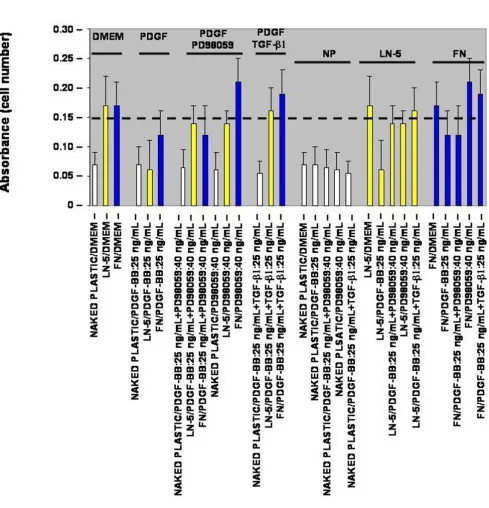
To determine whether or not the PDGF-BB-stimulated increase in rVSMC migration correlates with a reduction in rVSMC adhesion to laminin-5, thirty-minute in vitro adhesion assays were performed. In the absence of exoge-nous growth factor stimulation, laminin-5- and fibronec-tin-coated wells (20 µg/mL) sustained an approximate two and a half-fold increase in rVSMC adhesion com-pared with negative controls, as shown in Figure 2 (n = 24, p < 0.05). The addition of PDGF-BB, at the maximal migration-stimulating dose of 25 ng/mL, decreased the adhesion of rVSMC on laminin-5 by more than sixty-five (65) percent (n = 24, p < 0.005). The addition of exoge-nous PDGF-BB (25 ng/mL), however, decreased rVSMC adhesion on fibronectin by less than thirty (30) percent.
To investigate if the relationship between the PDGF-BB-stimulated increase in migration and corresponding decrease in adhesion of rVSMC on laminin-5 may be related to MAPK activation, cells were pre-treated with PD98059 (40 ng/mL) for twenty (20) minutes prior to assay and the adhesion media was supplemented with PD98059 (40 ng/mL). The addition of exogenous PD98059 (40 ng/mL) restored the PDGF-BB-stimulated reduction (25 ng/mL PDGF-BB) in rVSMC adhesion to laminin-5, to approximately eighty-five (85) percent of laminin-5 controls (n = 24, p < 0.05). The addition of PD98059, however, did not restore the thirty (30) percent PDGF-BB-stimulated reduction in rVSMC adhesion on fibronectin.
To determine if this modulation is restricted to MEK/ MAPK-inhibition, we tested the effects of adding exoge-nous transforming growth factor (TGF-β1). Our results indicated that the addition of TGF-β1 (25 ng/mL) was able to restore the PDGF-BB-stimulated reduction in rVSMC adhesion on laminin-5 by roughly the same level as PD98059, eighty two (82) percent versus eighty five (85) percent, respectively. In contrast to PD98059, the addition of TGF-β1 (25 ng/mL) was sufficient to restore the PDGF-BB-stimulated reduction in rVSMC adhesion on fibronectin.
Proliferation
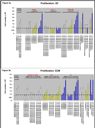
Based upon our observations of rVSMC migration and adhesion, we performed in vitro proliferation assays to determine the relative effects of the extracellular matrix (ECM) and exogenous growth factors described above. First, to test the effects of the ECM substrate on rVSMC proliferation in the absence of exogenous growth factors, quiescent cells were plated in cell-culture plates either coated with or without laminin-5 or fibronectin, in serum-free Dulbecco's Modified Eagle's Medium (DMEM) for one (1) to six (6) days.
To induce quiescence, rVSMC were incubated for forty eight (48) hours without mitogen (0% FCS, FBS) at 37°C and quiescence was verified by proliferation controls, cul-tured for forty eight (48) hours at 37°C with mitogen stimulus as shown in Figure 3a and 3b. Previous studies with VSMC report that greater than 95% of cells incubated in low-serum media (0.4% FCS or less) were arrested in G0(G1) between forty eight (48) and seventy two (72) hours as determined by flow cytometry and determina-tion of [3H] thymidine-labeled nuclei [15].
Our results suggest that culture of rVSMC plated on exog-enous laminin-5 (coated at a concentration of 20 µg/mL) did not significantly increase cellular proliferation com-pared with naked plastic controls over a period of four (4) days, as shown in Figure 3a (n = 24, p < 0.02). Culturing of rVSMC plated on exogenous fibronectin (coated at a concentration of 20 µg/mL) however, did significantly increase cellular proliferation over a period of four (4) days by nearly two-fold, as shown in Figures 3a and 3b.
Next, to test the modulating effects of exogenous growth factors on rVSMC proliferation when cultured on these ECM substrates, quiescent cells were plated in cell-culture plates either naked or coated with laminin-5 or fibronec-tin, in the presence of TGF-β1 or PDGF-BB, both at a con-centration of 25 ng/mL, from one (1) to six (6) days. Our results indicated that the addition of exogenous TGF-β1 (25 ng/mL) to the cell culture medium was sufficient to induce a suppressing effect on rVSMC proliferation on naked plastic, as well as laminin-5 and fibronectin, to lev-els approximating the quiescent growth controls.
Cell Communication and Signaling 2005, 3:2 http://www.biosignaling.com/content/3/1/2
PDGF-BB reduces rVSMC adhesion on laminin-5 adhesion in vitro Figure 2
The effect of growth factors on rVSMC proliferation in vitro Figure 3
Cell Communication and Signaling 2005, 3:2 http://www.biosignaling.com/content/3/1/2
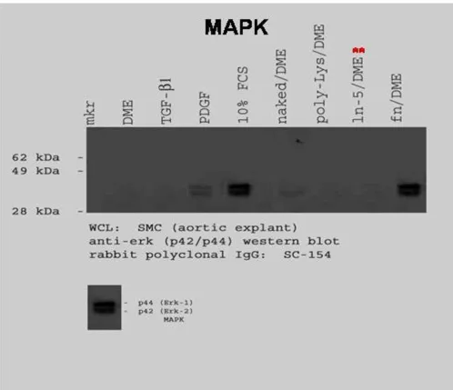
Mitogen Activated Protein Kinase (MAPK) Western Blot
To examine MAPK activation, rVSMC were plated onto laminin-5- or fibronectin-coated plates, then lysed after thirty minutes and prepared for immunoblotting. Our results indicated that laminin-5 did not induce a detecta-ble increase in MAPK activation over a thirty (30) minute time interval, as shown in Figure 4. rVSMC plated on fibronectin did exhibit a significant increase in MAPK lev-els after thirty (30) minutes.
The addition of growth factors, such as PDGF-BB and TGF-β1, have been associated with increases in p44/p42 (ERK1/2) or MAPK activation [16-18]. To determine the effects of PDGF-BB and TGF-β1 on rVSMC, subconfluent cell cultures were pre-treated with FCS (10%), PDGF-BB (25 ng/mL) or TGF-β1 (25 ng/mL) for thirty (30) minutes, then lysed and prepared for immunoblotting. p44/p42 phosphorylation levels were not detectable in DMEM-treated control cells, as shown in Figure 4. However, the The MAPK pathway in rVSMC is activated by different stimuli
Figure 4
addition of PDGF-BB, but not TGF-β1, was sufficient to induce measurable increases in MAPK activation levels.
Discussion
PDGF-BB is an in vitro VSMC mitogen and may be respon-sible for initiating the phenotypic changes in VSMC migration and proliferation during restenosis in vivo [19,20]. Our recent reports of low-level laminin-5 sion in the intima of normal arteries and increased expres-sion in the neointima of injured arteries suggest that laminin-5, in conjunction with soluble growth factors and mitogens, may determine VSMC phenotype during reste-nosis [11-13].
The current study augments the body of evidence that sug-gests VSMC growth is influenced by an ECM-VSMC inter-action [21] and that VSMC proliferation in different species respond differently to growth factor stimulus [15]. More specifically, this study explores evidence that lam-inin-5 and PDGF-BB may have combined and synergistic effects in determining rVSMC phenotype in vitro [6,8,10]. More specifically, our studies suggest that PDGF-BB strongly influences rVSMC behaviors such as migration. In addition, PDGF-BB significantly alters other cellular behaviors such as adhesion and proliferation on laminin-5, but only to a lesser extent on fibronectin.
Because PDGF-BB stimulation increases the overall levels of intracellular MAPK, as well as MAPK phosphorylation, we sought to explore this signaling cascade to determine its role in modulating these rVSMC behaviors on laminin-5 [22]. Specifically, the ERK1/2 form of MAPK mediates signaling by PDGF-AA, PDGF-AB, and PDGF-BB in VSMC, as well as signaling through laminin-5 binding integrins, and is therefore the most likely signaling molecule to modulate these cellular behaviors [23-25]. Our results suggest that rVSMC behaviors in vitro, driven by PDGF-BB responsiveness, can be blocked by MAPK inhibition (via MEK1 inhibitor: PD98059) on laminin-5, but not on fibronectin.
An additional signaling regulator, TGF-β1, has been implicated in the negative regulation and decreased rate of proliferation of VSMC stimulated with serum or PDGF [26-28]. Our results from this study indicate that TGF-β1, unlike the MEK1-inhibitor PD98059, was sufficient to block rVSMC behaviors on both laminin-5 and fibronec-tin. Specifically, the addition of TGF-β1 was able to reduce PDGF-BB-stimulated migration and proliferation of rVSMC on both ECM substrates and was also able to restore PDGF-BB-stimulated reductions in VSMC adhe-sion on these ECM.
Several lines of evidence now suggest that the anti-mitogenic effects of TGF-β1 may be dissociated from
inhi-bition of ERK1/2 signaling pathways [17,18]. These reports suggest that TGF-β1 inhibition of PDGF-BB may be temporally independent of other early signaling path-ways, such as MAPK, and is more likely to block VSMC behaviors, such as proliferation, by inhibiting events later in the G1 phase of mitosis.
Although our previous reports linked ERK1/2 to rVSMC adhesion and migration, these studies did not examine the possibility for differential signaling initiated by rVSMC binding to laminin-5 or fibronectin [12,13]. Expanding our original analysis of growth factor stimulation of MAPK to include ECM binding reveals that integrin binding of rVSMC to fibronectin strongly increases detectable MAPK activation levels, as does FCS and PDGF-BB stimulation, whereas binding to laminin-5 does not. These differences may help to explain the differ-ing effects on cellular behaviors of binddiffer-ing to these ECM ligands, as fibronectin and PDGF-BB may act in unison to activate intracellular signaling cascades that converge in MAPK activation, while laminin-5 may not.
Conclusions
Our results indicate that laminin-5 activates different intracellular signaling pathways from those of fibronectin and PDGF-BB in rVSMC and that binding to laminin-5 may modulate rVSMC behaviors that are distinctive from those modulated by fibronectin. Although binding of rVSMC to laminin-5 may not cause an initial increase in MAPK activation levels or proliferation, laminin-5 can augment PDGF-BB-stimulated proliferation and migra-tion of rVSMC in vitro. Based upon these findings, we pos-tulate that PDGF-BB and laminin-5 binding may initially activate different intracellular signaling cascades, causing rVSMC to be more responsive to the inhibition of MEK1 and MAPK on laminin-5 than those activated on fibronec-tin, as outlined in Figure 5.
In contrast to laminin-5, fibonectin and PDGF-BB may have parallel, reinforcing roles in MAPK activation. Our analysis of the effects of TGF-β1 demonstrated that
TGF-β1 does not strongly activate MAPK in rVSMC, but rather strongly inhibits the effects of fibronectin and PDGF-BB-stimulation on laminin-5. Our results support the previ-ous findings that TGF-β1 may inhibit mitogenesis and other VSMC behaviors via mechanisms independent of MAPK activation.
Cell Communication and Signaling 2005, 3:2 http://www.biosignaling.com/content/3/1/2
Integrins and growth factors activate intracellular signaling cascades in rVSMC
Figure 5
may help aid in the design of more effective therapies for the treatment of restenosis.
Methods
Cell cultureCells were maintained in 100 mm × 20 mm Corning tis-sue-culture dishes (Plainfield, NJ) at 37°C and 5% CO2 in humidified chambers. Cells were maintained in DMEM High Glucose, supplemented with 10% fetal bovine serum and 1% L-glutamine (29.2 mg/mL), penicillin G (10,000 U/mL), and streptomycin sulfate (10,000 mcg/ mL) (GPS) from Irvine Scientific (Santa Ana, CA). Rat aor-tic smooth muscle cell explants were a gift from RC Smith and were isolated and passaged as previously described [29]. Although greater than 95% quiescence, G0(G1) arrest can routinely be induced by incubation of cells for 72 h. in low-mitogen (0.5% FBS) medium [15,30], these authors suggest that incubation of VSMC for 48 h. without mitogen (0% FBS) is sufficient to induce quiescence. This was verified by proliferation control cells, cultured for 48 h. at 37°C with and without mitogen stimulus.
Migration assays
Cell migration assays were performed in Costar transwell filter plates either coated with purified matrix (laminin-5 or fibronectin) at a protein concentration of 20 µg/mL for one hour (60 min.) at room temperature, 25°C, and washed twice with phosphate-buffered saline 0.2% Tween-20 and 5% skim milk (PBST) prior to assay as pre-viously described [31,32]. Cells were seeded at a concen-tration of 1.2 × 10⁵ in each of 96-transwell chamber filters (100 µL of 1.2 × 10⁶ cells/mL solution) with and without ECM in the presence or absence of PDGF-BB at the indi-cated concentrations (5–25 ng/mL) and allowed to migrate for 18 hours at 37°C. Where applicable, the medium was supplemented with PD98059 (MEK1-inhib-itor) at the indicated concentration. Cells were counted at the end of an 18-hour interval as indicated, quantified with the following modification. 30 minutes prior to measuring migration, 5 µM calcein AM from Molecular Probes (Eugene, OR) was added to the migration wells at 37°C. To quantitate migration, cells were removed from the top of the filter with cotton-tipped applicators and flu-orescence of the incorporated calcein was measured from the bottom of the filter with a fluorescence plate reader. Relative fluorescence values for each experimental condi-tion are expressed relative to controls and untreated samples.
Adhesion assays
Cell adhesion assays were performed as previously described [31,32] using Costar 96-well cell culture cluster plates, coated with either laminin-5 or fibronectin solu-tion at a protein concentrasolu-tion of 20 µg/mL for 1 hour (60 min.) at room temperature, 25°C. Wells were then
washed twice with PBST prior to assay. Cells were seeded at a concentration of 1.2 × 10⁵ in each of 96-transwell chamber filters (100 µL of 1.2 × 10⁶cells/mL solution) with and without ECM-coating (described above) in the presence or absence of PDGF-BB (25 ng/mL), TGF-β1 (25 ng/mL), or both, and allowed to attach for 30 minutes at 37°C. Where applicable, cells were first incubated for 20 minutes with PD98059 (40 ng/mL), a MEK1 inhibitor from New England Biolabs (Beverly, MA) at 37°C and the adhesion assay culture medium was supplemented with PD98059 at 40 ng/mL. Following adhesion, non-adher-ent cells were removed by suspending plates upside down in a rotating tank of PBS for 10 minutes at room temper-ature, 25°C. Adherent cells were then fixed and stained and the relative absorbance was measured using a TECAN-SPECTRAFluor spectrophotometer (TECAN, Durham, NC) at 595 nm.
Proliferation assays
Tissue culture plates were coated with purified fibronectin from Calbiochem (La Jolla, CA) or laminin-5 from Demos (La Jolla, CA) at a 20 µg/mL protein concentration for 1 hour (60 min.) at room temperature, 25°C as previ-ously described [31]. Cells were seeded at a concentration of 1.2 × 10⁵ in 100 mm2 cell culture plates with and with-out fibronectin or laminin-5 and allowed to attach over-night (12 h.) at 37°C. Cells were then starved in serum-free DMEM for 48 hours to induce quiescence at 37°C, as outlined in Cell Culture Methods below. The medium was then replaced with fresh medium containing 25 ng/mL of TGF-β1 or PDGF-BB obtained from Calbiochem (La Jolla, CA) and incubated at 37°C. Cells were removed from cul-ture wells with trypsin/EDTA and counted using trypan blue stain from Gibco Life Technologies (Rockville, MD) and a VWR Scientific Counting Chamber (Plainfield, NJ) at 24 hour intervals, from 1 – 6 days.
Western Blot analysis
Cell Communication and Signaling 2005, 3:2 http://www.biosignaling.com/content/3/1/2
rabbit polyclonal IgG primary antibody and goat anti-rab-bit IgG-AP secondary antibody from Santa Cruz Biotech-nology (Santa Cruz, CA).
Statistics
The differences between untreated and treated cell popu-lations were measured using a t distribution. All samples were measured using two-tailed t tests as departure from normality can make more of a difference in a one-tailed than in a two-tailed t test. So long as the sample size is even moderate (>20) for each group, quite severe depar-tures from normality make little practical difference in the conclusions reached from these analyses [34].
Competing Interests
The author(s) declare that they have no competing interests.
Authors' contributions
KK carried out the migration, adhesion, and proliferation assays, the Western Blot analysis and assisted with experi-mental design. GEP conceived, monitored, and coordi-nated the experimental design. Both KK and GEP contributed equally to the writing of this manuscript.
Acknowledgements
KK and GEP thank Janice L. Huff and William L Rust for their assistance in reviewing experimental data, as well as Autumn Martinez and Christina Brown for their technical assistance in maintaining cell cultures and prepar-ing in vitro assays.
References
1. Abedi H, Zachary I: Signaling mechanisms in the regulation of vascular cell migration.Cardiovasc Res 1995, 30:544-556. 2. Grosenbaugh DA, Amoss MS, Hood DM, Morgan SJ, Williams JD:
Epidermal growth factor-mediated effects on equine vascu-lar smooth muscle cells.Am J Physiol 1988, 255:C447-C451. 3. Klagsbrun M, Edelman ER: Biological and biochemical
proper-ties of fibroblast growth factors: implications for the patho-genesis of atherosclerosis.Arteriosclerosis 1989, 9:269-278. 4. Fagin JA, Forrester JS: Growth factors, cytokines and vascular
injury.Trends Cardiovasc Med 1992, 2:90-94.
5. Intengan HD, Schiffrin EL: Structure and mechanical properties of resistance arteries in hypertension: role of adhesion mol-ecules and extracellular matrix determinants. Hypertension
2000, 36(3):312-318.
6. Morla AO, Mogford JE: Control of smooth muscle cell prolifer-ation and phenotype by integrin signaling through focal adhesion kinase.Biochem Biophys Res Commun 2000, 272:298-302. 7. Nguyen LL, D'Amore PA: Cellular interactions in vascular
growth and differentiation.Int Rev Cytol 2001, 204:1-48. 8. Lin CQ, Bissell MJ: Multi-faceted regulation of cell
differentia-tion by extracellular matrix.FASEB J 1993, 7:737-743.
9. Kanda S, Kuzuya M, Ramos MA, Koike T, Yoshino K, Ikeda S, Iguchi A: Matrix metalloproteinase and αVβ3 integrin-dependent vascular smooth muscle cell invasion through a type I colla-gen lattice. Arteriosclerosis, Thrombosis, and Vascular Biol 1999,
20:998-1005.
10. Raines EW, Koyama H, Carragher NO: The extracellular matrix dynamically regulates smooth muscle cell responsiveness to PDGF.Ann NY Acad Sci 2000, 902:39-51.
11. Kingsley K, Carroll K, Huff JL, Plopper GE: Photobleaching of arte-rial autofluorescence for immunofluorescence applications.
Biotechniques 2001, 30(4):794-797.
12. Kingsley K, Rust WL, Huff JL, Smith RC, Plopper GE: PDGF-BB enhances expression of, and reduces adhesion to, laminin-5 in vascular smooth muscle cells. Biochemical and Biophysical Research Communications 2002, 294:1017-1022.
13. Kingsley K, Huff JL, Rust WL, Carroll K, Martinez AM, Plopper GE:
ERK1/2 activation mediates PDGF-BB stimulated vascular smooth muscle cell proliferation and migration on laminin-5. Biochemical and Biophysical Research Communications 2002,
293:1000-1006.
14. Graves LM, Bornfeldt KE, Raines EW, Potts BC, Macdonald SG, Ross R, Krebs EG: Protein kinase A antagonizes platelet-derived growth factor-induced signaling by mitogen-activated pro-tein kinase in human arterial smooth muscle cells.Proc Natl Acad Sci 1993, 90(21):10300-10304.
15. Castellot JJ, Pukac LA, Caleb BL, Wright TC, Karnovsky MJ: Heparin selectively inhibits a protein kinase C-dependent mechanism of cell cycle progression in calf aortic smooth muscle cells.
Journal of Cell Biology109:3147-3155.
16. Servant MJ, Giasson E, Meloche S: Inhibition of growth factor-induced protein synthesis by a selective MEK inhibitor in aor-tic smooth muscle cells.J Biol Chem 1996, 271(27):16047-16052. 17. Cospedal R, Lobo M, Zachary I: Differential regulation of extra-cellular signal-regulated protein kinases (ERKs) 1 and 2 by cAMP and dissociation of ERK inhibition from anti-mitogenic effects in rabbit vascular smooth muscle cells.Biochem J 1990,
342:407-414.
18. Fu M, Zhang J, Lin Y, Zhu X, Zhao L, Ahmad M, Ehrengruber MU:
Early stimulation and late inhibition of peroxisome prolifer-ator-activated receptor γ (PPARγ) gene expression by trans-forming growth factor b in human aortic smooth muscle cells: a role of early growth-response factor-1 (Egf-1), activa-tor protein (AP1) and Smads.Biochem J 2003, 370:1019-1025. 19. Graf K, Xi XP, Yang D, Fleck E, Hsueh WA, Law RE:
Mitogen-acti-vated protein kinase activation is involved in platelet-derived growth factor-directed migration by vascular smooth mus-cle cells.Hypertension 1997, 29(2):334-339.
20. Pukac L, Huangpu J, Karnovsky MJ: Platelet-derived growth fac-tor-BB, insulin-like growth factor-1, and phorbol ester acti-vate different signaling pathways for stimulation of vascular smooth muscle cell migration.Exp Cell Res 1998, 242:548-560. 21. Herman IM, Catellot JJ: Regulation of vascular smooth muscle
cell growth by endothelia synthesized extracellular matrices.Arteriosclerosis7(5):463-9.
22. Yamaguchi H, Igarashi M, Hirata A, Susa S, Ohnuma H, Tominaga M, Daimon M, Kato T: Platelet-derived growth factor BB-induced p38 mitogen-activated protein kinase activation causes cell growth, but not apoptosis, in vascular smooth muscle cells.
Endocrinol J 2001, 48(4):433-442.
23. Kondo T, Konishi F, Inagami T: Differing signal transductions elicited by three isoforms of platelet-derived growth factor in vascular smooth muscle cells. J Biol Chem 1993,
268:4458-4464.
24. Inui H, Kitami Y, Tani M, Kondo T, Inagami T: Differences in signal transduction between platelet-derived growth factor AB receptors in vascular smooth muscle cells.J Biol Chem 1994,
269:30546-30552.
25. Jiang B, Yamamura S, Nelson PR, Mureebe L, Kent KC: Differential effects of platelet-derived growth factor isotypes on human smooth muscle cell proliferation and migration are medi-ated by distinct signaling pathways.Surgery 1996, 120:427-432. 26. Grainger DJ, Kemp PR, Witchell CM, Weissberg PL, Metcalfe JC:
Transforming growth factor beta decreases the rate of pro-liferation of rat vascular smooth muscle cells by extending the G2 phase of the cell cycle and delays the rise in cyclic AMP before into into M phase.Biochem J 1994, 299(1):227-235. 27. Grainer DJ, Kemp PR, Liu AC, Lawn RM, Metcalfe JC: Activation of transforming growth factor-beta is inhibited in transgenic apolipoprotein(a) mice.Nature 1994, 370(6489):460-462. 28. Kirschenlohr HL, Metcalfe JC, Weissberg PL, Grainger DJ:
Prolifer-ation of human aortic vascular smooth muscle cells in cul-ture is modulated by active TGF beta.Cardiovasc Res 1995,
29(6):848-855.
Publish with BioMed Central and every scientist can read your work free of charge "BioMed Central will be the most significant development for disseminating the results of biomedical researc h in our lifetime."
Sir Paul Nurse, Cancer Research UK
Your research papers will be:
available free of charge to the entire biomedical community
peer reviewed and published immediately upon acceptance
cited in PubMed and archived on PubMed Central
yours — you keep the copyright
Submit your manuscript here:
http://www.biomedcentral.com/info/publishing_adv.asp
BioMedcentral
30. Smith RC, Wills KN, Antelman D, Perlman H, Truong LN, Krasinski K, Walsh K: Adenoviral Constructs Encoding Phosphoryla-tion-Competent Full-length and Truncated Forms of the Human Retinoblastoma Protein Inhibit Myocyte Prolifera-tion and Neointima FormaProlifera-tion. Circulation 1997,
96(6):1899-1905.
31. Plopper GE, McNamee HP, Dike LE, Bojanowski K, Ingber DE: Con-vergence of integrin and growth factor receptor signaling pathways within the focal adhesion complex.Molecular Biology of the Cell 1995, 6(10):1349-1365.
32. Wagner JE, Huff JL, Rust WL, Kingsley K, Plopper GE: Perillyl Alco-hol Inhibits Breast Cell Migration without Affecting Cell Adhesion. Journal of Biomedicine and Biotechnology 2002,
2(3):136-140.
33. Spector DL, Goldman RD, Leinwand LA: Subcellular Localization of Genes and Their Products. In Cells: A Laboratory Manual Cold Spring Harb Lab Press, Cold Spring, NY; 1998:51.6-51.10.
34. Hays WL: Inferences about population means. Statistics