1. ENTOMOL Soc BRIT COLUMBIA 98. DECEMBER 2001 189
Implications of using development rates of blow fly (Diptera:
Calliphoridae) eggs to determine postmortem interval
SHERAH L. V ANLAERHOVEN,
1and GAIL S. ANDERSON/
CENTRE FOR PEST MANAGEMENT
DEPARTMENT OF BIOLOGICAL SCIENCES, SIMON FRASER UNIVERSITY,
8888 UNIVERSITY DRIVE; BURNABY, BC, CANADA VSA IS6
ABSTRACT
This research examined the eclosion times of blow fly eggs to determine whether eggs begin to develop at the time of oviposition, or in vivo. Eggs were obtained from laboratory colonies of Calliphora vic ina Robineau-Desvoidy, Phaenicia
sericala (Meigen) and Ellcalliphora lalifrons (Hough) and observed at 2-h intervals. All three species had eggs eclose earlier than the expected minimum of 22 h at 21°C. Precocious egg development occurred for 75% of the C vicina egg mass, while 100% of the E lal!/i'ons and P. sericala egg masses developed early.
Subsequently, we denied an oviposition medium to fresh C vicina and P. sericata colonies for 7 and 14 d and compared the eclosion times with that of eggs from colonies with a continual access to beef liver. In both species, no precocious egg development was observed as the eggs eclosed 3-4 h after the expected minimum time of eclosion in both treatments and control. Finally, we examined eclosion times of eggs laid by blow flies in the wild. Eggs laid in the wild by P sericala and
C vicina also took 1-3 h longer to eclose than the expected minimum time of eclosion. Our first experiment demonstrated that eggs laid by a single female at one time, can eclose at a wide variety of times, ranging from 2 h to the expected 22 h after oviposition at 21°C. Our inability to repeat the early eclosion in the laboratory with new colonies, despite the denial of oviposition media, or in the wild under natural conditions, is reassuring to those using egg development and eclosion to determine elapsed time since death. Clearly this phenomenon is not common, and may be explained as an ani fact of laboratory colonies that do not have a regular influx of wild blow flies.
Key words: forensic entomology, medico-legal entomology, elapsed time since death
INTRODUCTION
Forensic entomology, or the use of insects to determine the elapsed time since death of a homicide victim, is a technique that has been employed in many homicide investigations worldwide (Goff 1992; Leclercq and Vaillant 1992; Lord et al. 1994; Anderson 1995). It is the most accurate and often the only method available to determine elapsed time since death after 72 h. However, it also is used during the first 72 h after death, particularly in high profile crimes, to confirm pathological parameters, or when only a portion of the body has been recovered. Traditionally, medical parameters are used to determine time since death in the first hours after death, but these involve many variables (Henssge et al. 1995)
and pathologists are often reluctant to offer an opinion on time since death when more than
I Pacific Agri-Food Research Centre, Agassiz, BC
190 1. ENTOMOL Soc. BRIT. COLUMBIA 98, DECEMBER 200l
a few hours have passed. Thirty percent of forensic entomology cases in Canada in 1995 involved blow fly (Diptera: Calliphoridae) egg evidence alone and this trend has continued (Anderson and Cervenka 200 I). Although the cases were mainly homicide, they also included one poaching case where blow fly egg development evidence was vital in connecting time of death of bear cubs with the perpetrators at the scene (Anderson 1999).
Since blow flies usually arrive and begin laying eggs within minutes of death (Anderson and VanLaerhoven 1996), an analysis using the eclosion times of blow fly eggs will provide an estimate of the minimum time since death in the early postmortem interval. This method requires accurate research on the developmental rates of eggs. Previous research indicates that the time necessary for blow fly eggs to eclose depends on the species and the temperature (Kamal 1958; Nuorteva 1977; Greenberg 1993; Anderson 2000). However, these developmental rates and times of eclosion assume that egg development begins after oviposition and that the eggs do not begin to develop within the adult fly. Our laboratory research has indicated that this may not always be the case. If in vivo development does occur, this would change the estimate of elapsed time since death by as much as 24 h. We hypothesized that female flies which have a suitable oviposition medium available, will oviposit eggs which eclose after the normal length of time, after oviposition; whereas flies which are denied a suitable oviposition medium may have eggs developing in vivo, thereby decreasing the length of time between oviposition and eclosion.
The objectives of this research were to: determine whether insect eggs laid on a homicide victim begin to develop at the time of oviposition, or in vivo, as we have observed occasionally in the lab; and to determine whether early eclosion occurs in the wild or is an artifact of laboratory conditions.
MATERIALS AND METHODS
We examined egg eclosion under laboratory conditions at 21°C for three species of blow fly: Calliphora vicina Robineau-Desvoidy, Phaenicia sericala (Meigen) and
Eucalliphora latifi'ons (Hough). All three species were reared in laboratory colonies
descended from wild specimens collected locally in the Lower Mainland of British Columbia. They had been under laboratory conditions for approximately a year. On 5 March 1994, beef liver was presented to gravid females and after several hundred eggs were laid by -10 females over a 30 min period, the liver was removed from the cages. Each egg mass was examined for eclosion immediately after oviposition and at 2 h intervals until eclosion.
1. ENTOMOL SOC BRIT COLUMBIA 98. DECEMBER 200l 191
We also tested eggs laid by blow lly females in the wild. Petri dishes with
approximately 250 g of fresh beef liver were exposed in partially sunny locations in
eoquitlam, Be. The experiments were conducted between 17-25 September 1996 and 2-10 June 1997. At all times, blow llies were abundant in this mild region. The experiment was
replicated 15 times. After oviposition of at least 100 eggs in a 30-min period, the petri dishes were covered to prevent further oviposition and moved indoors. Each egg mass was examined for eclosion at 2-h intervals until eclosion.
Ambient temperature was recorded at 30-min intervals throughout each experiment using a double channel datalogger (Smal1Reader I ®, Young Environmental Systems, Richmond, BC). Temperatures cited are means of records from the time eggs were laid
until eclosion was complete.
RE
SULTS
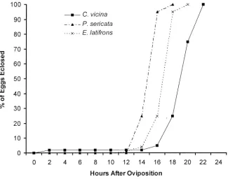
All three species had eggs eclose earlier than expected at 21 °e (Table I). Precocious
egg development occurred for 75% of the C. vicina egg mass, while 100% of the E.
tali/rons and P sericala egg masses developed early (Fig. I).
100 A .X
_____ C. vicina ~ X'
90 I
- .•. -P. sericata
80 ... x· .. E. latifrons
"0 70
Q) !II
60
.Q
C,,) W
!II 50 ;
Cl
Cl I
W 40
"- I
0 ~ 0
30
i
20 I
;<
10
.X 0
0 2 4 6 8 10 12 14 16 18 20 22 24
Hours After Oviposition
Figure 1. Percent of eggs eclosed from egg masses of three laboratory colonies of blow llies.
When new colonies of P. sericata and C. vicina were established, no precocious egg
development was observed (Table 2), despite the lack of ovipositional media. Phaenicia sericala and C. vic ina females took 3 d at 21 °e to develop mature eggs in their ovaries.
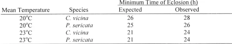
In the field experiments, no precocious egg development was observed for eggs laid by P. sericata and C. vicina (Table 3). The mean temperature was 200
192 J. ENTOMOL soc. BRIT. COLUMBIA 98. DECEMBER 200l
Table 1
Egg eclosion from egg masses of laboratory colonies of blow flies compared to expected
minimum times of eclosion at 21 °e (Anderson 2000).
Species
C vicina
P. sericata
E latif;-ons
Minimum Time of Eclosion (h)
Expected Observed
22 2
21 22
Table 2
14 12
Egg eclosion from egg masses of laboratory colonies of blow flies: held continuously with
oviposition media available; held 7 d without oviposition media; and held 14 d without oviposition media, compared to expected minimum times of eclosion at 21 °e (Anderson
2000).
Oviposition media
Available Available
7d
7d 14 d
14 d
Species
C vicina
P. sericala
C vicina P. sericala C vicina
P. sericala
Table 3
Minimum Time of Eclosion (h)
Expected Observed
22 26
21 22 21 22 21 24 26 24 26 24
Egg eclosion from egg masses of wild blow flies compared to expected minimum times of eclosion (Anderson 2000).
Mean Temperature 200
e 200
e
23°e
23°e
Species
C vicina
P. sericala
C vicina
P. ,I'erica/a
Minimum Time of Eclosion (h)
Expected Observed
26 28
25 21 21
DISCUSSION
26 24 24It is currently accepted that blow fly eggs do not generally develop in the female fly, but only begin to develop after oviposition. Therefore, a measure of the developmental
stage can be used to predict the age of the egg, and the time of eclosion can be used to
count backwards to determine the time of oviposition. However, our first laboratory
experiment demonstrated that eggs laid at the same time can eclose at a wide variety of times, ranging from 2 h to the expected 22 h after oviposition.
Early eclosion of blow fly eggs has been described in the literature, although it is rare (Auten 1934; Reiter 1984; Erzinclioglu 1990). It is possible that female flies may delay oviposition until a suitable site is found (Auten 1934), One recent study examined internal
egg development of Phormia regina (Meigen) and stated that only one developing egg can
1. ENTOMOL. Soc BRIT COLUMBIA 98. DECEMBER 2001 193
do not enter the oviduct until oviposition (Erzinclioglu 1990). Another study examined Calliphora lerraenovae Macquart, C. vomiloria (L.), C. vicina and P. .s·ericala (Meigen) and found precocious egg development of at least one egg within all four species (Wells and King 200 I).
The trigger for development within the female remains unknown. Our inability to repeat the early eclosion in the laboratory with new, wild-captured colonies, despite the denial of oviposition media, or in the wild under natural conditions, is reassuring to those using egg development and eclosion to determine elapsed time since death. Clearly this phenomenon is not conunon, and may be explained as an artifact of lab colonies that do not have a
regular influx of wild blow flies; it may even have been an artifact of those specific colonies, although this seems unlikely. In fact, in a large number of other experiments
conducted over several years, in which eggs were observed every 1-2 h until eclosion, not once was this phenomenon observed (Anderson 2000). As well, many other researchers who have performed similar experiments have not mentioned early eclosion (Melvin 1934; Kamal 1958; Nuorteva 1977; Nishida 1984; Greenberg 1993).
ACKNOWLEDGEMENTS
This research was supported by a research grant from the Pathology/Biology Section of
the American Academy of Forensic Sciences. We would like to thank Dr. Margaret Dogterom for the use of her property and Simon Fraser University for the use of its facilities. We would also like to extend our gratitude to Steve Halford for advice and
assistance, and to Hersimer Johl and Jasmine Wiles. We would like to thank Dr. Lisa Poirier for her advice and editorial comments.
REFERENCES
Anderson, G.S. 1995. The use of insects in death investigations: an analysis of forensic entomology in
British Columbia over a tive year period. Canadian Society of Forensic Sciences Journal 28: 277-292. Anderson, G.S. 1999. Wildlife forensic entomology: determining time of death in two illegally killed black
bear cubs, a case report. Journal of Forensic Sciences 44: 856-859.
Anderson, G.S. 2000. Minimum and maximum developmental rates of somc forensically important
Calliphoridae (Diptera). Journal of Forensic Sciences 45: 824-832.
Anderson, G.S. and V.J. Cervcnka. 200 I. Insects associated with the body: their usc and analyses. In: W.D. Haglund and M. Sorg, (Eds.) Forensic Taphonomy, Thc Postmortem Fate of I-Iuman Remains.
CRC Press, Boca Raton.
Anderson, G.S. and S.L. VanLaerhoven. 1996. Initial studies on insect succession on carrion in
southwestern British Columbia. Journal of Forensic Sciences 41: 617-625.
Auten, M. 1934. The early cmbryological development of Phormia regina: Diptera (Calliphoridae). Annals of the Entomological Society of America 27: 481-506.
Erzinclioglu, Y.Z. 1990. On the interpretation of maggot evidence in forensic cases. Medical Science and
Law 30: 65-66.
Erzinclioglu. Y.Z. 1996. Blowllies. Naturalist's Handbooks 23 Richmond Publishing Co. Ltd., Slough, UK.
Gore M.L. 1992. Problems in estimation of postmortem interval resulting from wrapping of the corpse -a
case study fi'om Hawaii. Journal of Agricultural Entomology 9: 237.
Greenberg, B. 1993. Different developmental strategies in two boreal blow Ilies (Diptera: Calliphoridae).
Journal of Medical Entomology 3: 481-484.
Henssge.
c..
B. Madea, 13. Knight. L. Nokes and T. Kroillpecher. 1995. The estilllation of the tillle sincedeath in the early postillorteill interval. 2"" Edition, Arnold. London.
Kalllal, A.S. 1958. COlllparative study of thirteen species of sarcosaprophagolls calliphoridae and sarcophagidae (Diptera) I. Bionoillics. Annals of the Entomological Society of America 51: 261-270. Leclercq, M. and F. Vaillant. 1992. Forensic Entomology: An original case. Annales De La Societe
194 1. ENTOMOL. SOc. BRIT. COLUMBIA 98, DECEMBER 2001
Lord, W. D., M.L. Goff, TR. Adkins and N.H. Haskell. 1994. The black soldier fly Hel'lI1elia illucens
(Diptera: Stratiomyidae) as a potential measure of human postmortcm interval: observations and casc
histories. Journal of Forensic Sciences 39: 215-222.
Melvin, R. 1934. Incubation period of eggs of certain muscoid !lies at different constant temperatures.
Annals of the Entomological Society of America 27: 406-410.
Nishida, K. 1984. Experimental studies on the estimation of postmortem intervals by means of tly larvae
infesting human cadavers. Japan Journal of Legal Medicine 38: 24-41.
Nuorteva, P. 1977. Sarcosaprophagous insects as forensic indicators. pp. 1072-1095 In: e.G. Tedeschi
(Ed.) Forensic medicine: a study in trauma and environmental hazards. Saunders, New York.
Reiter, e. 1984. Zum Wachtstumsverhalten der Maden der blauen Sehmeissfliege Calliphora vicina. Z.
Rechtsmed. 91: 295-308.