Doctoral School in Materials Science and Engineering – XXIV cycle
Capturing Complex Microenvironments for
Directed Stem Cell Differentiation
A Biomimetic Approach to Multifaceted, Complex
Biomaterial Design for Specific Tissue Regeneration
Michael Louis Floren
C
APTURING
C
OMPLEX
M
ICROENVIRONMENTS FOR
D
IRECTED
S
TEM
C
ELL
D
IFFERENTIATION
A
B
IOMIMETICA
PPROACH TOM
ULTIFACETED,
C
OMPLEXB
IOMATERIALD
ESIGN FORS
PECIFICT
ISSUER
EGENERATIONMichael Louis Floren
E-mail: [email protected]
Approved by:
Prof. - - - -, Advisor Department of - - - University of - - - -, Country.
Prof. - - - -, Department of - - - University of - - - -, Country.
Ph.D. Commission:
Prof. - - - -, Department of - - - University of - - - -, Country.
Prof. - - - -, Department of - - - University of - - - -, Country.
Prof. - - - -, Department of - - - University of - - - -, Country.
University of Trento,
Department of - - -
University of Trento - Department of - - -
Doctoral Thesis
Name - 2013
Published in Trento (Italy) – by University of Trento
Dedicated to:
Elizabeth Susan Floren
Abstract
Loss of vascular function associated with cardiovascular disease, such as arthrosclerosis, represents the leading medical epidemic in the United States and typically requires surgical intervention through synthetic or autologous vascular grafts. To overcome the limitations associated with adult cell sources, which are often restricted by supply or compromised by disease, mesenchymal stem cells (MSCs) have emerged as potential candidates for vascular tissue engineering. While evidence suggests the roles of several factors influencing MSC differentiation into vascular phenotypes, including matrix rigidity, geometry and chemistry, the phenomena associated with these events are still largely unknown. Further, the development of mature vascular phenotypes, such as vascular smooth muscle cells (vSMCs), with functional behavior remains elusive to the research community.
This thesis proposed to engineer and direct specific and mature vascular differentiation from MSCs by way of highly tailored matrices mimicking the vascular niche environment. Taking inspiration from natural organization, we contend that a biomimetic design approach to tissue scaffolds that display features of the natural cellular microenvironment whilst mimicking the bulk tissue properties may elicit highly specific differentiation of MSCs to vascular phenotypes. To validate our hypothesis, we employed a systemic approach incorporating physical and chemical microenvironmental cues, i.e. stiffness, biological ligands and chemical factors, with the aim to augment vascular phenotype expression, functionality, and final incorporation into a tailored biomaterial scaffolds.
First, we present a novel technique for the preparation of silk hydrogels directly from high pressure CO2 environments without the need for crosslinking agents or
additional additives such as surfactants or co-solvents. Through this novel method, we demonstrate the utility of CO2 as a volatile electrolyte, capable of sufficiently
influencing the sol-gel transition of silk proteins, resulting in the formation of stable hydrogels with properties suitable for biomedical applications.
address this, we investigated cellular differentiation on tunable SF hydrogels prepared using a solvent-free CO2 processing method. The focus of this portion of
the thesis is on exploiting the combined use of substrate stiffness and growth factor (TGF- β1) on SF matrices, with the aim of correlating the effects on the vascular commitment of human mesenchymal stem cells (hMSCs). Our data reveal that hMSC differentiation into mature SMCs can be achieved within modest culture periods (72 h) by combining appropriate SF hydrogel stiffness (33 kPa) with growth factor (TGF-β1). These findings advance our understanding of how complex multicomponent biomaterials, whereby mimicking the intricacy of natural tissue environments, can play a significant role in developing optimal stem cell differentiation protocols.
Third, we postulated that the presentation of ECM proteins on 3D matrices with tunable stiffness will augment the differentiation of MSCs to vascular lineages. To address this, we established a high-throughput ECM platform based on soft, fibrous PEG hydrogels meanwhile highly-tunable in stiffness and 3-dimensional geometry. Using this technique, we identified several microenvironments supporting MSC adhesion, spreading and differentiation toward early vascular lineages. This portion of the thesis supports the hypothesis that a complex milieu exists coupling protein functional behavior with substrate rigidity and that this phenomenon may potentially be exploited through proper application of high-throughput screening methodologies.
This thesis has provided fundamental insights into the effects of physiological stimuli on vascular differentiation of MSC in terms of the specificity and maturity of the final differentiated cells. Better understanding of such mechanisms will prove paramount in the sequential stages of MSC differentiation to mature vascular cells. Additionally, the findings of this thesis will help to better define the process of regenerating functional healthy vascular tissue from MSCs. Altogether, a combinatorial approach investigating the effects of matrix elasticity, biological ligands and growth factors on MSC differentiation in a 3D nanofiber culture will be critical towards understanding and recapitulating MSC differentiation in the in vivo vascular
Table of contents
Chapter I
Introduction ...15
1.1. Cardiovascular Disease: Motivation for New Therapies ...15
1.2. Tissue Engineering Therapies ...15
1.3. Stem Cells in Tissue Regeneration ...16
1.3.1. Vascular Regeneration ...16
1.4. Microenvironmental Cues Direct Stem Cell Fate Decisions 17
1.4.1. Biological Ligands Instruct Stem Cell Differentiation ...18
1.4.2. Soluble Factors as Critical Signals for MSC Differentiation ...18
1.5. Biomimetic Scaffolds for Stem Cells ...19
1.5.1. Scaffold Requirements ...19
1.5.2. Material Selection ...20
1.5.2.1. Synthetic Polymers ...20
1.5.2.2. Natural Polymers ...21
1.5.3. Fabrication Techniques ...22
1.6. Tailoring Microenvironments to Control Stem Cell Fate ...23
1.6.1. Current State of the Art ...23
1.6.2. Systemic Approaches to Multivariate Studies ...24
1.6.3. Translational Methods to Integrate Complex Stem Cell Environments ...25
1.7. Significance & Motivation for this Research ...25
Chapter II Specific Objectives ...27
2.1. Introduction ...27
2.2. Aims of this Thesis ...27
2.2.1. Aim 1 ...27
2.2.2. Aim 2 ...28
2.2.4. Aim 4 ...30
Chapter III Carbon Dioxide Induced Silk Protein Gelation for Biomedical Applications ...32
3.1. Introduction ...33
3.2. Materials & Methods ...36
3.2.1. Preparation of Silk Fibroin Solution ...36
3.2.2. Fabrication of Silk Hydrogels ...37
3.2.2.1. Atmospheric Conditions ...37
3.2.2.2. High Pressure Conditions ...37
3.2.2.3. Determination of Gelation Kinetics ...39
3.2.3. Gel Physical Properties ...39
3.2.3.1. Structural Analysis by Fourier Transform Infrared Spectroscopy ...39
3.2.3.2. Environmental Scanning Electron Microscopy ...39
3.2.3.3. Scanning Electron Microscopy ...40
3.2.3.4. Swelling Ratio ...40
3.2.3.5. Mechanical Properties in Compression ...40
3.2.4. Statistical Analysis ...41
3.3. Results & Discussion ...41
3.3.1. Gross Examination of Prepared Silk Hydrogels ....41
3.3.2. Gelation Kinetics ...42
3.3.2.1. In situ observation ...42
3.3.2.2. Gelation Screening ...42
3.3.3. Monitoring of Silk Structural Changes by FTIR ...44
3.3.4. Hydrogel Physical Characteristics ...46
3.3.4.1. Microstructure ...46
3.3.4.2. Hydrogel Swelling Behavior ...49
3.3.5. Properties in Compression ...51
3.3.6. Phenomenological Description ...54
3.3.6.1. Acidification Kinetics using Carbon Dioxide ...54
3.4. Conclusion ...64
Chapter IV Human MSCs Cultured on Silk Hydrogels with Variable Stiffness and Growth Factor Differentiate into Mature SMC Phenotype ...66
4.1. Introduction ...67
4.2. Materials & Methods ...69
4.2.1. SF Hydrogel Preparation ...69
4.2.2. Hydrogel Structural Characterization ...70
4.2.2.1. Thermal analysis by differential scanning calorimetry ...70
4.2.2.2. Fourier-Transform Infrared Spectroscopy (FTIR) analysis ...70
4.2.2.3. Morphology Assessment ...71
4.2.2.4. Mechanical Properties in Compression ...71
4.2.3. Cell Culture methods ...71
4.2.4. Cell Characterization methods ...72
4.2.4.1. Quantitative real-time polymerase chain reaction (qPCR) ...72
4.2.4.2. Immunofluorescent Staining ...73
4.2.4.3. Confocal Imaging ...73
4.2.5. Statistical analysis ...74
4.3. Results ...74
4.3.1. Hydrogel Fabrication and Characterization ...74
4.3.2. Hydrogel Pore Characteristics ...77
4.3.3. Hydrogel Compressive Properties ...78
4.3.4. Effect of SF Hydrogel Rigidity on hMSC attachment and spreading in Low-Serum or TGFβ-1 Fortified Media ...79
4.3.5. Effect of SF Hydrogel Rigidity and TGF-β1 on Vascular SMC Commitment of hMSCs ...81
4.4. Discussion ...84
Chapter V
Three-Dimensional, Soft Neotissue Arrays as High Throughput Platforms for the Interrogation of Engineered Tissue Environments
...90
5.1. Introduction ...91
5.2. Materials & Methods ...92
5.2.1. Materials ...92
5.2.2. Fabrication of PEGDM Soft Matrices ...93
5.2.3. Characterization of PEGDM Soft Matrices ...94
5.2.3.1. FTIR Analysis ...94
5.2.3.2. Scanning Electron Microscopy Imaging ...94
5.2.3.3. Fluorescent Imaging ...94
5.2.3.4. Rheology ...95
5.2.4. ECM Protein Array Preparation ...95
5.2.5. Cell Seeding and Cell Culture ...96
5.2.6. Immunofluorescent Staining ...97
5.2.7. Confocal Imaging ...97
5.2.8. Image & Statistical Analysis ...98
5.3. Results ...98
5.3.1. Characterization of 3-dimensional Fibrous Soft Hydrogel Matrix ...98
5.3.2. Design and Optimization of 3-dimensional Protein Microarray ...100
5.3.3. Stem Cell Adhesion and Spreading within 3-dimensional Neotissue Arrays ...103
5.3.4. Effect of Protein Environment and Matrix Elasticity on MSC Adhesion and Spreading ...104
5.3.5. Effects of Matrix Elasticity and Adhesive Protein Environment on MSC Fate Commitment in 3-dimensional Engineered Neotissues ...107
5.4. Discussion ...110
5.5. Conclusion ...116
Nanofibrous Photoclickable Hydrogel Microarrays for High-Throughput
Screening of Cellular Microenvironments ...117
6.1. Introduction ...118
6.2. Materials & Methods ...119
6.2.1. Macromer synthesis ...119
6.2.2. Electrospinning ...119
6.2.3. Scanning Electron Microscopy ...120
6.2.4. Peptide Synthesis ...120
6.2.5. Fluorescent Peptide Synthesis ...121
6.2.6. Engineered Peptide Array Preparation ...121
6.2.7. Cell Seeding & Cell Culture ...122
6.2.8. Immunofluorescent Staining ...122
6.2.9. Confocal Imaging ...122
6.2.10. Statistical analysis ...123
6.3. Results ...123
6.3.1. Characterization of 3-dimensional fibrous PEG thiol-ene hydrogel matrix ...123
6.3.2. Engineered Peptide Synthesis and Array Fabrication ...124
6.3.3. Primary Cell Adhesion and Spreading within Peptide Functionalized PEG thiol-ene Matrices ...125
6.3.4. Primary Cell Adhesion and Spreading within PEG thiol-ene Peptide Functionalized Arrays ...127
6.4. Discussion ...128
6.5. Conclusion ...129
Chapter VII Conclusion and Future Perspectives ...130
7.1. Conclusions ...130
List of abbreviation and acronyms ...136
References ...137
Scientific Production ...165
Participation to Congresses, Schools and Workshops ...166
Chapter I
Introduction & Background
1. Motivation
1.1. Cardiovascular Disease: Motivation for New Therapies
There is a pressing need to develop advanced strategies for the treatment of cardiovascular diseases (CVD). Cardiovascular disease (CVD) is the leading cause of death in the United States associated with 1 out of every 4 deaths as reported in 2009 (Kochanek, 2009). Current therapies rely on surgical intervention using autologous vascular bypass grafts such as the saphenous vein; however, these therapies are restricted in practice as they require surgical harvest and may be limited by a prior disease state or previous use (Weintraub, 1994). Approximately 1.5 million cases are alternatively treated with synthetic vascular grafts; however, these grafts commonly fail due to stenosis, an abnormal narrowing of the vessel, or thrombosis, formation of a blood clot in the vessel. These conditions arise because current synthetic vascular grafts fail to successfully integrate with the local vasculature at the implant site. Various recent studies have attempted to improve the biorecognition and integration of synthetic grafts by loading the substrates with primary vascular cells. Nonetheless, such methods still represent limited successful outcomes due to the need for invasive surgery and the limited expansion and functionality of these primary cells in vitro.
1.2. Tissue Engineering Therapies
tissues are highly organized, ranging from nanoscale matrix proteins, providing a medium for biological cues between adjacent cells, to macroscopic structures that influence the architecture and mechanical function of such vessels. This stunning organization found in nature at all size scales allows tissues to perform functions beyond the single cell. Vascular tissues rely on the viable and functional extra cellular matrix (ECM) to impart the necessary requisites of mammalian vasculature (Cleary, 2012). Taking inspiration from this natural organization, several reports have focused on preparing vascular tissue scaffolds that display features of the natural cellular environment whilst mimicking the bulk architecture; together mirroring the essential building blocks for functional vasculature.
1.3. Stem Cells in Tissue Regeneration
1.3.1. Vascular Regeneration
Stem cell therapy promises to revolutionize the treatment of vascular diseases with the potential to regenerate vascular tissues in vitro or in vivo (Krawiec, 2012). Mesenchymal stem cells (MSCs) are multipotent cells capable of differentiating into several tissues including vascular phenotypes, can be easily obtained and are rapidly expandable in vitro, thereby providing several advantages for their utility in vascular tissue regeneration (Kan, 2005). There is growing evidence that MSCs reside in a perivascular niche which is thought to interact largely with vascular signals in vivo and may play a significant role in local tissue regeneration and homeostatsis of the vasculature (da Silva Meirelles, 2008). Further, a recent study developed a protocol to produce functional vascular SMCs from induced pluripotent stem cells (iPSCs) via an MSC intermediate (Bajpai, 2012). The iPSC-derived MSC were found be both highly proliferative and capable of differentiating into highly contractile SMCs. The preceding citations lend credibility to the hypothesis that MSCs represent a potent cell source for the regeneration of highly specific and functional vascular tissues.
functional behavior. The vascular niche constitutes a complex microenvironment which conveys a variety of signals, eliciting specific biological responses and maintaining tissue function and homeostasis. Further resolution of the mechanisms and environmental signaling which regulate MSC differentiation towards vascular phenotypes has profound implications towards cell-based vascular tissue engineering and regeneration.
1.4. Microenvironmental Cues Direct Stem Cell Fate Decisions
There is abundant evidence to suggest that local signals from the tissue specific ECM microenvironments, through cell-ligand, cell-molecule or cell-matrix activation, significantly affect MSC differentiation and mature phenotype expression (Reilly, 2010; Suzuki, 2010; Engler, 2006) (Fig. 1). Substrate physical properties such as elasticity (Engler, 2006; Discher, 2005) and geometry (Cukierman, 2001; Huebsch, 2010) have been implicated to strongly influence MSC differentiation. Our
group has previously demonstrated that 3D nanofibrous PEG gel substrates with elasticity that mimics the natural vascular stiffness (E= 8 to 15 kPa) can direct MSC differentiation
toward vascular phenotypes within 24 hours (Wingate, 2012). Besides substrate physical properties, MSC differentiation and phenotype commitment are also greatly influenced by matrix chemical markers such as growth factors or matrix chemistry.
Together, these signals impart an instructional environment whereby MSC behavior and differentiation are directed in a highly specific manner. However, successful recreation of this complex milieu which directs MSCs to vascular phenotypes in vitro as well as the underlying mechanisms governing these events is still widely unknown. A biomimetic approach, incorporating several environmental signals, such as cell-matrix and cell-biomolecule interactions, will help to establish more robust and specific MSC differentiation protocols.
1.4.1. Biological Ligands Instruct Stem Cell Differentiation
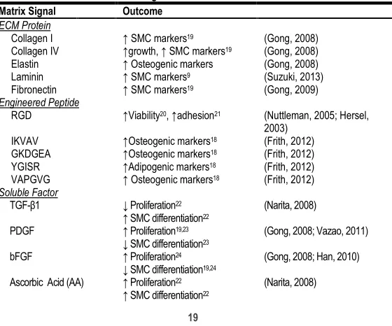
Cell-matrix and cell-biomolecule interactions play a critical role in diversity of biological events including cell adhesion, growth, differentiation, and apoptosis (Discher, 2009; Kuraitis, 2012). Growing evidence shows that that MSCs acquire tissue-specific characteristics when co-cultured with mature cells types or exposed to preformed biological matrices in vitro and that this instructive differentiation is elicited by the specific signals of the microenvironment (Philp, 2005). However, matrix and soluble factor (SF) signals are often observed independently to differentiate cells on 2D substrates, an environment vastly different from the way cells are presented naturally in vivo, i.e. a 3D tissue context which elicits multiple signal inputs to regulate cell fate. It is likely that crosstalk of these environmental factors on intracellular signaling molecules may be required to direct MSC differentiation toward desired cell type and function. Several reports have focused on the influence of different matrix signals such as ECM proteins, ECM-derived peptides and SFs on regulating MSC differentiation to vascular lineages (Table 1). A recent report demonstrated the potential of small peptides incorporated into 2D rigid substrates to guide MSC differentiation towards osteogenic or adipogenic lineages respectively (Frith, 2012). However, while this study provides insight into small peptide directed MSC differentiation; systematic studies of tailored peptides to instruct MSC differentiation to vascular phenotypes on soft 3D substrates are absent.
Recent evidence suggests that precise, complex SF regimens can result in highly specific and mature VSMCs from iPSCs (Lachaud, 2013; El-Mounayri, 2013). However, these protocols are often time consuming, utilizing multiple factors and requiring weeks to reach maturity. Studying the activity of early cell signaling events in the presence of SFs may help to refine current factor protocols. Several pathways mediating SF effects on VSMC differentiation from iPSCs have also been revealed recently. Though largely unexplored, the potential crosstalk sites between signaling induced by matrix adhesion and chemical signaling for vascular morphogenesis include ERK, AKT, RhoA or JNK (Discher, 2009; Martino, 2009; Park, 2011). Reports suggest inhibition of ERK activation in MSCs induces spontaneous VSMC differentiation, whereas RhoA mediating TGF-β signaling transduction or integrin mechanotransduction signaling leads to Smad activation for VSMC differentiation (Han, 2011). Despite considerable progress towards mechano-chemical signaling phenomena during cellular processes, questions remain as to how these mechanisms direct early instruction of MSC differentiation. Further, systematic studies evaluating the effect of growth factorregimens on early MSC differentiation processes is yet to be explored.
Table 1. Effect of Various Matrix Signals on hMSC Growth & Differentiation Matrix Signal Outcome
ECM Protein
Collagen I ↑ SMC markers19 (Gong, 2008) Collagen IV ↑growth, ↑ SMC markers19 (Gong, 2008) Elastin ↑ Osteogenic markers (Gong, 2008) Laminin ↑ SMC markers9 (Suzuki, 2013) Fibronectin ↑ SMC markers19 (Gong, 2009) Engineered Peptide
RGD ↑Viability20, ↑adhesion21 (Nuttleman, 2005; Hersel, 2003)
IKVAV ↑Osteogenic markers18 (Frith, 2012) GKDGEA ↑Osteogenic markers18 (Frith, 2012) YGISR ↑Adipogenic markers18 (Frith, 2012) VAPGVG ↑ Osteogenic markers18 (Frith, 2012) Soluble Factor
TGF-β1 ↓ Proliferation22 ↑ SMC differentiation22
(Narita, 2008)
PDGF ↑ Proliferation19,23
↓ SMC differentiation23 (Gong, 2008; Vazao, 2011) bFGF ↑ Proliferation24
↓ SMC differentiation19,24
(Gong, 2008; Han, 2010)
Ascorbic Acid (AA) ↑ Proliferation22 ↑ SMC differentiation22
Retionic Acid (RA) ↑ SMC differentiation23 (Vazao, 2011)
1.5. Biomimetic Scaffolds for Stem Cells
1.5.1. Scaffold Requirements
The design of 3D tissue scaffolds is essential to the success of an engineered tissue in order to permit cell adhesion, proliferation, differentiation, permeability for nutrients as well as structural support for tissue growth (Langer, 1993). To achieve this, a scaffold should include several criteria such as suitable porosity, pore size, permeability for nutrients, material biocompatibility and degradation, whilst mirroring the mechanical behavior of the intended tissue (Chua, 2001; Stevens, 2010). Ideally, a scaffold with high porosity improves cell infiltration as well nutrient diffusion. However, scaffold physical properties, such as porosity and cell geometry, influence the mechanical behavior of such constructs (Gibson, 1982). In this regard, a careful balance between mechanical function and elicited biological response must be considered when designing a tissue scaffold. Particularly with biodegradable polymers, as the material degrades mechanical properties diminish; thereby presenting the necessity to prepare scaffolds that maintain the required support for tissue growth before degradation. This concept is particularly significant for load bearing tissues, such as bone or vasculature, which provides appropriate mechanical properties due in part to its unique architecture and internal anisotropy (Fratzl, 2007).
1.5.2. Material Selection
tissue infilitration and tolerance of the foreign material are necessary to encourage new tissue formation. By adjusting the properties of the material utilized, the body’s response to foreign materials can be controlled such that cells can proliferate and infiltrate.
1.5.2.1. Synthetic Polymers
Synthetic polymers offer several advantages as materials for developing tissue engineering scaffolds including the ability to tailor mechanical properties and degradation kinetics to meet various applications. Specifically, synthetic polymers are attractive because they can be fabricated into various shapes with desired morphologies and features which can be permissive for cell maintence and in-growth (Gunatillake, 2003). For example, synthetic polymers can be produced reproducibily with specific molecular weights, block structures, degradable moities, and crosslinking mechanims. These properties in turn, govern material formation dynamics, crosslinking density, and material mechanical and degradation properties. PEO and poly(ethylene glycol) (PEG) are hydrophilic polymers that can be photocrosslinked by modifying each end of the polymer with either acrylates or methacrylates (Cruise, 1998; West, 1999; Mann, 2001). Hydrogels can then be prepared when the modified PEO or PEG is mixed with the appropriate photoinitiator and crosslinked via UV exposure (West, 1999; Bryant, 2001). Synthetic hydrogels are often attractive materials for thier inert properites since they lack cell adhesion receptors and proteins often do not readily absorb to them. Specifically, PEG has been used to prevent post-operative adhesions (West, 1996) and to prevent intimal thickening of arteries after damage (West, 1996). However, while synthetic materials are attractive for their cost, reproducible fabrication and facile manufacturing, their lack of cell-recognition sites as well as potential for toxic degradation products causing undesirable inflammation are often disadvantageous (Seo, 2013).
1.5.2.1. Natural Polymers
hierarchical organization, as well as providing anchoring sites for other extracellular matrix components. Among naturally derived fibrous proteins, silk fibroin, the primary structural protein of natural Bombyx Mori silk fibers, is a natural polymer that has been studied extensively for tissue engineering applications in part to its excellent mechanical properties, biocompatibility, and controllable degradation rate . Silk proteins have found significant utility in biomedicine due to their high biocompatibility, tunable biodegradability and material format versatility. Perhaps its earliest biomaterial rendition, natural silk fibers have been employed as sutures for wound ligation for centuries (Moy, 1991). Wound dressings have also been prepared using pure silk electrospun nets (Schneider, 2009) or with silk blends of PEG (Kweon, 2008) or carboxymethyl keratin (Lee, 1999), demonstrating improved inflammation response and wound healing kinetics. Silk fibroin coatings are also attractive for implant materials. Improved adhesion of human fibroblasts has been reported on silk fibroin coated polyurethane (Petrini, 2001) and polycarbonate (Chiarini, 2003) surfaces. Other adaptions of silk fibroin implant technologies include anterior cruciate ligament (ACL) reconstruction (Altman, 2002) knee meniscus repair (Mandal, 2011) and nerve guidance conduits (Yang, 2007).
1.5.3. Fabrication Techniques
nanofibers meshes is the absence of macroscopic structures which support porosity and interconnectivity, severely limiting cell infiltration and nutrient diffusion.
Recent studies have highlighted the importance of 3-dimensional, fibrous matrices to optimize stem cell niche environments (Carlson, 2012; Lim, 2009). One potential solution is the use of hydrogels which present both a fibrous microstructure reminiscent of the natural ECM combined with excellent nutrient diffusion. Hydrogels are networks of polymer chains that are stabilized either by chemical or physical crosslinking and dispersed throughout an immobilized water phase. The stability of the polymer network allows for the penetration and uptake of water (swelling) without dissolving, thus lending itself to a variety of attractive and practical applications. Hydrogels have become excellent material candidates for a wide selection of biomedical applications because of their high water content (allowing for efficient transport of biological molecules), improved biocompatibility and innate similarity to the physiochemical nature of natural tissues (Drury, 2003; Peppas, 2006).
1.6. Tailoring Microenvironments to Control Stem Cell Fate
1.6.1. Current State of the Art
2011). Here it was found that substrate elasticity could invoke different SC fate commitments in the presence of growth factor which alone was not sufficient. Expanding on this theory, Wingate et al. combined VEGF administration with a soft, three-dimensional substrate optimally designed for endothelial cells (ECs). The authors reported that the substrate combined with growth factor were significantly more effective at directing stem cell fate towards EC lineage than growth factor alone (Wingate, 2014). The two preceding citations provide compelling evidence that a synergistic role of individual environmental factors can assist in the MSC differentiation process. Challenges in the future will be in the discovery of multiple signal effectiveness for specific SC lineage commitment as well as platforms designed for end-user tunablility.
1.6.2. Systemic Approaches to Multivariate Studies
High through-put approaches have emerged in recent years to circumvent the limitations of traditional low through-put techniques (i.e. conventional cultureware), with the promise to develop complex platforms for combined biomolecule/substrate discovery. An early example of such technology, Flaim et al. developed an ECM microarrary to investigate the role of specific proteins on SC differentiation (Flaim, 2005). By spotting proteins onto acrylamide coated slides, the authors presented 32 different protein conditions in the form of discrete protein dots on the order of 200µm. The technology was reproducible and capable of producing multiple microarrays with significantly less protein requirement than traditional methods. The authors investigated the influence of the different protein conditions in the presence of both primary hepatocytes and mouse embryonic SCs. Interrogation of the respective cell
Table 2. Review of Biomaterials for Stem Cell Differentiation by Variable
Year Material Geometry Elasticity Biological Ligand Soluble Factor Ref
2006 2D planer soft - - Engler, 2006
2008 PGA mesh 3D scaffold stiff ECM proteins Various Gong, 2008
2010 PEGDM Gel 3D gel soft RGD - Huebsch, 2010
2011 Collagen I/PA gel 2D planer soft - TGF-β1 Park, 2011
2012 PS-PEO copolymer 2D planer stiff engineered peptides Frith, 2012
2012 PEGDM Nanofiber Gel 3D nanofibers soft - - Wingate, 2012
2013 Pegylated-Fibrin Gel 2D/3D gel soft - TGF-β1 Stowers, 2013
lines indicated several protein combinations which either maintained hepatocyte function or persuaded SC differentiation. While this report lends strong credibility to high through-put techniques, the incorporation of multiple signals within designed microarrays has proven to be a challenge within the field. An approach developed by Gobaa et al. has proven an important step as it provides the opportunity to analyze a greater number of variables and more rapidly than previous attempts (Gobaa, 2011). In particular, the authors designed individual hydrogel microwells, with both modular stiffness as well as variable protein composition. This report represents a paradigm shift in study of SC differentiation events from the traditional methods of single specimen culture ware to the sample robust high through-put capabilities. The development of complex high through-put platforms to investigate SC differentiation with combinatorial signaling will likely prove instrumental towards the design of future biomaterial platforms.
1.6.3. Translational Methods to Integrate Complex Stem Cell Environments
specialized with engineered vascular niche environments will be of considerable value to the research community.
1.7. Significance & Motivation for this Research
Chapter II
Specific Objectives
2.1. Introduction
phenotype expression and functionality, via relevant biomaterial scaffolds. We expand on these aims as follows:
2.2. Aims of this Thesis
2.2.1. Aim 1
The first objective of this thesis was to develop tunable hydrogel systems based on the natural polymer silk fibroin. Silk fibroin (SF), a natural protein extracted from Bombyx mori silkworms, is an attractive material for tissue engineering due to its excellent mechanical properties, biocompatibility, tunable degradation rate, and mild inflammatory response in vivo [Wang, 2008]. A diversity of regenerative tissues has been reported using SF-based constructs including bone [Meinel, 2005; Fini, 2005), cartilage (Wang, 2010), vascular (Soffer, 2008; Bondar, 2008; Bonani, 2011), skin (Unger, 2004), nervous (Yang, 2007), hepatic (Gotoh, 2004) and ocular (Lawrence, 2009) amongst others (Kundu, 2013). In light of these reports, the future and relevance of silk biomaterials for therapies catered to the biomedical community are believed to be great (Motta, 2012).
Hydrogels have become excellent material candidates for a wide selection of biomedical applications because of their high water content (allowing for efficient transport of biological molecules), improved biocompatibility and innate similarity to the physiochemical nature of natural tissues (Drury, 2003; Peppas, 2006). Recently, porous natural-based hydrogels have been chemically crosslinked under high pressure CO2 without the necessity for surfactants or co-solvents (Partap, 2006; Annabi, 2009; Annabi, 2010). Hydrogels produced using high pressure CO2 generally exhibit greater porosity and improved crosslinking, resulting in improved gel stiffness as well as an enhanced capacity to support cell and tissue infiltration (Annabi, 2010). The potential of CO2 to induce silk protein gelation without the need
for extensive chemical processes, circumventing complex materials fabrication protocols and avoiding complications to biological systems, would presumably be of considerable value to the biomaterials community.
Through this novel method, we demonstrate the utility of CO2 as a volatile electrolyte, capable of sufficiently influencing the sol-gel transition of silk proteins, resulting in the formation of stable hydrogels with properties suitable for biomedical applications. The influence of CO2 pressure, silk protein concentration and processing time were investigated in regard to the gelation kinetics, physical and mechanical properties of the prepared gels (Chapter III).
2.2.2. Aim 2
The second objective of this thesis was to study the influence of growth factor (TGF-β1) combined with silk fibroin hydrogels of varying stiffness, as prepared in Aim 1, on the differentiation of MSCs into a mature SMC phenotype.
Cell interactions with the local microenvironment are recognized in several important biological events including cell adhesion, growth, differentiation, and apoptosis [5, 6]. In particular, substrate biophysical properties such as rigidity (Engler, 2006; Discher, 2005), geometry (Cukierman, 2001; Huebsch, 2010) biological ligand (Suzuki, 2010; Gong, 2008), soluble factor (Narita, 2008), or combination thereof (Wingate, 2014) have been revealed to influence MSC differentiation events. However, integration of complex cellular signaling environments into biomaterial scaffolds presents a considerable challenge to the tissue engineering community (Kim, 2012). We previously developed a technique to produce porous, SF hydrogels with tunable stiffness and morphology using the green solvent, carbon dioxide (CO2), see Aim 1. Hydrogel elastic moduli approaching soft tissues (E = 6-30 kPa), combined with ease of fabrication and biocompatibility, motivated us to use these SF materials as a platform to instruct stem cell differentiation towards the vascular smooth muscle cell (SMC) lineage in a precise manner.
differentiation of stem cells (Narita, 2008). Therefore, the focus of this objective is on exploiting the combined use of substrate stiffness and growth factor (TGF- β1) on SF matrices, with the aim of correlating the effects on the vascular commitment of human mesenchymal stem cells (hMSCs).
2.2.3. Aim 3
Despite accumulated knowledge regarding individual and combined roles of various mechanochemical ECM signals in stem cell activities, the intricacy exhibited by cellular microenvironments poses a considerable challenge in resolving the mechanisms ascribed to stem cell behavior and fate determination processes. This complexity mandates a systemic approach whereby integrative studies must be expanded to capture a more comprehensive understanding of the determinants which direct stem cell differentiation toward desired cell type and function.
High through-put approaches have emerged in recent years to circumvent the limitations of traditional low through-put techniques (i.e. conventional cultureware), with the promise to develop complex platforms for combined biomolecule/substrate discovery. However, despite the versatility afforded by current microarray technologies, the incorporation of multiple signals within engineered microarrays remain limited. Meanwhile the integration of current combinatorial microarray technologies in three-dimensions, coupled with other biophysical properties, such as tunable stiffness and geometry, have yet to reach fruition.
2.2.4. Aim 4
Stem cell (SC) therapy promises to revolutionize the treatment of various diseases with the potential to regenerate functional tissues in vitro or in vivo. Several recent studies have demonstrated that cellular microenvironments such as ligand-activated cell-matrix interactions and/or matrix physical properties such as elasticity and geometry have significant role in directing the differentiation processes in stem cells (Lutolf, 2009; Wingate, 2012). Microarrays have emerged as an important tool for studying stem cell processes in a high-throughput manner (Gupta, 2010; Gobba, 2011). Nevertheless, most of the existing ECM arrays being either 2-D or shallow 3-D are not able to capture the effects of biophysical and chemical cues on stem-cell fate completely.
Chapter III
Carbon Dioxide Induced Silk Protein Gelation
for Biomedical Applications
Part of this chapter has been published in:
Michael L. Floren, Sara Spilimbergo, Antonella Motta, Claudio Migliaresi. “Carbon Dioxide Induced Silk Protein Gelation for Biomedical Applications”. Biomacromolecules, 13 (2012)
2060-2072.
Abstract: We present a novel method to fabricate silk fibroin hydrogels using high pressure carbon dioxide (CO2) as a volatile acid without the need for chemical
crosslinking agents or surfactants. The simple and efficient recovery of CO2 post
processing results in a remarkably clean production method offering tremendous benefit towards materials processing for biomedical applications. Further, with this novel technique we reveal that silk protein gelation can be considerably expedited under high pressure CO2 with the formation of extensive β-sheet structures and
stable hydrogels at processing times less than 2 hours. We report a significant influence of the high pressure CO2 processing environment on silk hydrogel physical
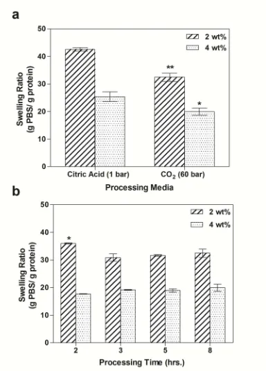
the less porous and heterogeneous structures of the citric acid control gels. The swelling ratios of silk hydrogels prepared under high pressure CO2 were significantly
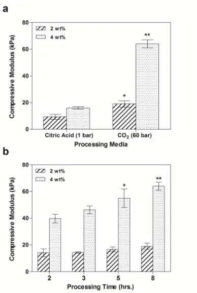
reduced compared to the citric acid control gels, which we attribute to enhanced physical crosslinking. Mechanical properties were found to increase significantly for the silk hydrogels prepared under high pressure CO2, with a 2 and 3 fold increase in
the compressive modulus of the 2 and 4 wt% silk hydrogels over the control gels, respectively. We adopted a semi-empirical theoretical model to elucidate the mechanism of silk protein gelation demonstrated here. Mechanistically, the rate of silk protein gelation is believed to be a function of the kinetics of solution acidification from absorbed CO2 and potentially accelerated by high pressure effects. The
attractive features of the method described here include the acceleration of stable silk hydrogel formation, free of residual mineral acids or chemical crosslinkers, reducing processing complexity, and avoiding adverse biological responses, whilst providing direct manipulation of hydrogel physical properties for tailoring toward specific biomedical applications.
3.1. Introduction
Hydrogels are networks of polymer chains that are stabilized either by chemical or physical crosslinking and dispersed throughout an immobilized water phase. The stability of the polymer network allows for the penetration and uptake of water (swelling) without dissolving, thus lending itself to a variety of attractive and practical applications. Hydrogels have become excellent material candidates for a wide selection of biomedical applications because of their high water content (allowing for efficient transport of biological molecules), improved biocompatibility and innate similarity to the physiochemical nature of natural tissues (Drury, 2003; Peppas, 2006).
counterparts. However, despite these limitations, natural polymers generally present improved biocompatibility and cellular interactivity, thereby enhancing their utility in the fabrication of biomaterials (Lee, 2001).
Among naturally derived proteins, silk fibroin, the primary structural protein of Bombyx mori (mulberry silkworm) silk fibers, is a natural polymer that has been studied extensively for biomedical applications due to its excellent mechanical properties, biocompatibility, and controllable degradation rate (Altman, 2003). The high content of hydrophobic domains in silk proteins allows for the self-assembly and formation of strong intra- and intermolecular β-sheet structures, providing the basis for the exceptional strength observed in natural silk fibers (Bini, 2004). The combination of the impressive mechanical performance in its natural form as well as excellent biocompatibility and controllable degradation highlights silk fibroin as an ideal biomaterial candidate meeting several therapeutic requirements. Silk proteins have been fashioned into several material formats such as films (Servoli, 2005; Motta, 2002; Motta, 2011), sponges (Nazarov, 2004; Kim, 2005; Li, 2001), fibrous networks (nano and micrometric) (Unger, 2004; Bondar, 2008; Silva, 2008; Kim, 2003; Jin, 2002), microspheres (Wang, 2007), and hydrogels (Ayub, 1993; Kim, 2004; Wang, 2008; Motta, 2004; Silva, 2008; Fini, 2005; Zhang, 2011). Remarkable progress has been reported toward the application of silk hydrogels for tissue engineering and drug delivery therapies. Fini et al. demonstrated the healing of confined critical-sized cancellous defects in the femoral condyles of New Zealand rabbits using injectable silk hydrogels (Fini, 2005). Silk hydrogels have also found value as drug delivery vehicles. Zhang et al. combined sonicated silk hydrogels with vascular endothelial growth factor (VEGF) and bone morphogenetic protein 2 (BMP-2) to successfully palliate defects in the rabbit maxillary sinus floor (Zhang, 2011). In light of these advances, the future and relevance of silk hydrogels for therapies catered to the biomedical community are believed to be great (Motta, 2012).
crosslinking and a stabilized hydrogel network. Conversely, at modest protein concentrations (<5 wt%) gelation is particularly time expensive, requiring days to reach complete gel formation. The sol-gel transition of aqueous silk fibroin solutions can be improved through the manipulation of the physiochemical environment such as protein concentration, temperature, ionic strength, low pH, or, alternatively, by physical methods such as sonication, shear vortexing and electrogelation (Ayub, 1993; Kim, 2004; Wang, 2008; Motta, 2004; Silva, 2008; Fini, 2005; Zhang, 2011; Motta, 2012; Matsumoto, 2006; Hanawa, 1995; Yucel, 2009; Servoli, 2008). For instance, reports have demonstrated a significant reduction in gelation time, from days to a matter of hours, at reduced solution pH near to the isoelectric point of silk fibroin (pH = 3.8-4.0) (Ayub, 1993; Matsumoto, 2006; Hanawa, 1995). However, the addition of solution electrolytes at non-physiological concentrations, such as mineral acids or metal ions, may have unfavorable effects on cellular activity and function when placed in a living subject; therefore, ideally the fabrication of such materials should proceed without the necessity for chemical processes.
High pressure carbon dioxide (CO2) has emerged in recent years as an
environmentally benign alternative to conventional organic solvents commonly employed for fashioning various biomaterials, such as facilitating porosity in tissue scaffolds as well as processing materials with thermally sensitive therapeutic agents (proteins) (Kazarian, 2000; Howdle, 2001; Floren, 2011). Recently, porous natural-based hydrogels have been chemically crosslinked under high pressure CO2 without
the necessity for surfactants or co-solvents (Partap, 2006; Annabi, 2009; Annabi, 2010). Hydrogels produced using high pressure CO2 generally exhibit greater
porosity and improved crosslinking, resulting in improved gel stiffness as well as an enhanced capacity to support cell and tissue infiltration (Annabi, 2010). Nonetheless, although these improvements are significant, it is important to note that residual crosslinking agents not recovered after processing or released during material degradation may result in adverse effects to biological systems (Jayakrishnan, 1996).
An alternative approach involves directly employing CO2 as a volatile
dioxide has been used extensively as a volatile electrolyte for the controlled isoelectric precipitation of food proteins such as soy and casein (Hofland, 2000; Hofland, 1999). Volatile acids are advantageous to conventional mineral acid processing, such as sulfuric and hydrochloric acids which are frequently employed by conventional precipitation processes, as their high volatility in atmospheric conditions allows for their efficient recovery (Thiering, 2001). Additionally, unlike conventional mineral acids, solution pH can be maintained simply by tuning the electrolyte solubility within solution, which here is a function of pressure and temperature. Appreciable pH drops of aqueous solutions have been reported at modest fractions of dissolved CO2 (Bortoluzzi, 2011). Li et al. reported the pH of
CO2-H2O systems approaching 4 at modest pressures (5 bar) (Li, 2007). The slow,
homogenous acidification of aqueous solutions by CO2 has generally been reported
to improve the precipitation and recovery of proteins or molecules which exhibit sensitivity to extreme drops in pH (Thiering, 2001). Thus, the ability to experience considerable drops in solution pH at relatively low fractions of absorbed CO2 permits
its utility as a viable alternative to conventional mineral acids. The potential of CO2
to induce silk protein gelation without the need for extensive chemical processes, circumventing complex materials fabrication protocols and avoiding complications to biological systems, would presumably be of considerable value to the biomaterials community.
Here, for the first time, we present a novel technique for the preparation of silk hydrogels directly from high pressure CO2 environments without the need for
crosslinking agents or additional additives such as surfactants or co-solvents. Through this novel method, we demonstrate the utility of CO2 as a volatile
electrolyte, capable of sufficiently influencing the sol-gel transition of silk proteins, resulting in the formation of stable hydrogels with properties suitable for biomedical applications. The influence of CO2 pressure, silk protein concentration and
processing time were investigated in regard to the gelation kinetics, physical and mechanical properties of the prepared gels. Gelation time could be significantly improved depending on the processing conditions employed. Moreover, the physical and mechanical properties of silk hydrogels prepared under high pressure CO2
conventional acid titration. Mechanistically, reduction in solution pH by CO2
absorption, governed by diffusion limitations, and potentially the high pressure environment, both influence the gelation of silk proteins as presented here. The use of high pressure CO2 as a volatile electrolyte to precisely control and accelerate silk
protein gelation, providing the direct tailoring of hydrogel physical properties, whilst being readily recoverable post processing, potentially offers tremendous benefit as a clean and efficient method toward silk hydrogel fabrication.
3.2. Materials & Methods
3.2.1. Preparation of Silk Fibroin Solution
Neat silk fibroin was obtained from cocoons of Bombyx mori silkworms and subsequently degummed to remove the exterior sericin proteins. Degumming was achieved by washing the extracted silk fibroin in boiling solutions of Na2CO3 at 1.1
g/L and 0.4 g/L both for 1.5 hours. Fibroin solution was prepared by dissolving the obtained fibers in 9.3M LiBr (Fluka Chemicals, Buchs, Switzerland) aqueous solution (10% w/v) at 65 °C for 4 hours and filtered to eliminate impurities. Filtered fibroin solution was then placed in Slide-A-Lyzer cassette (Pierce, 3500 Da MWCO) and dialyzed against distilled water for 3 days at room temperature to remove the residual salts. Following dialysis, solution protein concentration was determined using a NanoDrop ND-1000 spectrophotometer (Delaware). Solution volume was adjusted with distilled water to reach the desired concentration, namely 2 and 4 wt%.
3.2.2 Fabrication of Silk Hydrogels
3.2.2.1 Atmospheric Conditions
3.2.2.2 High Pressure Conditions
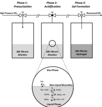
A schematic of the high pressure CO2 processing protocol is detailed in Figure
1. The high pressure apparatus consisted of two configurations: a large volume steel reactor (450 ml) custom fabricated in house for the preparation of large specimens, and a smaller volume (200 ml) high pressure quartz view cell reactor (Separex, France) which allowed for direct in situ observation during processing. Vessel temperature was controlled via an internal water-jacketed heat exchanger fed from an external water bath (MPM Instruments, Italy). The high pressure CO2
atmosphere was achieved via a HPLC pump (Gilson, mod 307 drive module with a maximum flow rate of 25 ml/min) set to the desired operating pressure. The high pressure vessel consisted of both a temperature and pressure transducer which allowed for accurate monitoring of both variables over the experimental times. Silk fibroin solution (5 ml) at different concentrations (2 & 4 wt%) were syringed into a 25 ml Teflon beaker ( h: 35 mm, Ø 30mm) and placed into the high pressure vessel. Once the system had been sealed and thermal equilibrium established (40 °C), CO2
was introduced into the vessel under various pressures, namely 5, 30, 60, 100 and 150 bar. Once pressure and temperature had reached stability, specimens were isolated and maintained at the set conditions for specific processing times (0-8 hours). Following processing, the system was depressurized slowly (approximately 30 minutes) to avoid sample rupture due to the high pressure release and maintain hydrogel integrity. Collected hydrogel specimens were immediately placed in distilled water and stored at 4 °C for future characterization.
Two sample preparations, hydrated and dry specimens, for the silk hydrogels were utilized for the measurements in this study. Hydrated samples were taken directly from the high pressure CO2 apparatus and characterized without further
Figure III - II. Schematic diagram of silk gelation using high pressure CO2. Phase 1: Pressurization, Phase 2: Acidification through carbonic acid formation, Phase 3: Protein
aggregation and gelation.
3.2.2.3 Determination of Gelation Kinetics
To establish the gelation time of silk fibroin solution under different conditions of high pressure carbon dioxide, 5 ml volumes of silk fibroin 2 and 4 wt% were pipetted into a 25 ml Teflon beaker and exposed to high pressure carbon dioxide (5-150 bar) at constant temperature 40 °C. Gelation time was determined upon examination of specimens removed from the high pressure environment at set pressures and processing times. Gelation was confirmed by the formation of a homogenous, opaque gel lacking sufficient viscosity to flow from an inverted beaker.
3.2.3 Gel Physical Properties
3.2.3.1 Structural Analysis by Fourier Transform Infrared Spectroscopy
transition during gelation, 5 ml volumes of 2 wt% silk solutions , 0.6cm thickness, were submitted to high pressure CO2 (60 bar) for 0-2 hours and collected every 15
minutes for structural characterization. A second set of samples was used to access the structural features of the silk hydrogels after 8 hours processing time. To preserve the secondary structure of the obtained silk hydrogels, specimens were immediately quenched in liquid nitrogen after treatment by high pressure CO2 and
subsequently lyophilized for 48 hours to obtain dry samples for characterization. We performed preliminary experiments comparing the spectra of lyophilized and hydrated gels obtained directly after processing in high pressure CO2 and found
similar data produced by both conditions. Structural data was acquired by loading lyophilized silk hydrogel specimens onto the IR apparatus and sample spectrums were collected as a mean of 32 acquisitions (between 4000 cm−1 and 400 cm−1) with a spectral resolution of 4 cm−1.
3.2.3.2 Environmental Scanning Electron Microscopy
To evaluate silk hydrogel surface morphology in the native state, wet specimens were examined using an environmental scanning electron microscope (ESEM, Philips XL 30 ESEM, Philips, Eindhoven, Netherlands) at a working distance 10-15 µm and voltage of 15 kV. Gel specimens were observed without any further treatment at a vacuum range (5-7 Torr) at constant temperature (5 °C).
3.2.3.3. Scanning Electron Microscopy
Dry imaging of hydrogel specimens was obtained using a scanning electron microscope (SEM) (Quanta 200 Scanning Electron Microscope – FE – operating mode: low vacuum, gaseous secondary electron GSE detector). Wet silk hydrogels were first quenched in liquid nitrogen for 5 minutes and subsequently lyophilized for 48 hours to prepare dry cross sections for imaging. Prior to imaging, lyophilized cross sections were sputter coated (Biorad SC500, Hemel Hempstead, UK) with a thin layer of gold to avoid charging of the sample.
3.2.3.4. Swelling Ratio
the swollen silk hydrogels was recorded (Wwet). Subsequently, the samples were quenched in liquid nitrogen for 5 minutes followed by lyophilization for 48 hours to obtain the dry mass (Wdry). The swelling ratio was then calculated from the following relation:
Swelling Ratio g PBS g protein⁄ = WwetW−Wdry
dry
3.2.3.5. Mechanical Properties in Compression
The mechanical performance of hydrated specimens was performed in the unconfined state by a uniaxial compression mechanical tester (Bose ELF3400) with a 50 N load cell. For testing specimens, cylindrical plugs (10 mm diameter, 6-7 mm height) were punched out of a large volume silk hydrogel (5 ml) and subsequently submersed in PBS at room temperature. Samples were let to stand no more than 1 hour prior to the mechanical testing. The mechanical properties of the gel samples were tested in the wet state, in PBS, at room temperature. Strain (mm) and load (N) were recorded using Wintest software at a cross speed of 20 µm/s up to 60% strain level. To ensure proper sample placement and flatness, samples were cyclically preconditioned at 1% strain for 10 cycles. From the collected data, a stress-strain plot was rendered and the compressive modulus extrapolated from the tangent slope at 10% strain of the stress-strain curve.
3.2.4. Statistical analysis
All tests were performed in triplicate. Statistical significance of collected data was determined at each condition using an independent Student’s t-test. Data are presented as mean ± standard deviation (SD) and was considered statistically significant at 95% confidence (p < 0.05).
3.3. Results and Discussion:
3.3.1. Gross Examination of Prepared Silk Hydrogels
The macrostructure of silk hydrogels prepared using high pressure CO2 and
soft, heterogeneous consistency of hydrogels prepared at atmospheric conditions using citric acid (Figure 2(a)). Mechanical integrity as well as elastic properties was also apparent from the 4 wt% silk hydrogels prepared under high pressure (Figure 2 (c,d)). Gel consistency and rigidity between the two protein concentrations was noticeably different, with higher concentrations (4 wt%) exhibiting more resilient mechanical stiffness compared to lower concentrations (2 wt%) which were often too soft for simple handling. The mechanical performance of silk hydrogels prepared under high pressure CO2 will be further evaluated in the mechanical properties
section of this paper.
Figure IIII - 2. Silk hydrogels at 2 wt% protein concentration prepared by conventional citric acid titration (a) and under high pressure CO2 (b). Demonstration of the handling properties of
a 4 wt% silk hydrogel prepared under high pressure CO2 (c,d).
3.3.2. Gelation Kinetics 3.3.2.1. In situ observation
Silk fibroin aqueous solutions at 2 and 4 wt% protein concentrations were processed under high pressure carbon dioxide at several pressures (5-150 bar) and processing times (2-8 hours). In situ gelation observations permitted from the high pressure quartz view cell reactor are presented in Figure 3. The introduction of high pressure CO2 resulted in a transition of the transparent silk solution to a slightly
into the solution under stagnant conditions can be considered slow even at elevated pressures (Thiering, 2001), the evidence of a turbid solution immediately upon submission to high pressure CO2 indicates the potential role of the hydrostatic
pressure environment, the effects of which will be discussed further in the phenomenological section of this paper.
Figure IV - 3. In situ observation of silk protein gelation induced under high pressure CO2. Conditions: 2 wt% silk fibroin solution, Pressure (CO2) = 60 bar.
3.3.2.2. Gelation Screening
Figure 4 reports the gelation time for silk solutions submitted to several CO2
pressures. To ascertain the role of solution concentration on gelation kinetics, two solution concentrations were prepared, 2 and 4 wt%, and evaluated under the various processing conditions. When pressure was increased, gelation time was markedly reduced from approximately 6-8 hours for samples prepared at atmosphere (citric acid control) to less than 2 hours at pressures above 60 bar regardless of the starting concentration. For each pressure step, gelation time reduced significantly up to 60 bar (p<0.05), after which the application of higher pressures was found to have no significant impact on gelation time. Interestingly, silk gelation could not be achieved at low CO2 pressures (5 bar, data not shown), which is likely a
consequence of the poor CO2 solubility in water at reduced pressures (Li, 2007).
concentrations when processed under high pressure CO2. Such an observation
indicates that other processing parameters, rather than protein concentration, impose a considerable influence on silk protein assembly. The mechanistic effects of high pressure CO2 processing on silk protein assembly will be elucidated further in
the phenomenological section of this paper.
Figure III – 4. Gelation kinetics of silk fibroin solutions (2 and 4 wt%) submitted to various pressures CO2. Constant sample volume of 5 ml and thickness of 0.6 cm were utilized. Values presented as mean ± standard deviation. Significance determined by performing a student t-test on all samples compared to 60 bar processing condition (*p < 0.05). Abbreviations: (a) citric acid control samples prepared at atmosphere.
3.3.3. Monitoring of Silk Structural Changes by FTIR
To investigate the structural transition of the silk proteins during the gelation process, sample silk solutions were collected periodically after processing under high pressure CO2 and studied by FTIR. Preliminary results comparing spectra of
hydrogels obtained from different protein concentrations produced similar data, thus the results presented here will be reserved to the 2 wt% silk samples. Figure 5 depicts the spectra obtained for 2 wt% silk solutions processed under high pressure CO2 (60 bar) from 0-8 hours and compared against the citric acid control. Here the
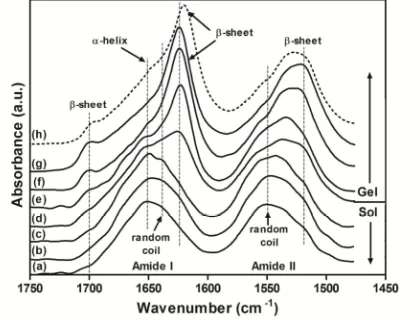
Hanawa, 1995; Hu, 2006). Several structural resonances have been identified for the amide I and amide II spectral domain including β-sheet structures (Hu, 2006; Hu, 2011) (1620-1630 cm-1, 1697-1700 cm-1, amide I; 1515 cm-1, amide II ), random coils (Hu, 2006, Um, 2001) (1640-1649 cm-1, amide I; 1540 cm-1, amide II), α-helix (Hu, 2006) (1652 cm-1, amide I), turns (Hu, 2006) (1663-1696 cm-1, amide I) and side chain residues (1605-1615 cm-1, amide I) (Hu, 2006). The presence of β-sheet structures is believed to play a critical role in the formation of silk hydrogels (Matsumoto, 2006). Initially, the unprocessed silk solution exhibited peaks centered at 1552 cm-1 (Amide II) and 1640 cm-1 (Amide I) which constitute random coil structures, and 1654 cm-1 (Amide I) which indicates α-helix structures. After submission to high pressure CO2, the formation of peaks centered at 1610-1630
cm-1 and cm-1700 cm-cm-1 (Amide I) appeared signifying the presence of β-sheet structures. Simultaneously, the 1510-1520 cm-1 (Amide II) peak increased indicating the dense packing of β-sheet structures (Hu, 2011). Concomitant to the formation of β-sheet structures, a decrease in the α-helix (1654 cm-1) and random coil (1552 cm-1, 1640 cm-1) peak contributions was realized. Based on the progressive augmentation of the β-sheet structure peaks, increasing processing time under high pressure CO2
encouraged the formation of β-sheet structures with the preparation of stable silk hydrogels attained after 90 minutes processing. Notably, the strong β-sheet absorbance peak centered at 1623 cm-1 attains the greatest intensity increase between 60 and 90 minutes processing time, with processing times longer than 2 hours resulting in only trivial changes to the absorbance intensity. These transitions are in agreement with the gelation data presented in the previous section, whereby stable silk hydrogels were only achievable after a minimum of 90 minutes processing under high pressure CO2 (60 bar). The structural transitions observed here are
consistent with previous studies on silk hydrogels produced in acidic environments (Ayub, 1993; Hanawa, 1995), which suggests that the transition to the stable β-sheet conformation under high pressure CO2 may proceed in a analogous fashion.
on the high pressure CO2 (<200 bar) treatment of aqueous α-elastin solutions,
whereby negligible changes to the protein secondary structure were reported both during and post high pressure processing (Dehghani, 2008). Thus, the application of pressure, at least at the magnitude applied in this study, does not seem to invoke significant modifications to the silk structural conformations. It is obvious that a more comprehensive study is necessary to establish these findings, in particular the probing of silk structural changes whilst submitted to high pressure environments. A high pressure study employing in situ spectroscopy techniques as recently described (Dehghani, 2008) may better elucidate the events during the gelation of silk proteins under high pressure CO2.
Figure III – 5. Dynamic β-sheet formation during silk gelation evaluated by FTIR for 2 wt% silk solutions submitted to high pressure CO2 (60 bar): (a) 0 min, (b) 30 min, (c) 60 min., (d) 75 min., (e) 90 min., (f) 2 hrs., (g) 8hrs., (h) citric acid control. Constant sample volume of 5 ml and thickness of 0.6 cm were utilized.
3.3.4. Hydrogel Physical Characteristics
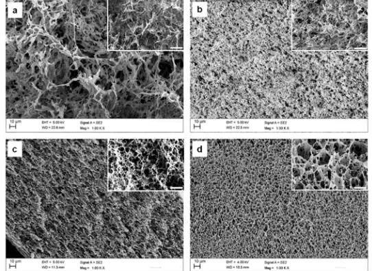
3.3.4.1. Microstructure
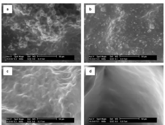
analysis demonstrated that silk hydrogels prepared under high pressure CO2 were
distinctly more homogenous when compared to the citric acid controls. By comparison, hydrogels prepared at atmospheric conditions with citric acid were considerably more heterogeneous, seemingly comprised predominantly of aggregate-like structures.
Figure III – 6. Environmental Scanning Electron Microscopy (ESEM) images of wet silk hydrogels prepared by citric acid titration (a) 2 wt% and (b) 4 wt% and under high pressure CO2 (c) 2 wt% and (d) 4 wt%.
Dry imaging was also performed using conventional scanning electron microscopy (SEM) techniques to better investigate the microstructures of the prepared hydrogels at various pressures (Figure 7). Comparison of the micrographs of silk hydrogels prepared by high pressure CO2 or by