ABSTRACT
LOVETT, MALLORYE DELORIS. Beta-lactoglobulin Complexed Vitamin D in an Aqueous Solution: Formulation and Bioavailability. (Under the direction of: Jonathan C. Allen).
Growing awareness of vitamin D status in the United States has resurfaced due to increasing reports of insufficiency and deficiency. Epidemiological research indicates that average intake of this nutrient is well below the RDA, and greater intake has been linked to optimal bone health, prevention of osteoporosis, osteomalacia, rickets, and other chronic diseases.
The first objective of this research was to test a water-soluble form of vitamin D as a fortifying ingredient in a flavored aqueous solution. Vitamin D was extracted by organic solvents and assayed by High Performance Liquid Chromatography (HPLC). The second objective was to test the ability of different vitamer sources (D2 versus D3) and quantitative amounts to increase serum vitamin D levels in a human intervention study. Vitamin D content of the complex was assessed by HPLC and VitaKit D™. Flavored beverages were formulated with 400 IU, 600 IU, and 1000 IU of vitamin D. Human subjects recruited from North Carolina State University (Raleigh, NC) participated in a blind randomized clinical trial designed to
initial treatment to final treatment. Subjects did not respond differently to drinks containing vitamin D2 or D3.
Beta-lactoglobulin Complexed Vitamin D in an Aqueous Solution: Formulation and Bioavailability
by
Mallorye Deloris Lovett
A dissertation submitted to the Graduate Faculty of North Carolina State University
in partial fulfillment of the requirements for the degree of
Doctor of Philosophy
Food Science
Raleigh, North Carolina 2010
APPROVED BY:
_______________________________ ______________________________
Jonathan C. Allen, PhD Brenda P. Alston-Mills, PhD
Committee Chair
________________________________ ________________________________
DEDICATION
This dissertation is dedicated to my parents, sister, grandmother, and in loving memory of my grandparents for their love, sacrifices, life lessons, encouragement, support and prayers, during
BIOGRAPHY
ACKNOWLEDGMENTS
The journey from conceptualization to ultimate completion of this degree and research study
has been a true odyssey of discovery. An odyssey of this nature and complexity could not have been accomplished without the help, support, and cooperation of many individuals.
Acknowledgements to individuals will be given in no particular order.
• This journey could have not been completed without the love and faithfulness of God.
“Great is thy faithfulness, Lord unto me” ~ Thomas O. Chisholm; these words have uplifted me through the trails, tribulations and joys of this journey. I am glad to behold His love, grace and faithfulness.
• Dr. Allen thank you for the opportunity to become a member of your lab group.
Thank you for your continued encouragement and guidance. This opportunity has
allowed me to grow in research and teaching.
• My committee members, thank you for your support and advice I am truly grateful.
• The Allen lab group, thank you for encouragement and research advice.
• Paige Luck, Jessica Childs, SAS consulting services personnel, Judy Cooper, Shirley
Lyles, and Sabrina Whitley-Ferrell thank you for assistance in equipment utilization,
development of computerized questionnaire, analysis of data and overall assistance. Each and everyone has been a tremendous support.
• To the participants who were involved in the research study, thank you. The
• To the Southeast Dairy Foods Research Center (SDFRC) and Dairy Management,
Inc., for their financial support.
• To the members of the Department of Food, Bioprocessing and Nutrition Sciences,
the Food Science Club, and the NCSU Chapter of MANRRS (Minorities in Agriculture, Natural Resources, and Related Science) thank you.
• Montreka, JeVelle, Tristan, Paula, Jeffrey, Jada, Mallory, Holly, Megan, Miniayah,
Carmel, Felita, Nhora, Lenese, YieHui, Lei, Lindsey, Wanida and Chellani thank you for all the good times. I will always remember the laughter and great conversations.
• My parents, words cannot express how thankful I am to have outstanding parents like
you. You have instilled in me the importance of education and achieving your heart’s desires. Mom thank you for your unremitting encouragement and assistance. Daddy,
thank you for continuous mantra of hope and reminding me that I am always loved. Thank you both for your constant love and support.
• My sister, whether near (USA) or far (another continent) you have always been my
number one cheerleader. You have encouraged me beyond measure to succeed and
never be afraid to go after your dreams. Thanks for your calming disposition and love.
“No man is an island, entire of itself; every man is a piece of the continent.” – John Donne To my continent of supporters, I say thank you for accompanying me on this journey. To each and every member of the following families and organizations you have contributed to
• Lovett, Durham, Nixon, Perry, Stiff, Young, Spinks, Crisp, Scott, Clegg, Johnson,
Bynum, Brown, Gause, Roberts, Cheatham, Myers, Sanders, Vanderkamp, Sult, Woodard, Hart, Wright, Kearney, Taylor, Flood, Little, friends and family at NCCU,
friends of UNC-G, First Baptist Church family, Alpha Lambda Fall 99-94 Degrees and Rising, Delta Sigma Theta Sorority, Inc., P&G RTCI (Research in Technical
Careers and Industry) conference cohorts, NC Health Careers – Wake AHEC, Delta Carousel, Top Teens of America, NC-MSEN Pre-College Program, ACTS, and Bennett College Intensive Summer Science Program (ISSP) . If any family or
TABLE OF CONTENTS
List of Tables ... xi
List Of Figures ... xiii
Chapter 1 - Introduction ... 1
1.1 Preface ... 2
References ... 5
Chapter 2 - Literature Review ... 7
2.1 Introduction ... 8
2.1.1 Water ... 8
2.1.2 Hydration Status ... 9
2.1.3 Hydration Status and Osteoporosis ... 11
2.1.4 Bottled Water Consumption ... 12
2.1.5 Flavored Fortified (Functional) Water Beverages ... 13
2.1.6 Benefits ... 14
2.2 History of Fortification ... 15
2.2.1 History ... 15
2.3 Vitamin D ... 16
2.3.1 Calcium Connection ... 16
2.3.3 Absorption and Digestion ... 22
2.3.4 Function ... 24
2.3.5 Effect of Vitamin D Deficiency ... 25
2.3.6 Effect of Vitamin D Insufficiency... 26
2.3.7 Vitamin D Fortification ... 27
2.4 Vitamin D Receptor (VDR) ... 29
2.5 Beta-lactoglobulin (BLG) ... 30
2.5.1 Source, Structure, and Synthesis ... 31
2.5.2 Beta-lactoglobulin Interaction with Vitamin D ... 33
2.6 High-Preformance Lipid Chromatography... 35
References ... 37
Chapter 3 - Preparation of Protein-Vitamin Complex Beverages ... 45
Abstract ... 46
3.1 Chapter Overview ... 47
3.2 Spray-drying of β-LG-vitamin Complexes ... 48
3.2.1 Introduction ... 48
3.2.2 Material and Methods ... 49
3.2.2.1 β-lactoglobulin and Vitamin D Complex Preparation ... 49
3.2.2.4 Vitamin D Analysis High Performance Liquid Chromatography (HPLC) ... 51
3.2.2.5 Vitamin D Analysis VitaKit D™ ... 52
3.2.2.6 Standard Curve and Recovery of Vitamin D from Complex ... 55
3.2.2.7 Assay of Vitamin D ... 57
3.2.2.7.1 Saponification ... 57
3.2.2.7.2 Extraction ... 57
3.4.3 Recovery of Vitamin D from BLG Powder ... 58
3.2.3 Results ... 60
3.2.4 Discussion and conclusion ... 61
3.3 Protein-vitamin binding - fluorescence study ... 64
3.3.1 Introduction ... 64
3.3.2 Materials and Methods ... 66
3.3.3 Results ... 67
3.3.4 Discussion and Conclusion ... 67
3.4 Shelf-Life and Milk Comparison Study ... 68
3.41 Introduction ... 68
3.4.2 Materials and Methods ... 69
3.4.3 Results ... 70
3.4.4 Discussion and Conclusion ... 71
Chapter 4 - Fortification of an queous solution with β-LG vitamin D complex: human
bioavailability ... 89
Abstract ... 90
4.1 Fortification of an Aqueous Solution with β-LG Vitamin D Complex: Human Study ... 92
4.1.1 Introduction ... 92
4.2 Materials and Methods ... 94
4.2.1 Analysis for Serum 25-OH Vitamin D3 ... 98
4.2.2 Statistical Analysis ... 99
4.3 Results ... 100
4.3.1 Bioavailability of Vitamin D in an Aqueous Solution ... 104
4.4 Discussion and Conclusion ... 106
References ... 137
Chapter 5 - Summary and conclusion ... 141
5.1 Summary and conclusion ... 142
References ... 146
Appendices ... 148
Appendix A Vitamin D Intervention Study-Recruitment ... 149
Appendix B Anonymous Computerized Questionnaire to Determine Eligibility ... 150
LIST OF TABLES
Table 2.1 Recommended Adequate Intake by the IOM for Calcium ... 18
Table 2.2 Selected Food Sources of Vitamin D ... 21
Table 2.3 1997 RDI Adequate Intakes for Vitamin D ... 28
Table 2.4 Composition of Whey Protein in Bovine Milk ... 31
Table 2.5 Advantages and Limitation of HPLC ... 36
Table 3.1 HPLC Program Flow ... 55
Table 3.2 Vitamin D3 (Cholecalcifrol) Standards ... 56
Table 3.3 Retention Time and AUC for Vitamin D2 and D3 ... 59
Table 3.4 Protein-Vitamin Binding - Trehalose versus Lactose Comparison ... 76
Table 3.5 Comparative Analyses High Performance Liquid Chromatography (HPLC) versus VitaKit D™ ... 77
Table 3.6 Shelf-Life Vitamin D Content after 7-Day Storage ... 80
Table 3.7 Milk Comparisons - Vitamin D Content of Commerically Available Milk versus Research Beverages ... 81
Table 4.1 Modified Food Frequency Questionnaire for Assessing Dietary Intakes of Calcium and Vitamin D in Beverages ... 102
Table 4.2 Modified Food Frequency Questionnaire for Assessing Dietary Intakes of Calcium and Vitamin D in Food Items ... 103
Table 4.3 Extrapolated Daily Dietary Intake of Calcium and Vitamin D ... 104
Table 4.5 GLM Procedure: Repeated Measures Analysis of Variance Test of Hypotheses for between Subject Effects ... 112 Table 4.6 Change in Mean Vitamin D Status ... 113 Table 4.7 Comparison of Mean Serum 25(OH)D Between Ethnic Groups ... 114
LIST OF FIGURES
Figure 2.1 Osteoporosis Risk Factors ... 12
Figure 2.2 Structures of Vitamin D2 and D3 ... 22
Figure 2.3 Production, Metabolism and Biological Functions of Vitamin D3 ... 24
Figure 2.4 Structure of Beta-lactoglobulin ... 34
Figure 3.1 HPLC Analysis of vitamin D in BLG - viramin D complex ... 55
Figure 3.2 HPLC Chrromatogram of Vitamin D ... 59
Figure 3.3 Vitamin D Transport and Binding by Beta-lactoglobulin ... 65
Figure 3.4 Vitamin D Standard Curve ... 74
Figure 3.5 Vitamin D Standard Curve VitakitD™ ... 75
Figure 3.6 Flourescence emission spectrum of BLG in the absence(a) and presence(b) of D2 powder, (c) D3 powder, (d) D2 liquid, and (e) D3 liquid ... 78
Figure 3.7 Fluorescence emission spectrum of BLG in the absence (a) and presence (b) of D2 powder, (c) D3 powder (top panel); and fluorescence emission spectrum of BLG in the absence (a) and presence (d) D2 liquid, and D3 liquid ... 79
Figure 3.8 Aerobic Bacteria – β-LG Vitamin D Complex ... 82
Figure 3.9 Aerobic Bacteria - Stock Solution and Non-Spray Dried Beverage Complete ... 83
Figure 3.10 Aerobic Bacteria Spray Dried versus Non Spray Dried and 7-day Storage ... 84
Figure 4.2 Mean Serum 25(OH)D Concentration in Subjects Consuming Control and Fortified
Aqueous 400 IU Vitamin D Beverage ... 116
Figure 4.2 Participant 002: Serum 25(OH)D ... 117
Figure 4.4 Participant 004: Serum 25(OH)D ... 118
Figure 4.5 Participant 005: Serum 25(OH)D ... 119
Figure 4.6 Participant 006: Serum 25(OH)D ... 120
Figure 4.7 Participant 007: Serum 25(OH)D ... 121
Figure 4.8 Participant 008: Serum 25(OH)D ... 122
Figure 4.9 Participant 009: Serum 25(OH)D ... 123
Figure 4.10 Participant 012: Serum 25(OH)D ... 124
Figure 4.11 Participant 011: Serum 25(OH)D ... 125
Figure 4.12 Participant 015: Serum 25(OH)D ... 126
Figure 4.13 Participant 016: Serum 25(OH)D ... 127
Figure 4.14 Participant 017: Serum 25(OH)D ... 128
Figure 4.15 Participant 018: Serum 25(OH)D ... 129
Figure4.16 Participant 019: Serum 25(OH)D ... 130
Figure 4.17 Participant 024: Serum 25(OH)D ... 131
Figure 4.18 Participant 027: Serum 25(OH)D ... 132
Figure 4.19 Participant 028: Serum 25(OH)D ... 133
Figure 4.20 Participant 029: Serum 25(OH)D ... 134
CHAPTER 1 Introduction
Mallorye D. Lovett
1.1 Preface
Vitamin D is an essential micronutrient that regulates calcium and phosphorus metabolism to achieve homeostasis and is required for growth, development, and structural integrity of the skeleton. Moreover, vitamin D has non-calciotropic autocrine and paracrine functions that have been shown to play a possible role in cancer and other chronic disease such as diabetes and cardiovascular disease (Holick, 2003). Vitamin D is referred to as the “sunshine vitamin” due to the production vitamin D from cholesterol via photosynthesis when skin is exposed to
Beta-lactoglobulin (β-LG) has many functional properties including the capacity to bind fat-soluble compounds including vitamins A and D. Lipophilic vitamins bind to the hydrophobic core of the β-LG molecule. Therefore using β-LG as a carrier for fortification of low or non-fat solutions could structurally protect vitamins A and D and improved their solubility and stability. Wang et al. (1997) illustrated via fluorescence spectroscopy that β-LG is capable of binding at approximately two moles of retinyl palmitate and one mole of vitamin D per mole of protein. Furthermore, previous studies have suggested that β-LG has a higher affinity for vitamin D than for other lipophilic vitamins.
A possible use for the complex formed between β-LG and vitamin D is fortification of bottled water and similar rehydration products. The bottled water market continues to grow at a faster rate than other beverage sectors. Consumers are aware of the health benefits associated with daily consumption of fluid water. Fortification of bottled water with nutrients (β-LG and vitamin D), non-caloric sweetener, and flavor may increase the bottled water market.
In this study, the bioavailability of β-LG-complexed vitamin D in an aqueous solution was investigated to justify the potential for using β-LG as a stable and protective carrier for vitamin D. The purpose of this study was to produce a beverage fortified with vitamin D in a β-LG
complex, demonstrate that the β-LG and vitamin D remain in association with each other in the aqueous environment, and to assess the capacity the protein-based aqueous solution to elevate serum vitamin D status in a human feeding trial.
REFERENCES
Holick, M.F. (1992). The vitamin content of fortified milk and infant formula. New England Journal of Medicine,326,1178.
Holick, M. F. (1995). Environmental factors that influence the cutaneous production of vitamin D. The American Journal of Clinical Nutrition, 61(3), 638S.
Holick, M. F. (1999). Vitamin D: Molecular biology, physiology, and clinical applications. Totowa, N.J.: Humana Press.
Holick, M. (2003). Vitamin D: A millenium perspective. Journal of Cellular Biochemistry, 88(2), 296.
Holick, M. (2003). Evolution and function of vitamin D. Recent Results in Cancer Research, 164, 3.
Institute of Medicine. Standing Committee on the Scientific Evaluation of Dietary Reference Intakes. (1997). DRI, dietary reference intakes: For calcium, phosphorus, magnesium, vitamin D, and fluoride. Washington, D.C.: National Academy Press. Retrieved from
http://www.nap.edu.www.lib.ncsu.edu:2048/books/0309063507/html/
Office of Dietary supplements (2008). Dietary supplement fact sheet: vitamin D. Bethesda, M.D.: Office of Dietary Supplements National Institutes of Health. Retrieved from http://dietary-supplements.info.nih.gov/factsheets/vitamind.asp
CHAPTER 2 Literature Review
Mallorye D. Lovett
2.1 Introduction 2.1.1 Water
The most abundant constituent in the human body is water. The body is comprised of an estimated 60 to 75 percent water (ranging from about 50% in obese individuals and 70% in lean individuals). The human brain (95%), lungs (90%), and blood (82%) are composed mainly of water. Water is the cornerstone in the maintenance of good health. Water provides
fundamental nutrients necessary to sustain cellular homeostasis and life. Homeostasis in all of the major organ systems in the human body depend on water for the following functions:
• Moistens tissues such as those in the mouth, eyes, and nose
• Protects the body’s organs and tissues
• Aids in digestion
• Helps prevent constipation
• Helps dissolve minerals and other nutrients to increase bioavailability to the body
• Regulates body temperature
• Lubricates joints
• Lessens the burden on the kidneys and liver by flushing out waste products
• Carries nutrients and oxygen to cell
facilitates the movement of nutrients, hormones, antibodies, and oxygen throughout the blood stream and lymphatic system (WHO, 2006). Water utilized by the body is obtained from various sources including drinking water, water in beverages, water obtained from food, and water produced by metabolic reactions. Previous research studies have found an association between low intake of total water and some chronic diseases including breast cancer, prostate cancer, and cardiovascular disease (IOM, 2004). The Institute of Medicine advised that men consume approximately 3 liters (about 13 cups) and women consume 2.2 liters (about 9 cups) of total beverages daily to obtain the beneficial effects of water. For example, the DRI (Dietary
Reference Intake) has an AI (Adequate Intake) for total water intake for young men and women (ages 19 to 30 years) of 3.7 L and 2.7 L per day, respectively. These amounts represent
approximately 81% of total water intake in the US. Even though the essential benefits of water have been clearly defined, the current United States Department of Agriculture (USDA)
MyPyramid does not include water as a component of many foods in their database (WHO, 2005). Although it has been clearly defined that water is the most abundant component in the human body, and despite knowledge of health hazards associated with lack of water, the
scientific community is unable to clearly define the definition of optimal water requirement and establish intake recommendations (Ritz, 2005). However, the body of research has established that water is essential to sustain life (IOM, 2004).
2.1.2 Hydration Status
loss from the skin, the respiratory system and the gastrointestinal tract is under less regulatory control. Additionally, hormonal mechanisms regulating thirst play a significant role in
2.1.3 Hydration Status and Osteoporosis
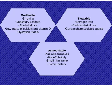
Osteoporosis is defined as a condition characterized by a decrease in bone mass and density that leads to increased bone fragility (Ringe, 2004). The prevalence of osteoporosis increases with age. Osteoporosis is a multifactorial disease in which nutrition plays a vital role. The risk factors of osteoporosis are classified as modifiable, unmodifiable, and treatable (Figure 2.1). Osteoporosis is characterized by continued calcium deficiency that leads to negative
Modifiable •Smoking •Sedentary Lifestyle
•Alcohol abuse
•Low intake of calcium and vitamin D •Hydration Status
Unmodifiable •Age at menopause
•Race/Ethnicity •Small, thin frame
•Family history
Treatable •Estrogen loss •Corticisisteriod use •Certain pharmacologic agents
Figure 2.1 – Osteoporosis Risk Factors
2.1.4 Bottled Water Consumption
However, even with the aforementioned slight reduction in growth; bottled water continues to be a key component in the beverage sector. Moreover, in 2005 bottled water volume exceeded 7.5 billion gallons, a 10.7% advance over the previous year. These findings suggest that the typical American consumes an average of 26.1 gallons annually. The average consumption of bottled water surpasses any other beverage, except for carbonated soft drinks (CSDs). Even though CSDs continue to be the most consumed beverage by volume, the soft drink market has been struggling recently due to competition from the bottled water sector (Decker, 2006). From 2002-07, CSBs grew a mere 5%, while non-carbonated beverages (milk, fruit juices, and functional beverages) increased 30%. This increase can be attributed to the heightened concerns about health (Dairy Foods, 2008). As non-carbonated beverages become more than a commodity purchase, industries are trying to expand this market sector by adding vitamins, minerals, sweeteners, flavor, etc. These line extensions have found niche markets that have increased sales in this beverage segment. Consumers’ perceive that these new products are a healthier replacement for traditional CSDs (Decker, 2003).
2.1.5 Flavored Fortified (Functional) Water Beverages
overall wellness has contributed to the proliferation of this sector. Functional waters that have been fortified with vitamins and minerals are becoming increasingly accepted by consumers. Functional beverages are projected to increase from a $10 billion in 2004, to a $12.8 billion industry by 2009 (Decker, 2006).
2.1.6 Benefits
Popkin et al., (2006) recently proposed guidelines for beverage consumption after
completing an in-depth review of research investigating effects of fortified beverages on health. The authors stressed the importance of a healthy diet including adequate intake of water for metabolism and normal physiological function (Popkin et al., 2006). The overwhelming
evidence that current American diet is inadequate in good quality nutrition and fluid water fuels the flame that functional beverages have the ability to fill in some nutritional gaps (Kleiner, 1999 and Popkin et al., 2006). Functional foods, foods or food ingredients, have the ability to prevent or reduce the development of symptoms or diseases associated with nutritional inadequacies (Milner, 2000). Functional food formulation has progressed to the development and
implementation of dietary supplements. For example, a current functional food utilized is probiotic and prebiotics in dairy products that aid in maintaining intestinal health
2.2 History of Fortification 2.2.1 History
Fortification is defined as the addition of nutrients to food constituents to maintain or improve the overall nutritional quality of the food (Yetley, 2004). In 1924, voluntary
fortification of salt with iodine was a result of the increased prevalence of goiter among the US population. Iodization of salt led to a decrease in prevalence of goiter from 38.6% to 9%. Continued use of iodine fortified salt nearly led to the elimination of goiter in the 1930s (Backstrand, 2002). Similarly, in 1933 vitamin D was utilized as a fortifier for fluid milk. Milk was fortified to combat the increasing prevalence of rickets, a deficiency of vitamin D that may cause abnormal bone formation (IOM, 2003).
The United States continued to address the role of fortification as systematic approach to alleviate nutritional deficiencies. In addition, throughout World War II the enlistees
frequently had poor nutritional status. To combat the issues of poor nutritional status and nutritional deficiencies, President Roosevelt assembled the National Nutrition Conference for Defense in 1941 which is currently referred to as the Food and Nutrition Board (FNB). A major result of this meeting was the recommendation for enrichment of flour and bread. Moreover, this coincided with the first presentation of the Recommended Daily Allowances (RDAs). RDAs were established for various vitamins and minerals and some of these components were approved as fortifiers. Thiamin, niacin, riboflavin, calcium, vitamin D and vitamin A were approved as fortifiers for such food products as flour, bread, milk and
• The intake of the nutrient, in the absence of fortification, is below the desirable level in diets of
a significant number of people.
• The food from which the nutrient is to be derived is likely to be consumed in quantities that
will make a significant contribution to the diet of the population in need.
• The addition of the nutrient is unlikely to create an imbalance of essential nutrients.
• The nutrient added is stable under proper conditions of storage and use.
• The nutrient is physiologically available from the food to which it will be added.
• There is a reasonable assurance against intake sufficiently in excess to be toxic (IOM,
2003).
2.3 Vitamin D
2.3.1 Calcium Connection
An intimate relationship exists between vitamin D and calcium. The synergistic
interactions between calcium and vitamin D have been well documented (Iwamoto et al., 2004). Adequate amounts of calcium and vitamin D throughout life helps reduce the risk of abnormal bone formation, osteoporosis, high blood pressure, obesity and colon cancer (Calvo et al., 2005). However, to truly understand the biological functions of vitamin D, an investigation of the evolutionary relationship between the above aforementioned vitamin and mineral must be addressed.
variety of cellular metabolic events. However, when the vertebrates transitioned to land the new environment was insufficient in calcium. Therefore an adaptation occurred among the
vertebrate species to allow for utilization of sunlight and synthesis of vitamin D to regulate intestinal absorption of calcium. This transformation allowed better usage calcium from the diet and had a positive impact on skeletal mineralization (Holick, 1999).
The role of calcium in mammalian species is well understood. Calcium is a major constituent of mammalian mineralization. Calcium aids in the normal growth and development of bone (Bronner, 1998). The majority of calcium (~99%), an essential nutrient, is found in the bones and teeth. The remaining 1% is contained in the serum, extravascular fluid, muscle, and other tissues (Committee of Nutrition, 1999). This nutrient is regulated by several hormones including the Parathyroid Hormone (PTH), calcitonin, and vitamin D. These regulators, when operating appropriately, maintain calcium balance within narrow limits. A low intake of calcium stimulates the breakdown of bone to supply the needed calcium for normal physiological functions. Continued release of calcium from the bone causes a reduction in bone mass and increases the risk of osteoporosis. Good sources of calcium are abundant throughout the U.S. Calcium can be obtained from calcium rich foods such as milk, dairy products, and other
Table 2.1 Recommended Adequate Intake by the IOM for Calcium
Male and Female Age Calcium (mg/day) Pregnancy & Lactation
0 to 6 months 210 N/A
7 to 12 months 270 N/A
1 to 3 years 500 N/A
4 to 8 years 800 N/A
9 to 13 years 1300 N/A
14 to 18 years 1300 1300
19 to 50 years 1000 1000
51+ years 1200 N/A
(Institute of Medicine, 1997)
Calcium is transported by active transport and passive diffusion. Active transport is defined as the movement of calcium, entry, intracellular diffusion, and extrusion which requires metabolic energy. Active transport is the primary mechanism of calcium
absorption at low and moderate intake and is highly regulated by 1, 25 dihydroxyvitamin D, the active form of vitamin D. Conversely, passive diffusion is imperative with high intakes of calcium diffusion across the intestinal mucosa (Bronner, 1998; IOM, 1997). Calcium is absorbed in the small intestine (duodenum) with favorable low pH (<6) (IOM, 1997).
2.3.2 Source, Structure, and Synthesis
Previously little was known about the evolutionary perspective of vitamin D (calciferol). In 1919, a scientist named Edward Mellanby discovered the importance of vitamin D on
development, growth and maintenance of a healthy skeletal system when investigating the prevalence of rickets (Holick, 2003). In addition, throughout the years of 1924 - 1927
and zooplankton. These scientists were able to demonstrate that seasonal variation in vitamin D could be noticed in oily fish and fish liver, with the highest amounts of vitamin D during the summer months and a lower content of vitamin D during the winter months (Holick, 2003, Hollis, 2005).
As illustrated in Figure 1.2, vitamin D comes in various forms, but the two most prominent forms are ergocalciferol (vitamin D2) and cholecaliferol (vitamin D3) (IOM, 1997). Vitamin D2 is derived from yeast and plant sterol, and is the usual form of vitamin D employed during fortification. On the other hand, vitamin D3 is obtained when radiant energy (UV light) from the sun interacts with 7-dehydrocholesterol, a precursor of cholesterol, found in the skin. During exposure to sunlight, provitamin D is thermally isomerized to vitamin D and
translocated throughout the body via vitamin D binding protein. Sufficient sun exposure can generate adequate active vitamin D for biological functions. However, seasonal, geographical latitude, time of day, cloud cover, environmental smog, skin melanin content (increased
pigmentation), and use of sunscreen to prevent development of skin cancer are among some of the factors that affect vitamin D synthesis via UV radiation exposure. Due to the fact that the body is capable of producing cholecalciferol, vitamin D is not defined as the classical vitamin. A more accurate description of vitamin D is a calciotropic prohormone; which indicates that the vitamin is metabolized to a biologically active form that functions as a steroid hormone. Nonetheless, since the importance of vitamin D has been discovered it has been classified among the lipid-soluble vitamins (IOM, 1997, Lipkin, 2006). The primary sources of vitamin D in nature are very few foods including fish liver oils (manufactured through ultraviolet
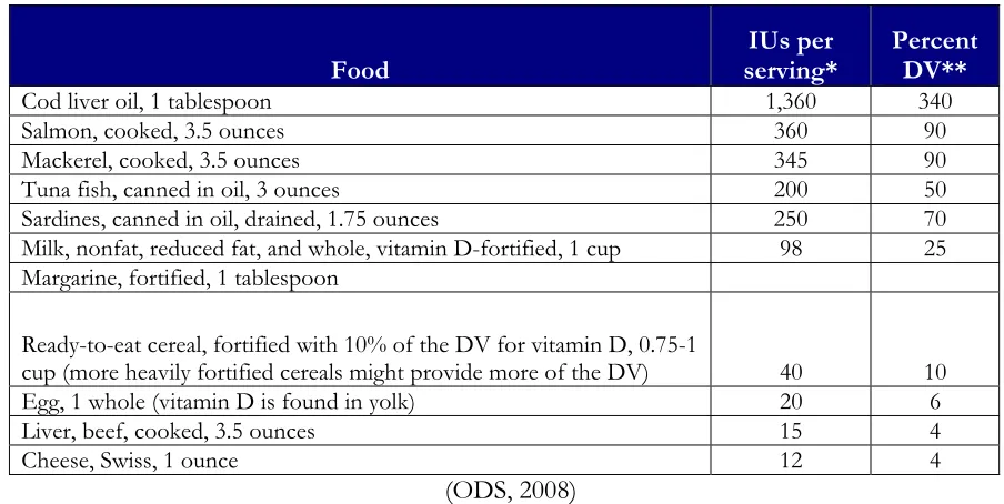
Table 2.2 Selected Food Sources of Vitamin D
Food serving* IUs per Percent DV**
Cod liver oil, 1 tablespoon 1,360 340
Salmon, cooked, 3.5 ounces 360 90
Mackerel, cooked, 3.5 ounces 345 90
Tuna fish, canned in oil, 3 ounces 200 50
Sardines, canned in oil, drained, 1.75 ounces 250 70 Milk, nonfat, reduced fat, and whole, vitamin D-fortified, 1 cup 98 25 Margarine, fortified, 1 tablespoon
Ready-to-eat cereal, fortified with 10% of the DV for vitamin D, 0.75-1
cup (more heavily fortified cereals might provide more of the DV) 40 10
Egg, 1 whole (vitamin D is found in yolk) 20 6
Liver, beef, cooked, 3.5 ounces 15 4
Cheese, Swiss, 1 ounce 12 4
(ODS, 2008)
*IUs = International Units.
Figure 2.2 Structures of Vitamin D2 and D3 Vitamin D2 top, Vitamin D3 bottom (IOM, 1997)
2.3.3 Absorption and Digestion
Enzymes in the liver and kidney convert the prohormone form of vitamin D to the active form of vitamin D (Figure 1.3). When adequate amounts of vitamin D are ingested via food, approximately 80% vitamin D is incorporated into the micelles of the small intestine and absorbed along with the dietary fat in the intestine. Exposure to sunlight converts 7-dehydro-cholestrol to vitamin D. Vitamin D binds to the protein carrier in the blood and is transported to the liver by chylomicrons via the lymphatic system. While in the liver, vitamin D is
hormone form 1, 25 dihydroxy vitamin D by the kidney; the active vitamin D is transported to the target tissue in the body. It has been determined that tissues other than the kidney have the ability to produce 1,25 dihydroxyl vitamin D. Many tissues in the body, including the prostate, colon, breast, skin, osteoblasts have the ability to express 1-α-hydrozylase and have the ability to synthesis 1,25(OH)2 D3 (Tangpricha, 2001). The aforementioned process, may be causal event for the prevention of development of certain chronic diseases. Moreover, the metabolism of vitamin D is feasible due to a specific protein carrier. Vitamin D that is circulated throughout the body is bound to a specific protein carrier, vitamin D binding protein (DBP) (Rowling, 2006). DBP binds the metabolites of vitamin D with different affinities: 25(0H)D >
Figure 2.3- Production, Metabolism and Biological Functions of Vitamin D3 (Holick, 2003)
2.3.4 Function
Moreover, vitamin D plays a significant role in the maintenance of serum calcium homeostasis and phosphorus homeostasis within the range that supports normal neuromuscular function, bone calcification and other cellular processes. Additionally, calcium and phosphorus work synergistically to promote normal bone mineralization. Maintenance of calcium and phosphorus within narrow limits by vitamin D is very important for all living organisms (IOM, 1997).
2.3.5 Effect of Vitamin D Deficiency
Historically, rickets, first discovered in 1645, was of profound concern at the turn of the 20th century in northern Europe and North America (Eitenmiller et al., 2008). Industrialization of many cities during this period provided an environment that obscured sunlight, which decreased the photosynthesis of vitamin D in the skin. The lack of exposure to sunlight limited skin’s production of vitamin D and the low availability of naturally vitamin D containing foods led to suboptimal vitamin D levels within the body. Low levels of vitamin D throughout the bloodstream have been associated with low or low-normal blood levels of calcium and
phosphorous. Inadequate vitamin D in the body elevates serum parathyroid hormone (PTH), which facilities an increase in bone resorption. The condition of prolonged insufficient vitamin D is termed rickets in children, which was first described in 1645, and osteomalacia in the adult population. These conditions are common in children and women who have inadequate amount of vitamin D in the body (Holick, 2005).
2009). However, researchers have reported that the majority of US citizens are exposed to suboptimal levels of sunlight, mainly during the winter months (Holick, 1992). For optimal conversion of 7-dehydrocholesterol to vitamin D, UVB irradiation from sun exposure of 20 mJ/cm2 is required. In the northern United States above latitude 40°, sun exposure levels do not meet the requirements of 20 mJ/cm2 (Hollis, 2005). Rickets and osteomalacia may occur in breastfed infants who do not receive vitamin D supplementation, women who have inadequate vitamin D intake from food and/or lack of exposure to ultraviolet light for photosynthesis of vitamin D, (Vieth, 2001) and older individuals with age-related synthesis of vitamin D
(Eitenmiller, 2008).
2.3.6 Effect of Vitamin D Insufficiency
Recommended nutrient intakes are intended to prevent the development of nutrient deficiencies and insufficiencies (Yates, 1998). Current vitamin D recommendations have been shown to prevent vitamin D deficiency as it relates to the traditional bone and teeth health. However, the identification of other non-calciotropic autocrine and paracrine functions of vitamin D has led the scientific community to reassess current vitamin D recommendations. These suggestions have led to the additional classification of vitamin D ‘insufficiency’ (Cranney et al., 2007). Vitamin D insufficiency is a condition when serum 25-hydroxyvitamin
al., 1997). Numerous cross-sectional research studies have found a direct link between vitamin D insufficiency and increased risk of developing cancer, cardiovascular disease, diabetes mellitus, hip fractures, and osteoporosis (Calvo et al., 2005). As stated earlier, vitamin D and calcium work together synergistically. However, calcium may not be needed for vitamin D to work with VDR-RXR (retinoic acid receptor) molecules to regulate cellular proliferation, differentiation, and function. When the above relationship does not exist, vitamin D causes cellular dysfunction and increase risk of developing certain chronic disease (Peterlik et al., 2009).
2.3.7 Vitamin D Fortification
In 1941, President Roosevelt issued a request for fortification of food products with vitamin D. His goal was to ensure that consumers had a sufficient amount of vitamin D in the diet to alleviate the epidemic of vitamin D deficiency in industrialized communities. This outcry from President Roosevelt resulted in vitamin D-fortified milk. Currently, there are many other food items fortified with vitamin D including yogurts, cereals, orange juice, and nutritional bars. However, with additional fortification of certain food products with vitamin D, research studies continuously report that the current fortification is inadequate or not found in enough different food products to prevent vitamin D deficiency (IOM, 2003).
Due to the concern of adequate vitamin D intake and challenges associated with vitamin D fortification beyond the traditional media, it is apparent that research must address these issues. One study in particular, conducted by Johnson et al. (2005) investigated the
difference in serum levels among those who consumed the fortified processed cheese. These findings lead to further investigation of vitamin D bioavailability in different foodstuff. The second part of the study compared bioavailability of fortified cheese (5880 IU D2/56.7g) versus water (containing 32,750 IU/250 mL) among different age groups (older versus younger) subjects who received an acute amount of vitamin D on serum vitamin D levels. These results showed that serum levels were significantly lower with consumption of fortified water than with fortified processed cheese. In addition, the peak serum vitamin D levels were similar among both age groups. Moreover, the acute amount of vitamin D in study two was indeed bioavailable when compared to the processed cheese consumed in study 1, which had no significant effect on blood serum levels. This study also highlighted the primary concern in the fortification of foods which is recovery of vitamin D after processing by various methods. In conclusion, research studies must continue to investigate vitamin D fortification techniques and quantification in various food sources (Johnson et al., 2005).
Table 2.3 – 1997 RDI Adequate Intakes for Vitamin D
RDI for Vitamin D µg/day (5µg = 200IU)
Babies
Birth - 12 months 5
Children
1-13 years 5
Adults
14-50 years 5
51 - 70 years 10
Over 70 years 15
Pregnant Women 5
Nursing Women 5
2.4 Vitamin D Receptor (VDR)
Vitamin D nutrition affects health beyond bone density. Vitamin D is able to function in various capacities via a signaling mechanism with serum vitamin D and VDR, a soluble high-affinity receptor protein (Vieth, 2001). A widespread distribution exists for VDR in the body including the pancreas, lymph nodes, adrenal medulla, excreta; the vitamin D is dispersed into the cell membrane and transferred via the cytoplasm to the nucleus where the binding occurs (Holick, 2003).
VDR, a nuclear receptor for 1α,25(OH)2D3, the active form of vitamin D3, plays a critical role in bone formation. VDR is responsible for activation of calbindin, a calcium
transport protein in the small intestine. Moreover, the distribution of VDR throughout the body demonstrates the various functions of 1α,25(OH)2D3 beyond mineral and skeletal homeostasis (Holick, 2003 and Veith 2001). Vitamin D3 is responsible for cell proliferation and inhibition or maturation of normal and tumor cells. Tanaka et al, (1982) investigated the effect of vitamin D on preleukemic cells in rats that were vitamin D sufficient. The researchers found that
2.5 Beta-lactoglobulin (BLG)
Beta-lactoglobulin (β-LG ), a small soluble protein that is extremely acid stable,is the major whey protein present in milk (Table 1.4). Whey protein is one of the highest quality
commercially available proteins (Perez, 1990). Whey protein is comprised of a higher
concentration of branched-chain amino acids (BCAA) and essential amino acids than most other protein sources. In addition, other components of whey, such as β-LG and peptides have been shown to promote increased protein synthesis, weight loss, body fat loss, and decreased plasma insulin and triglyceride profile. Therefore, the protein components of whey have been
Table 2.4 Composition of Whey Protein in Bovine Milk
Protein Approximate % of Whey
Protein
Benefits
Beta-lactoglobulin 50-55% Source of essential and
branched chain amino acids
Alpha-lactalbumin 20-25% Primary protein found in
human breast milk. Source of essential and branched chain amino acids
Immunoglobulins 10-15% Primary protein found in
colostrum immune modulating benefits
Bovine Serum Albumin 5-10% Source of essential amino acids
Large protein Glycomacropeptide
(GMP)
2-5% Source of branched chain
amino acids
Lacks the aromatic amino acids phenylalanine, tryptophan, and tyrosine
Lactoferrin 1-2% Antioxidant
Antibacterial, antiviral, and antifungal
Promotes growth of beneficial bacteria
Naturally occurs in breast milk, tears, saliva, bile, blood, and mucus
(Marshall, 2004 and Onwulata et al., 2008) 2.5.1 Source, Structure, and Synthesis
Beta-lactoglobulin production occurs in the mammary gland with inclusion in milk. β-LG
This purification of β-lactoglobulin by Palmer has provided the framework for investigating the physico-chemical properties and potential uses of this protein (Sawyer et al., 1999).
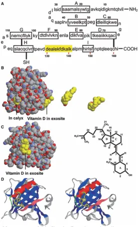
Although it was previously believed that β-LG was only found naturally in ruminant species, the protein is present in the milk of most mammalian species, excluding rodents and primates. Therefore, β-LG has been characterized in most detail from ruminants. This model has provided insight into the nature of the protein. Within this order, β-LG exists as a dimer of subunit with a relative molecular weight approximately 18,400 daltons for generic variant A and 18, 276 daltons for B (Papiz et al., 1986). Each monomer contains 162 amino acids, with one free cysteine and two disulphide bridges. The complete amino acid sequence of β-lactoglobulin genetic variants A and B differ in two amino acid residues – Asp 64 and Val 118 in variant A are replaced in variant B with Gly and Ala (Wong et al., 1996). The crystalline structure of β-LG is similar to retinol-binding protein and other proteins in the lipocalin family, which are generally noted to transport small hydrophobic molecules (Kontopidis, et al., 2002). The configuration of the protein is a β-barrel with eight antiparallel β-strands to form a conical central cavity in which the hydrophobic ligand is located. On the outer surface of the β-barrel there is a three-turn α -helix. The secondary structure of the protein is 15% α-helix, 50% β-sheet, and 15-20% of reverse turn. This configuration plays a significant role in the binding ability of β-lactoglobulin (Wu et al., 1999).
through the stomach. The protein is most heat sensitive near pH 4.0, with the maximum stability at pH 6.0 and decreasing stability in the higher pH range (Resch, 2004).
2.5.2 Beta-lactoglobulin Interaction with Vitamin D
β-Lactoglobulin has been shown to have the ability to bind in vitro to certain hydrophobic molecules such as retinoids, retinyl palmitate, vitamin D, cholesterol and some other fatty acids. The β-lactoglobulin molecule has been shown to function in binding and/or transport of small hydrophobic molecules (Kontopodis et al., 2004). Research studies have investigated the binding ability of vitamin D to β-LG, and found that vitamin D can bind to β -LG. The β-LG molecule structurally has two hydrophobic pockets that are capable of binding lipopholic molecules. The sites of binding are the calyx formed by the β-barrel and the other between the α-helix and the surface of the barrel. The binding site of lipopholic molecules is still controversial, but most scientific research finds vitamin D binds near the monomer contact surface of the dimer. Fluorescence spectroscopy has denoted a change in tryptophanyl
Figure 2.5- Structure of Beta-Lactoglobulin
2.6 High-Performance Lipid Chromatography
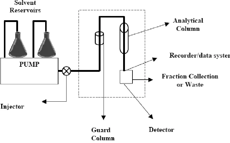
High-performance lipid chromatography (HPLC) is a predominant method for separation and quantification of various compounds including pharmaceuticals, biomolecules, polymers, and other organic and ionic compounds (Dong, 2006). HPLC is divided into several types however the two most common are normal-phase chromatography (NPC) and reverse-phase chromatography (RPC). Normal phase separates analytes based on adsorption/desorption (typically silica or silica modified with CN and NH2 groups) and polarity (typically silica or alumina). The mobile phase utilizes non-polar solvents hexane with addition of small amount of more polar solvent including iso-propanol, ethanol or chloroform. This type of chromatography is particularly useful for separation of small non-polar and cis/trans compounds. A main
disadvantage of normal phase chromatography is the ease of contamination of the instrument’s column by sample components. Whereas RPC is the most common separation mode,
accounting for approximately 70% of all HPLC analyses. RPC separates based on a polar mobile phase and a hydrophobic (non-polar) stationary phase. It is used in many
pharmaceutical, biochemical, and analytical chemistry applications (Dong, 2006). Separation occurs primarily because of solvophonic or hydrophobic interaction. The non-polar column is typically made from C18 or C8 material, and a more polar molecule. RPC is advantageous to the scientific community because it is the most stable HPLC method. Conversely, disadvantages for RPC include an increased time needed for analysis and re-equilibration of the column.
and disadvantages (Table 2.5), continues to be the primary tool for quantitation of various samples including vitamin D from most matrices (Mata-Granados, et al., 2009).
Research has shown that both NPC and RPC can be utilized to quantify vitamin D in solutions. The two official methods for vitamin D determination are AOAC international official method 981.17 for fortified milk and milk powder and AOAC international official method 995.05 for infant formulas and enteral products. Both of these methods rely on normal-phase HPLC to determine total vitamin D in a system, but neither allow for the vitamers to be used as an internal standard. RPC is more readily employed because it has the ability to separate the vitamers (i.e. D2 vs. D3) and allow use of an internal standard. NPC is unable to identify the amount of D2 compared to D3 . However this procedure is able to separate vitamin D, 25-hydroxyvitamin D2, and 25-hydroxyvitamin D3 and other hydroxylated metabolites (Perales et al., 2005). Taking the factors of this discussion into account RPC, was the method utilized for this research.
Table 2.5 Advantages and Limitations of HPLC
Advantages:
• Rapid and precise quantitative analysis
• Automated operation
• High-sensitivity detection
• Quantitative sample recovery
• Amenable to diverse samples Limitations:
• No universal detector
• Less separation efficiency than capillary GC
• More difficult for novices
REFERENCES
Armas, L. A. G. (2004). Vitamin D2 is much less effective than vitamin D3 in humans. The Journal of Clinical Endocrinology and Metabolism, 89(11), 5387.
Backstrand, J.R . (2002). The history and future of food fortification in the United States: A public health perspective. Nutrition Reviews, 60(1), 15.
Bossingham, M., Carnell, N., & Campbell, W. (2005). Water balance, hydration status, and fat-free mass hydration in younger and older adults. The American Journal of Clinical Nutrition, 81(6), 1342.
Bronner, F , & Pansu, D . (1999). Nutritional aspects of calcium absorption. The Journal of Nutrition, 129(1), 9.
Calvo, M., Whiting, S., & Barton, C. (2005). Vitamin D intake: A global perspective of current status. The Journal of Nutrition, 135(2), 310.
Chapuy, M.C. (1997). Prevalence of vitamin D insufficiency in an adult normal population.
Osteoporosis International, 7(5), 439.
Committee on Nutrition. (1999). Calcium requirements of infants, children, and adolescents.
Pediatrics. 104:1152.
Cranney, A., Horsley, T., O'Donnell, S., Weiler, H., Puil, L., Ooi, D., et al. (2007). Effectiveness and safety of vitamin D in relation to bone health. Evidence report/technology Assessment, (158), 1.
Cranney, A., University of Ottawa Evidence-based Practice Center, & United States. Agency for Healthcare Research and Quality. (2007). Effectiveness and safety of vitamin D in relation to bone health. Rockville, Md.: Agency for Healthcare Research and Quality. Retrieved from http://purl.access.gpo.gov.www.lib.ncsu.edu:2048/GPO/LPS86588
Dairy Foods (2008). State of the industry beverages: changes in the big beverage market. Dairy Foods, November 80-88.
D'Anci, KE . (2006). Hydration and cognitive function in children. Nutrition Reviews, 64(10), 457.
Decker, K.J. (2003). Wonder waters: fortifies and flavored waters. Food Product Design Application. August 57-74.
Decker, K.J. (2006). Bottled water continues as number 2 in 2005. Food Product Design Application. August 60-64.
Dong, M. W. Modern HPLC for practicing scientists Hoboken, N.J. : Wiley-Interscience, c2006. Retrieved from http://search.trln.org/search?id=NCSU1929745
Dusso, A.S. (2005). Fluid and Electrolyte Physiology, American Journal of Physiology 289(1), F8.
Eitenmiller, R. R., Ye, L., & Landen, W. O. (2008). Vitamin analysis for the health and food sciences
(2nd ed.). Boca Raton: CRC Press.
Gleick, P . (2004). The world's water, 2004-2005: The biennial report on freshwater resources. Island Press, Washington, D.C.
Holick, M.F. (1992). The vitamin content of fortified milk and infant formula. New England Journal of Medicine,326,1178.
Holick, M. F. (1999). Vitamin D: Molecular biology, physiology, and clinical applications. Totowa, N.J.: Humana Press.
Holick, M. (2003). Vitamin D: A millenium perspective. Journal of Cellular Biochemistry, 88(2), 296.
Holick, M. (2003). Evolution and function of vitamin D. Recent Results in Cancer Research, 164, 3.
Holick, M. F. (2007). Vitamin D deficiency. New England Journal of Medicine, 357(3), 266.
Hollis, B. (2005). Circulating 25-hydroxyvitamin D levels indicative of vitamin D sufficiency: Implications for establishing a new effective dietary intake recommendation for vitamin D.
Journal of Nutrition, the, 135(2), 317.
Institute of Medicine. Standing Committee on the Scientific Evaluation of Dietary Reference Intakes. (1997). DRI, dietary reference intakes: For calcium, phosphorus, magnesium, vitamin D, and fluoride. Washington, D.C.: National Academy Press. Retrieved from
http://www.nap.edu.www.lib.ncsu.edu:2048/books/0309063507/html/
Institute of Medicine (2003). Dietary reference intakes: guiding principles for nutrition labeling and fortification. National Academy Press, Washington, DC.
Institute of Medicine (2004). Dietary reference intakes: Guiding principles for nutrition labeling and fortification.(2004). Nutrition Reviews, 62(2), 73-79. Retrieved from
http://search.ebscohost.com.www.lib.ncsu.edu:2048/login.aspx?direct=true&db=aph&AN =12412277&site=ehost-live&scope=site
Iwamoto, J. (2004). Effects of vitamin D supplementation on calcium balance and bone growth in young rats fed normal or low calcium diet. Hormone Research, 61(6), 293.
Johnson, J.L, Mistry, V.V., Vukovich M.D., et al. (2005). Bioavailability of vitamin D from fortified process cheese and effects on vitamin D status in the elderly. J Dairy Sci, 88, 2295.
Kleiner, S. M. (1999). Water an essential but overlooked nutrient. Journal of the American Dietetic Association, 99(2), 200.
Kontopidis, G. , Holt, C. , & Sawyer, L. (2004). Invited review: Beta-lactoglobulin: Binding properties, structure, and function. Journal of Dairy Science, 87(4), 785.
Lapp, J. L. (2009). Vitamin D: Bone health and beyond. American Journal of Lifestyle Medicine, 3(5), 386.
Lipkin, M. (2006). Mechanisms of action of vitamin D: Recent findings and new questions.
Journal of Medicinal Food, 9(2), 135.
Marshall, K. (2004). Therapeutic applications of whey protein. Alternative Medicine Review, 9(2), 136.
Mata-Granados, J., Gomez, J., Luque de Castro, M. (2009). Fully automatic method for the determination of fat soluble vitamins and vitamin D metabolites in serum. Clinica Chimica Acta, 403(1/2), 126.
Milner, J. A. (2000). Functional foods: The US perspective. The American Journal of Clinical Nutrition, 71(6), 1654S.
Office of Dietary Supplements. National Institutes Health (2008). Dietary supplement fact sheet: vitamin D. http://ods.od.nih.gov/factssheets/vitamind.asp
Onwulata, C., & Huth, P. (2008). Whey processing, functionality and health benefits (1st ed.). Ames, Iowa: Wiley-Blackwell.
Papiz, M. Z. (1986). The structure of β-lactoglobulin and its similarity to plasma retinol-binding protein. Nature, 324(6095), 383.
Perales, A., Alegria, A., Barbera, R., & Farre, R. (2005). Review: Determination of vitamin D in dairy products by high performance liquid chromatography. Food Science and Technology International, 11(6), 451.
Perez, M. D. (1995). Interaction of {beta}-lactoglobulin with retinol and fatty acids and its role as a possible biological function for this protein: A review. Journal of Dairy Science, 78(5), 978.
Peterlik, M. (2009). Vitamin D and calcium insufficiency-related chronic diseases: Molecular and cellular pathophysiology. European Journal of Clinical Nutrition, 63(12), 1377.
Popkin, B., Armstrong, L., Bray, G., Caballero, B., Frei, B., & Willett, W. (2006). A new proposed guidance system for beverage consumption in the United States. The American Journal of Clinical Nutrition, 83(3), 529.
Ringe, J.D. (2004). Prevention and therapy of osteoporosis: The roles of plain vitamin D and alfacalcidol. Rheumatology International, 24(4)
Ritchie, R.F . (2007). Patient hydration: A major source of laboratory uncertainty. Clinical Chemistry and Laboratory Medicine, 45(2), 158.
Ritz, P. (2005). The importance of good hydration for day-to-day health. Nutrition Reviews, 63(6), 6.
Rowling, M. J. (2006). Megalin-mediated endocytosis of vitamin D binding protein correlates with 25-hydroxycholecalciferol actions in human mammary cells. The Journal of Nutrition, 136(11), 2754.
Sawyer, L. (1999). β-Lactoglobulin-a three-dimensional perspective. International Journal of Food Science Technology, 34(5-6), 409.
Stechschulte, S.A. (2009). Vitamin D: Bone and beyond, rationale and recommendations for supplementation. The American Journal of Medicine, 122(9), 793.
Tanaka, H., Abe, E., Miyaura, C., Kuribayashi, T., Konno, K., Nishii, Y., Suda, T. (1982) 1 ,25-Dihydroxycholecalciferol and a human myeloid leukaemia cell line (HL-60). The presence of a cytosol receptor and induction of differentiation. Biochem. J 204(713-719).
Trang, H.M. (1998). Evidence that vitamin D3 increases serum 25-hydroxyvitamin D more efficiently than does vitamin D2. American Journal of Clinical Nutrition, 68(854-858).
Vieth,R . (2001). Vitamin D nutrition and its potential health benefits for bone, cancer and other conditions. Journal of Nutritional Environmental Medicine, 11(4), 275.
Wang,Q , Allen , J.C , & Swaisgood, H.E. (1997). Binding of vitamin D and cholesterol to beta-lactoglobulin. Journal of Dairy Science, 80(6), 1054.
Wong, D. W., Camirand, W. M., & Pavlath, A. E. (1996). Structures and functionalities of milk proteins. Critical Reviews in Food Science and Nutrition, 36(8), 807.
World Health Organization (WHO). (2005). Water. http://www.who.int/topics/water/en
World Health Organization (WHO). (2006). Meeting of the experts on the possible protective effect of hard water against cardiovascular disease. Washington, DC. 27-28 April, 2006. Printed by the WHO Document Product Services, Geneva, Switzerland.
Wu, S. Y. (1999). Beta-lactoglobulin binds palmitate within its central cavity. The Journal of Biological Chemistry, 274(1), 170.
Yates, A A.(1998). Process and development of dietary reference intakes: Basis, need, and application of recommended dietary allowances. Nutrition Reviews.56(4)S5-9.
CHAPTER 3
Preparation of Protein-Vitamin Complex Beverages
Mallorye D. Lovett
ABSTRACT
Beta-lactoglobulin (β-LG ) is a major component of whey protein. It is a member of the lipocalin family, and has been reported capable of binding hydrophobic compounds. β-LG has the ability to bind retinol (vitamin A), fatty acids, cholesterol, and vitamin D. This binding ability has shown importance because β-LG could serve as a carrier of hydrophobic compounds, more specifically vitamin D, without the presence of the fat in which they are normally
associated. In order to justify the potential of β-LGas a transporter, stabilizer, and protective carrier for vitamin D, stability of β-lactoglobulin complexed vitamin D in an aqueous solution was investigated.
In this study, a β-LG vitamin D complex was created via spray drying. The process of spray drying has the potential to denature the protein, which may result in low retention of vitamin D due to exposure to high temperature (heat) during the procedure. In addition, certain sugars such lactose and trehalose, have been found to exhibit characteristics that allow for the stabilization of whey protein during spray drying. The objective of this research was to observe the stability of β-LG vitamin D complex in an aqueous solution with the use of either lactose or trehalose. The stability of the complex was observed by High Performance Liquid
Chromatography (HPLC) and an Enzyme-Linked Immunosorbent Assay (ELISA) based procedure. Results showed that there was no difference in binding when comparing different sugars. Additionally, β-LG vitamin D complex remained stable in an aqueous solution.
3.1 Chapter Overview
As a member of the lipocalins family, beta-lactoglobulin (BLG), like bovine serum albumin, has many functional properties including binding and transportation of many hydrophobic molecules such as vitamins. β-LG ’s structure has the ability to tightly bind to retinol (vitamin A), cholesterol and vitamin D (Kontopidis, 2004). The aforementioned
properties have continued to fuel research and the interest in use of β-LG in the food industry. The spray drying process is a commonly used method for dehydration of fluids. This process has been applicable with processing of milk, whey, buttermilk, butter, ice cream mixes, and other dairy products. Food and pharmaceutical industries are investigating spray-drying as a means of incorporating vitamins and medication into various application systems (Ameri, 2006). However, with these applications the heat from the spray drying equipment is of concern. The structure and solubility of β-LG and other whey proteins are interrelated and affected by most heat treatments including spray drying. Heat can lead to denaturation of the β-lactoglobulin protein and cause a complex to dissociate. In order to produce a β-LG –vitamin complex, heat must be controlled to prevent denaturation (DeWitt et al., 1984). Even though, denaturation of
β-LG may be a disadvantage (control point) during processing there are many advantageous reasons to utilize spray drying as a mechanism to increase functionality in dairy products, pharmaceutical agents, and other processing avenues.
Therefore, the purpose of the study was to spray-dry a solution containing a complex of
experiments to demonstrate binding of the particular “water soluble” form of vitamin D used in this study to β-LG, and a shelf-life study to demonstrate the stability of the vitamin-D fortified beverage in comparison with vitamin-D milk.
3.2 Spray-drying of β-LG -vitamin complexes 3.2.1 Introduction
Although many drying methods are available, spray drying is one of the most abundantly used drying methods in both the food and pharmaceutical industries. The method of spray drying has been used since the 1950s in procedures such as encapsulation of flavor oils to protect against degradation and oxidation, preservation of vitamins, and the traditional conversion of a liquid to a powder (Desai et al., 2009). Typically this method results in a dry powder from a liquid, suspension, or emulsion by rapidly drying a given solution with hot gas pumped through an atomizer (Tratnig et al., 2009). The atomizer is a rotating wheel or nozzle that disperses the liquid into a high-velocity stream of dry hot air, which results in the
production of droplets (Desai et al., 2006). The spray dyer takes a liquid stream and separates the solution into solute phase as a suspension that forms droplets and the solvent into vapor phase. The droplets formed pass through the controlled heated atmosphere by either co-current or counter-current flow, while simultaneously rapidly evaporating the moisture. Solid material is formed as the liquid is quickly vaporized, and the large particles fall to the bottom of the
vaporization. These conditions create a protective film around the droplets so that the particle is kept at the saturation temperature. As a result of this phenomenon, heat-sensitive products, such as beta-lactoglobulin and vitamins can be spray dried at relatively high air temperature if the solids do not approach or exceed the dryer outlet temperature (DeWitt et al., 1984).
Sugars such as lactose and trehalose have been shown to play an important role in the stabilization of the β-LG conformation when drying whey protein. Sucrose and trehalose are both disaccharides and have been shown to stabilize protein structure during processing
(Richards et al., 2002 and Liao et al., 2002). Studies have shown that trehalose may play an even greater role in maintaining the conformation of the protein and increase binding of the ligand (Liao et al., 2002). The purposes of this experiment were a) determine the effectiveness of lactose verses trehalose in preserving the β-LG configuration, that is favorable for vitamin D binding; b) to spray-dry the β-LG –vitamin complex solutions and produce stable and convenient fortifiers of vitamins for a brief storage study and c) compare the accuracy of the traditional HPLC vitamin analysis with a new ELISA.
3.2.2 Material and Methods
3.2.2.1 β-lactoglobulin and Vitamin D Complex Preparation
reagents, were also utilized in analysis. Trehalose was provided by Hayashibara International Inc., (Broomfield, CO). Four hundred mL of 2% (w/v) of β-lactoglobulin (~8 g) in DI water was prepared. The solution was mixed on a magnetic stirring plate at low speed (2) to prevent foaming until a homogenous clear solution was obtained. This technique was employed to prevent the denaturation of β-LG. When β-LG is exposed to high speeds there is an increase in protein breakdown and also reduction in the binding capacity of β-LG to vitamin D.
Cholecalciferol (D3) and Ergocalciferol (D2) in the amount of 0.18 g were dissolved by 400 µL absolute ethanol, the above solutions were pipetted into the beta-lactoglobulin solution until 1:1 molar ratio of vitamin to monomer of protein was reached. The protein-vitamin solution was incubated at 40°C for 2 hours according to the method used by Kontopidis et al. (2004). For the complex with lactose and trehalose, 5:1 weight ratio of lactose/trehalose to protein was added to the solution after the incubation and the mixture was allowed to sit at room
temperature until a homogenous clear solution resulted. Work conducted with vitamin D was performed under dim yellow light and amber containers enclosed in aluminum foil to prevent degradation of vitamin D from direct light exposure.
3.2.2.2 Spray Drying β-lactoglobulin and Vitamin D Complex
The complex was spray dried on a pilot scale dryer (Annhydro, Denmark). The
pumped into the machine at a flow rate ~2 mL/min while continuously observing the inlet and outlet temperatures to ensure that heat denaturation would not occur.
3.2.2.3 Powder Recovery
To ensure adequate recovery, the powder was weighed prior to flushing the system with deionized water. Simultaneously the system was turned off to cool and air flow was ceased to prevent loss of powder. After the powder was weighed and flushed with nitrogen, it was stored in an amber vial with aluminum foil surrounding the exterior and placed in the freezer (-20°C) for further analyses.
3.2.2.5 Vitamin D Analysis VitaKit D™
The vitamin D content of beta-lactoglobulin –vitamin D complex was determined by VitaKit D™ (SciMed Laboratories Inc., Alberta, Canada). VitaKit D™ is a rapid ELISA, (Enzyme-Linked-Immunosorbernt Serologic Assay) which is based on the principle of a competitive binding enzyme immunoassay. The assay reduces analysis of vitamin D content from a 2-5-day procedure in typical HPLC analysis to a 2-3 hours procedure, by utilizing a fixed number of vitamin D3 molecules immobilized on a solid phase. The unknown vitamin D molecules contained in milk samples or other liquids containing vitamin D compete for a fixed number of binding sites on the enzyme-labeled monoclonal antibodies. An inverse relationship exists between amount of vitamin D in the solution and the amount of enzyme – labeled antibodies contained within the assay (SciMed, 2007).
tubes were placed on a shaker at 200 rpm for 2 minutes at room temperature. After completion of the shake period, 2 mL of 90% potassium hydroxide was added to each tube and samples were capped. The samples were returned to the shaker at 400 rpm for 2 minutes at room temperature. The samples then were incubated at 45°C for 10 minutes in a water bath. Samples were once again returned to the shaker at 400 rpm for 2 minutes, incubated an additional 10 minutes at 45°C in water bath, and followed by an additional incubation period of 10 minutes at room temperature. The samples were return to the shaker at 400 rpm for 2 minutes at room temperature. Once the samples were removed from the shaker, 2 mL of hexane was added to each test tube. The samples were then placed on the shaker at 400 rpm for 2 minutes at room temperature. The samples were then centrifuged at 2500 rpm for 5 minutes at room
temperature. Two-hundred µL of the organic layer was removed and placed in a small labeled amber vial. The samples were tightly capped and the ELISA assay procedure immediately followed.