Usability testing of a smartphone-based retinal camera
among first-time users in the primary care setting
Patrick Li1, BA, Yannis M. Paulus, MD, FACS1,4; Jose R. Davila, MD2; John Gosbee, MD3,4; Todd Margolis, MD PhD5; Daniel A. Fletcher, PhD6; Tyson N. Kim MD, PhD1*
1 Department of Ophthalmology and Visual Sciences, University of Michigan School of Medicine, Ann Arbor, MI, USA
2 Department of Ophthalmology, Stanford University School of Medicine, Palo Alto, CA, USA 3 Department of Internal Medicine, University of Michigan School of Medicine, Ann Arbor, MI, USA
4 Department of Biomedical Engineering, University of Michigan, Ann Arbor, MI, USA 5 Department of Ophthalmology and Visual Sciences, Washington University School of Medicine in St. Louis, St. Louis, MO, USA
6 Department of Bioengineering and Biophysics Program, University of California, Berkeley, Berkeley, CA, USA
*Correspondence: Tyson Kim, MD, PhD University of Michigan 1000 Wall Street Ann Arbor, MI 48105 [email protected] 415-246-2705
ABSTRACT:
Smartphone-based retinal photography is a promising method for increasing accessibility of retinal screening in the primary care and community settings. Recent work has focused on validating its use in detection of diabetic retinopathy. However, retinal imaging can be
technically challenging and additional work is needed to improve ease of retinal imaging in the primary care setting. We therefore performed usability testing of a smartphone-based retinal camera, RetinaScope, among medical assistants in primary care who had never performed retinal imaging. A total of 24 medical assistants performed first-time imaging in a total of five rounds of testing, and iterative improvements to the device were made between test rounds based on the results. The time to acquire a single ~50 degree image of the posterior pole of a model eye decreased from 283 ± 60 seconds to 34 ± 17 seconds (p < 0.01) for first-time users. The time to acquire 5 overlapping images of the retina decreased from 325 ± 60 seconds to 118 ± 26 seconds (p = 0.02) for first-time users. Testing in the human eye demonstrated that a single wide-view retinal image could be captured in 65 ± 7 seconds and 5 overlapping images in 229 ± 114 seconds. Users reported high Systems Usability Scores of 86 ± 13 throughout the rounds, reflecting a high level of comfort in first-time operation of the device. Our study demonstrates that smartphone-based retinal photography has the potential to be very easy to perform among medical assistants in the primary care setting.
INTRODUCTION:
Diabetes affects more than 346 million people worldwide and is predicted to afflict 552 million people by 2030.[1] Diabetic retinopathy (DR) is the most common microvascular complication of diabetes and is the leading cause of vision loss in working-age adults aged 20 to 74 in the United States.[2-5] Effective treatments exist for DR, and patients have better vision outcomes with early detection and management.[6,7,8] Despite well-established guidelines,[9] nearly half of adults with diabetes in the USA do not receive regular recommended screening, and African-American and Hispanic populations have estimated screening rates as low as 10-20% per year. [10] Barriers to obtaining eye care are multi-factorial and range from limitations in health literacy, cultural differences, socioeconomic status, and access to transportation.[11,12,13-15] Telehealth is a promising approach for increasing the accessibility of retinal screening. Retinal photography combined with remote evaluation by an eye specialist has been validated as an effective approach in screening for DR in a cost-effective manner.[3,4,6] Even though routine retinal screening has been a focus as a measurement of quality of care as part of the Health Employers Data and Information Set (HEDIS), eye examination rates have been unacceptably low7. There are ongoing efforts to bring this approach to the primary care setting.[8-10]
However, wide-spread implementation is hindered by the relatively high cost of benchtop retinal cameras and the need for skilled operators.
factor for implementation of diabetic retinopathy screening in the primary care setting and community,[11,18] and ideally may be performed by medical assistants or technicians. The amount of time and training needed to implement retinal photography is likely a key factor for the adoption of smart-phone based fundus photography as screening tool.[19,20]
To our knowledge, no studies have directly evaluated ease-of-use of smartphone retinal photography in the primary care setting.[21] We therefore conducted usability testing of a smartphone-based retinal camera, RetinaScope, among medical assistants in primary care who had never performed retinal imaging. RetinaScope is capable of capturing high-quality, wide-field images of the retina and utilizes software intelligence and automation to simplify imaging for the novice user.[16,23] The goal of this study was to evaluate the time on task, errors made, and subjective preferences of primary care medical assistants and technicians using the
RetinaScope for the first time, and to see if improvement could be realized based on iterative end-user feedback.
MATERIAL AND METHODS:
Medical assistants and technicians were recruited for a total of five rounds of usability testing using the RetinaScope in conjunction with an iPhone 5S (Apple Inc., Cupertino, CA, USA). The RetinaScope consists of a handheld apparatus with a light-emitting diode (LED) illumination and an organic light-emitting diode (OLED) display that attaches to the RetinaScope apparatus using magnets (so that it can be moved from one side of the apparatus to the other for imaging of both eyes). When positioned in front of the eye, the RetinaScope captures ~50 degree images of the retina, and the external display helps direct the patient’s gaze for acquisition of multiple
overlapping images to generate a ~100-degree montage of the retina with 52.3 pixels per retinal degree (Figure 1). The entire operation is controlled by a custom iPhone application, which is automated to simplify operations. RetinaScope was designed to meet minimum guidelines for photographic-based screening of diabetic retinopathy including at least 30 pixels per degree described by the National Health Service and greater than 90 degree view of the retina as established by the Early Treatment Diabetic Retinopathy study24. None of the study participants had prior experience with ophthalmic imaging. Participants were given a brief ~5 minute standardized tutorial on how to use the smartphone camera and software application to take a picture of the retina. Instructions consisted of how to attach the iPhone onto the device, turn the device on, open the software application on the iPhone, enter patient identifying information, and capture an image through the custom software. Participants were shown a sample retinal image and asked to capture a similar photograph of the retina inside a model eye. Instructions were then given on how to attach the external fixation screen onto the device. The study facilitator rotated the gaze of the model eye in the direction indicated on the external fixation screen to mimic a patient’s gaze, and participants captured five fields of the retina (central, superior, inferior, temporal, nasal).
Failure to complete any of the steps were noted. Image capture duration was measured from the moment the user took the device out of the box to the time took to acquire an image of the retina. Users self-determined the quality of their photographs from an initial sample retinal
photograph, and re-captured images when they deemed it was necessary. Immediately
afterwards, users filled out a Systems Usability Survey (SUS), an industry standard 10 question questionnaire where users rate various aspects of their experience using the device on a 5-point scale. Users subsequently gave feedback about the device. Software, hardware, and/or
instructional modifications were made in accordance with their feedback prior to the next round. This portion of the study was exempt from the University of Michigan Institutional Review Board approval because it did not involve patients or protected health information (PHI).
The first round of testing was conducted with ophthalmic technicians at the University of Michigan Kellogg Eye Center (n = 7). This was considered an appropriate initial test group because they have some familiarity with eye care, but no previous experience with ophthalmic imaging. Users were given the instructions a few days before their testing, and then asked to capture images without guidance the day of the trial. For the second round and beyond, the testing was transitioned into a primary care setting (Dexter Health Center and Ypsilanti Health Center, both affiliated with the University of Michigan Medical School) with medical assistants who were naïve to both ophthalmology and retinal photography (n = 17). Instructions were given immediately prior to testing for these rounds. Based on user feedback, software graphic user interface (GUI) adjustments were made, including a reminder to turn the device Bluetooth on, an alert if the external fixation screen was attached on the wrong side, and double-click image capture capabilities. The modifications made for the fourth round included improved handlebar grip to enhance the device ergonomics and improved device illumination for image capture. After the fourth round, when user timing stabilized and there was no new user feedback, a final, a proof-of-concept test was performed with a different group of participants (N = 7), with the same skill level as those in previous rounds, on the dilated eye of a human volunteer (TK). The final round retinal images (N=35) were de-identified and graded by a retinal specialist (YP) using a 5-point scale previously validated for retinal imaging.[25]
Data was analyzed using statistical methods comparing image capturing time across trials and against reference standards. An independent T-test was done comparing the image capture time from the first trial with the image capture times in the 4th trial. One sample T-test was used to compare the time on task in the human trial against generally accepted ophthalmoscopy image duration standards. All data analysis was conducted using Statistical Package for the Social Sciences (SPSS) version 24 (Armonk, NY, USA). In addition, the SUS scores were tabulated according to the preset formula, and a score out of one hundred was calculated. SUS scores were calculated with only the group of medical assistants, and not in the group of ophthalmic
technicians, in order to maintain consistency in the score across trials. All data was collected using Microsoft Excel version 15.16 (Redmond, Washington, USA).
Table 1: Tasks to be completed during RetinaScope usability testing
1. Attaching device to smartphone
2. Turning the device on/iPhone Bluetooth 3. Open App and navigate to capture screen 4. Attach eye target on the correct side 5. Select imaging field
RESULTS:
A total of 24 medical assistants and technicians were recruited to participate. The average age of the participants was 34.1 years (standard deviation = 7, minimum = 21, maximum = 50). 42% of the participants were female, 58% were male. All users self-reported not having any experience with ophthalmic imaging. All reported having experience using their smartphones to take pictures.
There was an overall decrease in average image capture time after each round. (Trial 1 (N = 7): 260 ± 60 seconds for 1 image, 325 ± 60 seconds for 5 images; Trial 2 (N = 5): 55 ± 20 seconds for 1 image, 121 ± 41 seconds for 5 images; Trial 3 (N = 6): 43 ± 16 seconds for 1 image, 108 ± 13 for 5 images; Trial 4 (N = 5): 34 ± 17 seconds for 1 image, 119 ± 26 seconds for 5 images). There was a statistically significant difference in 1 image and 5 image capturing times between the first trial and the last trial (p = 0.01 for 1 image; p = 0.02 for 5 images). For the proof-of-concept test on a human volunteer (N=7), the average time to capture 1 image was 66 ± 7 seconds, and 229 ± 114 seconds to capture 5 images. One sample T-test comparing time to capture one image using the RetinaScope compared with existing standards showed statistically significant decrease in time (P < 0.02) (Figure 2).[18]
The user-reported SUS grades remained consistently high throughout all of the trials. In Trial 2, the average rating was 80 ± 14.5. In trial 3, the average rating was 78 ± 15.1. In trial 4, the average rating was 90 ± 10.4. Finally, in the human trial, the average rating was 93 ± 5.1.
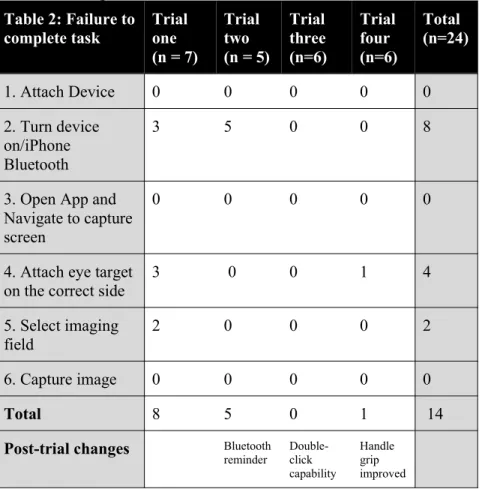
All of the steps were closely observed with an established checklist of required steps and failure to complete them were tallied (Table 2). Attaching the device and opening the iPhone application to navigate to the capture screen did not cause any problems for any of the participants in all the trials. Turning the device
on and syncing it with the iPhone’s Bluetooth was a problem with 33.3% (8/24) of the participants involved in the first 2 trials. After a software modification to remind users to turn the device on for trial 3, none of the subsequent participants experienced difficulties with that step. 16.6% (4/24) of participants made errors in attaching the external fixation screen by either forgetting to attach the screen or putting it on the wrong side. After the software adjustment was made in
Table 2: Failure to
complete task Trial one (n = 7)
Trial two (n = 5)
Trial three (n=6) Trial four (n=6) Total (n=24)
1. Attach Device 0 0 0 0 0
2. Turn device on/iPhone Bluetooth
3 5 0 0 8
3. Open App and Navigate to capture screen
0 0 0 0 0
4. Attach eye target on the correct side
3 0 0 1 4
5. Select imaging field
2 0 0 0 2
6. Capture image 0 0 0 0 0
Total 8 5 0 1 14
Post-trial changes Bluetooth
round three to prompt users to adjust when the screen was attached to the wrong side, only one participant made an error. About 33.3% (8/24) of participants reported afterwards that the device felt heavy (Table 2).
There was also an overall decrease in the errors made across trials. In total, eight errors were made in trial one, five errors in trial two, zero errors in trial three, and one error in trial four (Table 2).
In terms of image quality of the photograph from the human eye trial, the grader could exclude all emergent findings in 94% (33/35) of the photos. The grader could exclude subtle findings in 51% (18/35) images.
DISCUSSION:
The inherent qualities of smartphones make them well suited for primary care based diabetic retinopathy screening. They are portable, affordable, and have high resolution cameras and powerful computer processing capabilities to capture and transfer photographs electronically. Such telemedicine-based approaches for diabetic retinopathy screening have been effective in decreasing the rates of blindness in countries such as the United Kingdom and Ireland.[26] While smartphone retinal imaging is a promising tool for retinal screening, published studies have shown significant variability in image quality and our own early testing demonstrated that smartphone retinal imaging can be quite variable.[14,23] We therefore incorporated user feedback when designing the RetinaScope to make it intuitive to use. Taking this approach, usability testing is a valuable means for assessing the effectiveness, efficiency, and satisfaction of a product.[22] It has proven to be an effective way to tailor the design of a product to the user’s preferred way of work and to reduce the time needed for user training and support. [22,27,28] Nielsen and Landaeur have shown that 4-5 users maximized the cost-to-benefit ratio of detecting usability problems.[29] Nielson has also stated that iterative design maximizes the utility of usability because it can detect a greater percentage of usability problems.[30] The goal of this study was to utilize usability testing to evaluate the time on task, errors,[22] and
subjective preferences of primary care medical assistants and technicians using the RetinaScope and to see if improvement can be made through iterative end-user feedback. This is the first study to test the usability of a smartphone ophthalmoscope by non eye-care specialists.[13]
It is worth noting that the ability to capture 5-field images is important in the screening accuracy of diabetic retinopathy, as single-field images may not be adequate to determine the severity of DR.[31-33] However, capturing multiple fields is technically challenging. The RetinaScope was specifically designed and tested to simplify the process of capturing wide-field images.
.
The consistently high SUS metrics, ranging from 80.5 to 89.6 throughout the trials, suggest a general ease-of-use and high level of user satisfaction with the RetinaScope.[34]
consistently demonstrated that the time required to perform a photograph of the eye was critical for the adoption of this technology and that the time needed to be less than 5 minutes.
RetinaScope was able to meet this demand with a single photograph captured in 66 seconds and five images in under 4 minutes. The number of errors made was also reduced across the trials from 8 in trial one to 1 in trial four. These changes, including the Bluetooth notification, handle-grip improvement and double-click image capture capability, over the course of the trials suggest that the adjustments made throughout the trials resulted in improved usability of the device.
The proof-of-concept round demonstrated that ophthalmic technicians and medical assistants without an ophthalmic background can use the RetinaScope to capture an image of a human retina in significantly less time (p < 0.02) than existing devices after a brief training session. The average image capture time of one human eye with the RetinaScope in the last trial was 66 ± 7 seconds for 1 image and 229 ± 114 seconds for 5 images. The image quality was good, with the ability to detect over 94% of emergent findings. This was defined as optic disc edema, optic disc pallor, retinal vascular occlusion, intraocular hemorrhages, and grade III/IV retinopathy.[25] There have been no other reports of retinal image capture time using smartphone
ophthalmoscopy, so our comparisons are with other non-smartphone fundus cameras. The DigiScope, a well-accepted fundus camera for teleretinal imaging, reported one-eye imaging times of 5.6 ± 2.4 minutes.[18] Other DR telehealth imaging technologies, such as nonmydriatic fundus photography (NMFP) and ultra-wide field retinal imaging (UWFI) have reported similar image times of 12.8 minutes and 9.2 minutes per patient respectively.[35] RetinaScope requires minimal training and can capture retinal images more rapidly than existing imaging modalities (Figure 2).
We postulate that users’ familiarity with the smartphone interface contributes to the ease-of-use of the RetinaScope device. Our results indicate that the steps associated with conventional smartphone imaging were easier to perform than those additional steps not part of standard smartphone photography. In trial 1, where there was a 3-4 day delay between the giving of the instructions and the testing of the device, the tasks that the most users struggled with were those not shared with smartphone picture-taking, for example turning on the RetinaScope Bluetooth button, attaching the external fixation, and selecting the imaging fields (Table 2). Of all the documented errors across the usability studies, 93.3% (14/15) were steps that were outside of normal smartphone imaging, which includes turning the device Bluetooth on, attaching the external fixation screen, and selecting the imaging field (Table 2). The functions that resembled everyday smartphone picture-taking, such as opening the smartphone application and navigating to the capture screen were successfully completed by all users. When the time between
instructions and device use was removed in trial 2, the number of users who completed tasks not related to smartphone photography increased. This suggests that the tasks themselves are
There were several limitations in our study. Our data focused primarily on image capture using a model eye for 4 rounds of testing with limited capture on a human eye only in one final round of testing, which may not be representative of clinical usage on patients with varying anatomy. In addition, participants using the device immediately after receiving directions may not be
representative of a true clinical encounter. Our data was limited by a relatively small sample size per iteration, and were not directly compared with other devices. All participants in our study reported familiarity with using a smartphone to take photos, and thus it is unclear if people without this familiarity will be able to use the RetinaScope equally well and rapidly. However, with the growing ubiquity of smartphones in the general population worldwide, this likely is not a major issue. In addition, our subject was pharmacologically dilated, and photographing through an undilated pupil could pose greater challenges to medical assistants. However, this study serves as a proof of concept study for larger usability studies of the RetinaScope.
Despite these limitations, our study demonstrated the usability of the RetinaScope by ancillary health workers without prior retinal experience to capture quality retinal photos in a timely manner. Future studies will involve usability testing on human eyes directly comparing the different types of existing retinal cameras with a larger group of users.
CONCLUSION:
Smartphone-based retinal photography is a promising method for increasing accessibility of retinal screening in the community. This study demonstrates that usability testing and feedback-driven engineering is capable of rapidly improving smartphone-based retinal imaging among novice users. RetinaScope enables high-quality retinal photography among medical assistants in primary care within minutes of first-time operation. Further investigation is required to validate screening of retinal disease in the primary care setting using RetinaScope operated by medical assistants and non-ophthalmic personnel.
ACKNOWLEDGEMENTS:
Thank you to the faculty and staff at the Briarwood Family Medicine Center and Ypsilanti Health Center at the University of Michigan for your enthusiasm and general support for this project.
FINANCIAL SUPPORT:
This work was supported by the University of Michigan Translational Research and Commercialization for Life Sciences (TNK, YMP), the University of Michigan Center for Entrepreneurship Dean’s Engineering Translational Prototype Research Fund (TNK, YMP), the National Eye Institute grant 4K12EY022299 (YMP), the University of Michigan Department of Ophthalmology and Visual Sciences, and unrestricted departmental support from Research to Prevent Blindness (University of Michigan and Washington University)
DISCLOSURES:
References:
1. Murchison AP, Haller JA, Mayro E, et al. Reaching the Unreachable: Novel Approaches to Telemedicine Screening of Underserved Populations for Vitreoretinal Disease. Curr Eye Res. 2017;42(7):963-970. doi:10.1080/02713683.2017.1297463
2. Gower EW, Silverman E, Cassard SD, Williams SK, Baldonado K, Friedman DS. Barriers to Attending an Eye Examination after Vision Screening Referral within a Vulnerable Population. J Health Care Poor Underserved. 2013;24(3):1042-1052.
doi:10.1353/hpu.2013.0134
3. Li Z, Wu C, Olayiwola JN, Hilaire DS, Huang JJ. Telemedicine-based digital retinal imaging vs standard ophthalmologic evaluation for the assessment of diabetic retinopathy. Conn Med. 2012;76(2):85-90. http://www.ncbi.nlm.nih.gov/pubmed/22670358. Accessed April 24, 2018.
4. Pasquel FJ, Hendrick AM, Ryan M, Cason E, Ali MK, Narayan KMV. Cost-effectiveness of Different Diabetic Retinopathy Screening Modalities. J Diabetes Sci Technol.
2016;10(2):301-307. doi:10.1177/1932296815624109
5. Lord RK, Shah VA, San Filippo AN, Krishna R. Novel Uses of Smartphones in Ophthalmology. Ophthalmology. 2010;117(6):2008-2011.
doi:10.1016/j.ophtha.2010.01.001
6. MacCuish AC. Early detection and screening for diabetic retinopathy. Eye. 1993;7(2):254-259. doi:10.1038/eye.1993.59
7. Hazin R, Colyer M, Lum F, Barazi MK. Revisiting Diabetes 2000: Challenges in Establishing Nationwide Diabetic Retinopathy Prevention Programs. Am J Ophthalmol. 2011;152(5):723-729. doi:10.1016/j.ajo.2011.06.022
8. Garg S, Jani PD, Kshirsagar A V., King B, Chaum E. Telemedicine and Retinal Imaging for Improving Diabetic Retinopathy Evaluation. Arch Intern Med. 2012;172(21):1677. doi:10.1001/archinternmed.2012.4372
9. Romero P, Sagarra R, Ferrer J, Fernández-Ballart J, Baget M. The incorporation of family physicians in the assessment of diabetic retinopathy by non-mydriatic fundus camera. Diabetes Res Clin Pract. 2010;88(2):184-188. doi:10.1016/j.diabres.2010.02.001
10. Zimmer-Galler I, Zeimer R. Results of Implementation of the DigiScope for Diabetic Retinopathy Assessment in the Primary Care Environment. Telemed e-Health. 2006;12(2):89-98. doi:10.1089/tmj.2006.12.89
11. Bolster NM, Giardini ME, Bastawrous A. The Diabetic Retinopathy Screening Workflow: Potential for Smartphone Imaging. J Diabetes Sci Technol. 2015;10(2):318-324. doi:http:// dx.doi.org/10.1177/1932296815617969
ophthalmic imaging adapter: User feasibility studies in Hyderabad, India. Indian J Ophthalmol. 2016;64(3):191-200. doi:10.4103/0301-4738.181742
13. Ludwig CA, Newsom MR, Jais A, Myung DJ, Murthy SI, Chang RT. Training time and quality of smartphone-based anterior segment screening in rural India. Clin Ophthalmol. 2017;11:1301-1307. doi:10.2147/OPTH.S134656
14. Ryan ME, Rajalakshmi R, Prathiba V, et al. Comparison among methods of retinopathy assessment (CAMRA) study: Smartphone, nonmydriatic, and mydriatic photography. Ophthalmology. 2015;122(10):2038-2043. doi:10.1016/j.ophtha.2015.06.011
15. Rajalakshmi R, Arulmalar S, Usha M, et al. Validation of smartphone based retinal photography for diabetic retinopathy screening. PLoS One. 2015;10(9):1-10. doi:10.1371/journal.pone.0138285
16. Maamari RN, Keenan JD, Fletcher DA, Margolis TP. A mobile phone-based retinal camera for portable wide field imaging. Br J Ophthalmol. 2014;98(4):438-441. doi:10.1136/bjophthalmol-2013-303797
17. Burgess PI, Msukwa G, Beare NA, et al. Diabetic retinopathy in sub-Saharan Africa: meeting the challenges of an emerging epidemic. BMC Med. 2013;11(1):157.
doi:10.1186/1741-7015-11-157
18. Zeimer R, Zou S, Meeder T, Quinn K, Vitale S. A fundus camera dedicated to the screening of diabetic retinopathy in the primary-care physician’s office. Investig Ophthalmol Vis Sci. 2002;43(5):1581-1587.
19. Crabtree BF, Miller WL, Cohen DJ. Delivery of Clinical Preventive Services in Family Medicine Offi ces. Ann Fam Med. 2005:430-435. doi:10.1370/afm.345.INTRODUCTION
20. Yarnall KSH, Pollak KI, Østbye T, Krause KM, Michener JL. Primary Care : Is There Enough Time for Prevention ? 2003;93(4):635-641.
21. Darma S, Zantvoord F, Verbraak FD. The quality and usability of smartphone and hand-held fundus photography, compared to standard fundus photography. Acta Ophthalmol. 2015;93(4):e310-e311. doi:10.1111/aos.12632
22. Liljegren E. Usability in a medical technology context assessment of methods for usability evaluation of medical equipment. Int J Ind Ergon. 2006;36(4):345-352.
doi:10.1016/J.ERGON.2005.10.004
23. Kim T, Myers F, Reber C, et al. A Smartphone-Based Tool for Rapid, Portable, and Automated Wide-Field Retinal Imaging.; 2018. doi:10.1101/364265
25. Lamirel C, Bruce BB, Wright DW, Delaney KP, Newman NJ, Biousse V. Quality of nonmydriatic digital fundus photography obtained by nurse practitioners in the emergency department: The FOTO-ED study. Ophthalmology. 2012;119(3):617-624.
doi:10.1016/j.ophtha.2011.09.013
26. Tozer K, Woodward MA, Newman-Casey PA. Telemedicine and Diabetic Retinopathy: Review of Published Screening Programs. J Endocrinol diabetes. 2015;2(4).
http://www.ncbi.nlm.nih.gov/pubmed/27430019. Accessed September 11, 2018.
27. Jaspers MWM. A comparison of usability methods for testing interactive health technologies: Methodological aspects and empirical evidence. Int J Med Inform. 2009;78(5):340-353. doi:10.1016/j.ijmedinf.2008.10.002
28. Nielsen J. Usability Engineering. San Diego: Academic Press; 1993.
29. Nielsen J, Landauer TK. A Mathematical Model of the Finding of Usability Problems. Transport. 1910:206-213. doi:10.1145/169059.169166
30. Nielsen J. Why You Only Need to Test with 5 Users. Alertbox. 2000;19(September 23):1-4. doi:http://www.useit.com/alertbox/20000319.html
31. Vujosevic S, Benetti E, Massignan F, et al. Screening for Diabetic Retinopathy: 1 and 3 Nonmydriatic 45-degree Digital Fundus Photographs vs 7 Standard Early Treatment Diabetic Retinopathy Study Fields. Am J Ophthalmol. 2009;148(1):111-118.
doi:10.1016/j.ajo.2009.02.031
32. Aptel F, Denis P, Rouberol F, Thivolet C. Screening of diabetic retinopathy: effect of field number and mydriasis on sensitivity and specificity of digital fundus photography.
Diabetes Metab. 2008;34(3):290-293. doi:10.1016/j.diabet.2007.12.007
33. Murgatroyd H, Ellingford A, Cox A, et al. Effect of mydriasis and different field strategies on digital image screening of diabetic eye disease. Br J Ophthalmol. 2004;88(7):920-924. doi:10.1136/bjo.2003.026385
34. Bangor A, Kortum PT, Miller JT. An Empirical Evaluation of the System Usability Scale. Int J Hum Comput Interact. 2008;24(6):574-594. doi:10.1080/10447310802205776