Conservative management of displaced
paediatric supracondylar
fractures: A systematic review
Authors: Yeomans, D1 Graham, S.M2 Mkandawire, N3,4,5 Harrison, W.J6 Perry, D.C7 1. Oxford University Hospitals NHS Foundation Trust, Oxford, UK
2. Aintree University Hospital, Liverpool, UK
3. Beit CURE International Hospital, Blantyre, Malawi
4. The Department of Surgery, College of Medicine, University of Malawi, Malawi 5. School of Medicine, Faculty of Health Sciences, Flinders University, Australia 6. Countess of Chester Hospital, Chester, UK
7. Alder Hey Hospital, Liverpool, UK
Corresponding Author: Daniel Yeomans Email: danyeomans@gmail.com
Postal Address: John Radcliffe Hospital
Headley Way Headington
OX3 9DU
Funding: This research received no specific grant from any funding agency in the public, commercial or not-for-profit sectors.
Conflict of Interest: Yeomans, D, Graham, S.M, Mkandawire, N, Harrison, W.J, Perry, D declare they have no conflict of interest.
Key Words: Casting, Conservative, Paediatric, Supracondylar, Traction
Acknowledgements: We would like to thank Oliver Hardcastle for the illustration of figure 1. 1
Abstract:
Background: In high-income countries, displaced supracondylar fractures are managed with K-wire
fixation. Alternatively, in low-income countries where surgical expertise and resources are limited,
these injuries are managed with traction or closed reduction and casting. The aim of our study is to
systematically present the published evidence regarding outcomes of conservatively managed
displaced supracondylar fractures.
Methods: A systematic review of the literature was performed identifying studies evaluating the
outcome of displaced supracondylar fractures managed non-operatively.
Results: 46 papers examined the outcome of displaced supracondylar fractures managed
conservatively. Our results show management by traction is equivalent to percutaneous pinning,
whereas outcomes following closed reduction and casting were inconsistent.
Conclusion: Closed reduction and casting is inferior to traction and operative intervention in the
management of displaced supracondylar fractures. Traction remains a viable option in low income
countries (LIC). However, at present there is little data from LICs, limiting the transferability of our
conclusions. 33
34
35
1. Background
Supracondylar fractures of the distal humerus are the most common injury in children under the age
of 7 years, and constitute 18% of the fractures sustained by those under 16 age group [1].
Classification of such injuries is based on a system initially described by Gartland in the 1950’s,
summarised in figure 1 [2]. However current management is based on the modified Gartland
classification [3].Undisplaced Gartland type I fractures are typically managed with cast
immobilisation, resulting in good functional outcomes and are not the focus of this study [3, 4].
In the absence of neurovascular injury, displaced closed supracondylar fractures are either managed
conservatively or surgically [3]. Surgical treatment options include open reduction and internal
fixation or open/closed reduction with percutaneous kirschner wires (k-wire) fixation. The American
Association of Orthopaedic Surgeons (AAOS) published guidance in 2011 recommending closed
reduction with pin fixation for all patients with displaced injuries [3]. Furthermore, the British
Orthopaedic Association Standards for Trauma (BOAST 11) recommend early surgical treatment for
these injuries [5]. However, these two guidelines are based on expert opinion and case-series.
Options for non-operative management comprise either traction (skin/skeletal) or casting with closed
reduction. 54
55 56 57
58
Historically, closed reduction and casting provided the mainstay of treatment for displaced injuries.
However, rates of Volkmanns ischaemic contracture were high [6]. In the 1920’s Dunlop began
treating displaced supracondylar fractures with traction [3]. In doing so, successfully reduced the
frequency of serious complications [7]. However, long hospital stays and the inherent associated
costs lead to a shift in favour of operative management[8].
In many low and middle income countries (LMICs) surgeons treating supracondylar fractures often
lack access to the resources or expertise to manage supracondylar fractures operatively [9]. As a
result, many institutes continue to treat displaced supracondylar fractures conservatively [10]. In our
institute at Queen Elizabeth Central Hospital, Blantyre, Malawi, straight-arm traction remains the
mainstay of treatment for such injuries, with few observed complications.
There is no Level-I evidence available comparing the outcomes of operative versus conservatively
managed supracondylar fractures. The aim of this review is to systematically present the published
evidence regarding the outcomes of conservatively managed displaced supracondylar fractures.
2. Methods
A review of the literature was performed using the Preferred Reporting Items for Systematic
Reviews and Meta-analysis (PRISMA) checklist and algorithm [11]. A search was conducted using
Medline, EMBASE and Cochrane computerised literature databases in July 2016. Inclusion and
exclusion criteria are displayed in table 1. No year of publication limits were applied and all English
language studies were included. 76
77 78 79 80 81 82 83 84 85 86 87 88 89 90 91
92
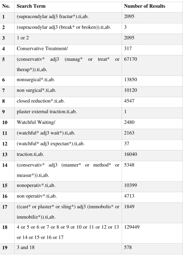
Table 2 shows an example of our data search in Medline, when run in Embase, we retrieved 751
results. The flow diagram of papers included in our review is outlined in figure 21. Following review
of abstracts, 35 papers were selected for full text review. Full text review of the remaining papers
excluded a further 3; one examining only humeral shaft fractures [12], a second lateral condyle
fractures [13], and one contained no conservative intervention [14]. A further 14 articles meeting our
inclusion criteria were identified through review of the reference lists. Using Cochrane guidance, a
data extraction table was formulated, which was used to collate relevant information from each full
text included in the review including assessment of the risk of bias. [15]
Outcome was difficult to assess due to the heterogeneity of the studies, however Flynn’s criteria was
the most widely used and comparable outcome measure. All results were tabulated and grouped by
method of intervention for ease of comparison. Two authors (DY, SG) performed both review of
abstracts and data extraction of the included studies. A total of 46 papers were included for final
review (figure 21).
3.0 Results
A summary of the 46 papers included in our review are summarised in appendices 1 to 4. Countries
income level is based on their World Bank classification as of 1st January 2017 [16].
98 99 100 101 102 103 104 105 106 107 108 109 110 111 112 113
114
115
3.1 Closed reduction and casting
28 studies examined outcome data of closed reduction and casting for displaced supracondylar
fractures [17–44]. This included any method of immobilisation whereby plaster of paris was applied
to the injured arm, set under any degree of flexion. Closed reduction was performed and where
specified, manipulation of the displaced injury was performed under general anaesthetic, local block,
or sedation.
The most commonly used outcome measure was Flynn’s criteria (Figure 1) which evaluates both
cosmetic and functional outcomes. The remainder used other functional criteria, radiographic
evaluation or re-operation rate following failure of plaster cast immobilisation. Due to the
heterogeneity of the outcomes amongst the studies in question, we did not to compare them
quantitatively.
Four studies found casting to be viable first line management for displaced supracondylar fractures
[25, 27, 29, 44]. However all acknowledge a proportion would require delayed pinning in the event
reduction was lost, this was not found to affect long term outcomes. These papers did not
differentiate between Gartland types II and II.
Overall, inconsistent outcomes following closed reduction and casting makes it challenging to draw
broad conclusions from the data in our review. Although a number of papers continue to advocate
the method, especially in a low-income setting [44] others suggest operative fixation provides 119
120
superior outcomes [33, 38]. In two papers, when a distinction was drawn between Gartland type II
and III injuries, casting was found to provide unsatisfactory outcome in type III, but good results in
type II [33, 38].
3.2 Traction
Our review of the literature found 24 studies presenting outcome data following traction as
management of displaced supracondylar fracture [8, 18, 22, 29–31, 37, 40, 42, 43, 45–60]. Outcomes
of overhead skeletal traction straight-arm lateral traction, skin traction, side arm traction, and brace
with traction, were all examined and results displayed in appendices 1, 2 and 3.
When compared directly to operative intervention for comparison, traction resulted in equivalent
outcomes [18, 29, 37, 42, 43, 56, 57]. Sutton et al. presented a retrospective case series (n=65)
directly comparing traction with percutaneous pinning [56]. Using Flynn’s criteria as their principle
outcome measure, the paper concludes no statistical difference when comparing the two methods
[56]. However the paper did not state the grading of Gartland fracture between the two treatment
groups. Where cost of treatment was assessed, traction was more expensive than operative
intervention, hence two papers concluding percutaneous pinning to be their preferred option [8, 56].
A total of sixteen papers studied the outcomes of skeletal traction, of these, eight used Flynn’s
criteria as their primary outcome measure. Eight studies examined straight arm traction, two of 141
142 143 144
145
146
which used Finn’s criteria. There was no statistically significant difference when compared directly.
One paper did not specify the method of traction used [42].
One paper concluded traction to have superior outcomes to casting, pinning and ORIF [22].
All eleven studies examining traction without operative intervention found it to provide excellent
outcomes in severely displaced or swollen injuries. No patients managed with traction required
subsequent operative intervention [47–52, 54, 55, 58-60].
4.0 Conclusion
Our review has shown that current evidence for the management of displaced supracondylar
fractures is inconclusive. It appears that closed reduction and casting may be utilised in the first
instance with positive results, with the option of percutaneous pinning in the event of failed
reduction. Whereas outcomes following traction appear to be equivalent to that of percutaneous
pinning, although this conclusion is drawn from a limited number of studies. Despite this, the trend
of managing all displaced injuries operatively within high-income setting remains unchallenged.
Where resources allow, operative intervention is now regarded as the gold standard management for
Gartland II and III injuries [3]. The British Orthopaedic Association Standards for Trauma state that
displaced supracondylar fractures “…require early surgical treatment; ideally on the day of
admission… surgical stabilisation should be with bicortical wire fixation.” [5] However, these are 162
163 164 165 166 167 168 169 170 171
172
guidelines and cannot be translated to healthcare provision in resource-limited settings. The results of
this review suggest that where surgical intervention is unavailable, such as in Malawi, traction
remains the preferred management.
Anatomical reduction is required for percutaneous pinning to succeed [3]. Hence attempting the
procedure without the aid of intraoperative fluroscopy would be hazardous, limiting its use to
environments where such resources are available. Complications such as ulnar nerve injury, pin
migration and pin tract infection are reported in the literature with rates between 1.8% and 4.7% [61–
63]. O’Hara et al. report rates of cubitus varus deformity of up to 32% when protocol and x-ray is not
strictly followed with K-wire insertion [34]. There is therefore a trade-off between conservative
management, where possible mild mal-union would result in normal function but a potential
cosmetic issue, and operative intervention where the complications intra and post-operative
complications can be significantly worse.
It is not only the access to surgical skills and equipment that limit the use of operative intervention in
LIC’s. Access to anaesthesia is a problem throughout sub-Saharan Africa, where facilities to deliver
safe anaesthesia to a child have been reported to be as low as 13% [62]. This additional risk of
operative intervention provides us with further insight as to why traction remains the preferred
method of treatment in many countries. 184
There were no cases of Volkmann’s ischaemic contracture in the papers included within our review.
Loss of reduction was the only indication reported in the four studies recommending initial closed
reduction with subsequent operative intervention.
The disparity between outcome measures used gives our data limited transferability. Although
Flynns criteria is used the most frequently, outcome measures in the literature are wide ranging [63].
Flynn provides a method of analysis whereby results can be easily compared using change in
carrying angle and range of motion. However, this clinical outcome is not patient reported and can be
prone to measurement bias. Changes in carrying angle, associated with a poorer score of Flynn’s
criteria, may not always equate to a worse functional outcome. There is a need for a validated
functional outcome measure in children, encompassing patient reported outcome measures.
One limitation of many papers in our review is the lack of transparency when allocating patients to
treatment groups. It was frequently unclear how patients had been selected to be managed
conservatively or operatively. Indeed, in retrospective case series, this cannot accurately be
measured. Recruitment bias may well therefore have confounded several authors conclusions.
Cubitus varus is widely considered a cosmetic problem, usually only evident when standing in the
anatomical position [64]. Review of patients with residual cubitus varus following supracondylar
fracture found no functional deficit and can be corrected via planned geometric osteotomy at a later
date if required [65]. The incidence and long-term consequences of cubitus varus deformity in
low-income countries have not been investigated in the current literature. 204
Traction is well documented to result in a longer hospital stay than operative intervention. In our
review, a total of thirteen papers specify the length of hospital stay when managed by traction. If not
otherwise stated, the total duration of traction was taken as the length of hospital stay. Duration of
inpatient stay ranged from 11 – 22 days with a median of 19 days. Two papers used length of stay as
contributing factors of their cost analysis, both concluding traction was considerably more expensive
than pinning [8, 56]. When considering duration of stay, theatre fees, anaesthetic fees, recovery room
fees and radiography fees Sutton et al. and Piretto et al. calculated traction was more expensive by
142% and 179% respectively. However, both papers were based in high income countries, where
costs of both equipment and service provision make calculations non-transferable to less
economically developed nations.
The use of traction for Gartland types II and III supracondylar fractures provides a safe and effective
alternative to percutaneous wire fixation in the resource poor setting. In countries where few
specialist centres are managing increasingly high volume of trauma, the benefit of such surgical
intervention remains to be proven. With the correct expertise, traction can be safely applied in a local
setting, avoiding the need for long-distance transfer and associated financial cost. All papers in our
review analysing long term outcome measures of traction alone, support this premise.
Currently there is no level one evidence comparing percutaneous pin fixation with traction for
displaced supracondylar fractures of the distal humerus in children. Drawing on conclusions from the 226
equipoise. Our review also highlights the lack of data from LICs on this topic, which would improve
the transferability of our conclusions.
Figure Legends:
Figure 1: Lateral views of supracondylar fractures according to the Gartland Classification (i) Type 1 (undisplaced) (ii) Type 2 and (iii) Type 3
Figure 21: Flow diagram outlining studies included for review
Inclusion Criteria Exclusion Criteria
1. Paper written in English 1. Undisplaced supracondylar fractures included in study
2. Level I, II, III or IV study design by Journal of Bone and Joint Surgery criteria
3. Series reporting on supracondylar fractures of Gartland types II or III (displaced)
4. Conservative (non-operative) management in one or all arms of the study
5. Assessment of outcome (functional, anatomical or radiological)
6. All patients in the study were <18 years old
Table 1: An outline of inclusion and exclusion criteria used in our review 248
249 250 251 252 253 254 255 256 257 258
No. Search Term Number of Results 1 (supracondylar adj3 fractur*).ti,ab. 2095
2 (supracondylar adj3 (break* or broken)).ti,ab. 3
3 1 or 2 2095
4 Conservative Treatment/ 317 5 (conservativ* adj3 (manag* or treat* or
therap*)).ti,ab.
67170
6 nonsurgical*.ti,ab. 13850
7 non surgical*.ti,ab. 10120
8 closed reduction*.ti,ab. 4547 9 plaster external traction.ti,ab. 1
10 Watchful Waiting/ 2480
11 (watchful* adj3 wait*).ti,ab. 2163 12 (watchful* adj3 expectan*).ti,ab. 37
13 traction.ti,ab. 16040
14 (conservativ* adj3 (manner* or method* or measur*)).ti,ab.
5348
15 nonoperativ*.ti,ab. 10399
16 non operativ*.ti,ab. 4713
17 ((cast* or plaster* or sling*) adj3 (immobolis* or immobiliz*)).ti,ab.
1849
18 4 or 5 or 6 or 7 or 8 or 9 or 10 or 11 or 12 or 13 or 14 or 15 or 16 or 17
129449
19 3 and 18 578
References
[1] J. C. Cheng, B. K. Ng, S. Y. Ying, and P. K. Lam. A 10-year study of the changes in the pattern and treatment of 6,493 fractures. J. Pediatr. Orthop. 1999, 19:344–350
[2] J. J. Gartland. Management of supracondylar fractures of the humerus in children. Surg. Gynecol. Obstet. 1959, 109:145–154
[3] E. S. Paxton, J. L. Matzon, A. C. Narzikul, P. K. Beredjiklian, and J. A. Abboud. Agreement Among ASES Members on the AAOS Clinical Practice Guidelines. Orthopedics. 2015, 38:169–177
[4] M. S. Ballal, N. K. Garg, A. Bass, and C. E. Bruce. Comparison between collar and cuffs and above elbow back slabs in the initial treatment of Gartland type I supracondylar humerus fractures. J. Pediatr. Orthop. 2008, 17:57–60
[5] British Orthopaedic Association, ‘British Orthopaedic Association Standards for Trauma (BOAST) - Supracondylar fractures of the humerus in children’ . [Cited 2017 July 25] Available From: https://www.boa.ac.uk/wp-content/uploads/2014/12/BOAST-11.pdf..
[6] S. J. Mubarak and N. C. Carroll. Volkmann’s contracture in children: aetiology and prevention. J. Bone Joint Surg. Br. 1979, 61–B:285–293
[7] H. S. Dodge. Displaced Supracondylar Fractures of the Humerus in Children-Treatment by Dunlop’s Traction. J Bone Jt. Surg Am. 1972, 54:1408–1418
[8] C. A. Prietto. Supracondylar fractures of the humerus. A comparative study of Dunlop’s traction versus percutaneous pinning. J. Bone Joint Surg. Am. 1979, 61:425–428
[9] K. E. Wilkins. Nonoperative Management of Pediatric Upper Extremity Fractures or Don't Throw Away the Cast. Tech. Orthop. 2005, 20:115–141
[10] A. Chaudhuri, S. Datta, B. Sirdar, D. Roy, S. Dharmadevan, and S. Ghosh. Management of displaced supracondylar fracture of the humerus in children. Saudi J. Sports Med. 2015, 15:193 [11] D. Moher, A. Liberati, J. Tetzlaff, D. G. Altman, and T. P. Group. Preferred Reporting Items for
Systematic Reviews and Meta-Analyses: The PRISMA Statement. PLOS Med. 2009, 6: e1000097
[12] N. Osman, C. Touam, E. Masmejean, H. Asfazadourian, and J. Y. Alnot. Results of non-operative and non-operative treatment of humeral shaft fractures. A series of 104 cases. Chir. Main 1998. 17:195–206
[13] P.-S. Marcheix, V. Vacquerie, B. Longis, P. Peyrou, L. Fourcade, and D. Moulies. Distal humerus lateral condyle fracture in children: when is the conservative treatment a valid option? Orthop. Traumatol. Surg. Res. 2011, 97:304–307
[14] Y.-L. Wang, W.-N. Chang, C.-J. Hsu, S.-F. Sun, J.-L. Wang, and C.-Y. Wong. The recovery of elbow range of motion after treatment of supracondylar and lateral condylar fractures of the distal humerus in children. J. Orthop. Trauma. 2009, 23:120–125
[15] Cochrane, ‘The Cochrane Public Health Group Data Extraction and Assessment Template’[Cited 2017 July 08]. Available from:
[16] World Bank Group, ‘World Bank Classification of Countries [Cited 2016 December 10] Available from: http://data.worldbank.org/country
[17] M. Ababneh, A. Shannak, S. Agabi, and S. Hadidi. The treatment of displaced supracondylar fractures of the humerus in children. A comparison of three methods. Int. Orthop. 1998, 22:263–265
[18] I. Arnala, H. Paananen, and L. Lindell-Iwan. Supracondylar fractures of the humerus in children. Eur. J. Pediatr. Surg. 1991, 1:27–29
[19] I. U. Babar, N. Shinwari, M. R. Bangash, and M. S. Khan. Management of supracondylar fracture of humerus in children by close reduction and immobilization of the elbow in extension and supination. J Ayub Med Coll Abbottabad. 2009, 21:159–61
[20] J. Bender and C. A. Busch. Results of treatment of supracondylar fractures of the humerus in children with special reference to the cause and prevention of cubitus varus. Arch. Chir. Neerl. 1978, 30:29–41
[21] T. Camus, B. MacLellan, P. C. Cook, J. L. Leahey, J. C. Hyndman, and R. El-Hawary.
Extension type II pediatric supracondylar humerus fractures: a radiographic outcomes study of closed reduction and cast immobilization. J. Pediatr. Orthop. 2011, 31:366–371
[22] O. Celiker, F. I. Pestilci, and M. Tuzuner. Supracondylar fractures of the humerus in children: analysis of the results in 142 patients. J. Orthop. Trauma. 1990, 4:265–269
[23] R. S. Chen, C. B. Liu, X. S. Lin, X. M. Feng, J. M. Zhu, and F. Q. Ye. Supracondylar extension fracture of the humerus in children manipulative reduction, immobilisation ans fixation using a U-shapes plaster slab with the elbow in full extension. J. Bone Joint Surg. Br. 2001, 83:883– 887
[24] J. W. Colaris, T. M. Horn, E. D. van den Ende, J. H. Allema, and J. W. S. Merkus.
Supracondylar fractures of the humerus in children. Comparison of results in two treatment periods. Acta Chir. Belg. 2008, 108:715–719
[25] S. V. Dharmadevan, S. Ghosh, A. Chaudhuri, S. Datta, B. K. Sirdar, and D. S. Roy.
Management of displaced supracondylar fracture of the humerus in children. Saudi J. Sports Med. 2015, 15:193
[26] A. M. Eid. Reduction of displaced supracondylar fracture of the humerus in children by manipulation in flexion. Acta Orthop. Scand. 1978, 49:39–45
[27] P. G. Fitzgibbons, B. Bruce, C. Got, S. Reinert, P. Solga, J. Katarincic, C. Eberson. Predictors of failure of nonoperative treatment for type-2 supracondylar humerus fractures. J. Pediatr. Orthop. 2011, 31:372–376
[28] H. W. Grant, L. E. Wilson, and W. H. Bisset. A long-term follow-up study of children with supracondylar fractures of the humerus. Eur. J. Pediatr. Surg. 1993, 3:284–286
[29] A. T. Hadlow, P. Devane, and R. O. Nicol. A selective treatment approach to supracondylar fracture of the humerus in children. J. Pediatr. Orthop. 1996, 16:104–106
[30] R. Hagen. Skin-traction Treatment of Supracondylar fractures of the humerus in children. A ten-year review. Acta Orthop. Scand. 1964, 35:138–148
[32] M. S. Khan, S. Sultan, M. A. Ali, A. Khan, and M. Younis. Comparison of percutaneous pinning with casting in supracondylar humeral fractures in children. J. Ayub Med. Coll. Abbottabad. 2005, 17:33–36
[33] I. Miranda, P. Sánchez-Arteaga, V. G. Marrachelli, F. J. Miranda, and M. Salom. Orthopedic versus surgical treatment of Gartland type II supracondylar humerus fracture in children. J. Pediatr. Orthop. 2014, 23:93–99
[34] L. J. O’Hara, J. W. Barlow, and N. M. Clarke. Displaced supracondylar fractures of the humerus in children. Audit changes practice. J. Bone Joint Surg. Br. 2000, 82:204–210 [35] S. N. Parikh, E. J. Wall, S. Foad, B. Wiersema, and B. Nolte. Displaced type II extension
supracondylar humerus fractures: do they all need pinning? J. Pediatr. Orthop. 2004, 24:380– 384
[36] P. Persiani, M. Di Domenica, M. Gurzi, L. Martini, R. Lanzone, and C. Villani. Adequacy of treatment, bone remodeling, and clinical outcome in pediatric supracondylar humeral fractures. J. Pediatr. Orthop. 2012, 21:115–120
[37] A. M. Pirone, H. K. Graham, and J. I. Krajbich. Management of displaced extension-type supracondylar fractures of the humerus in children. J. Bone Joint Surg. Am. 1988, 70:641–650 [38] M. Shoaib, A. Hussain, H. Kamran, and J. Ali. Outcome of closed reduction and casting in
displaced supracondylar fracture of humerus in children. J. Ayub Med. Coll. Abbottabad 2003, 15:23–25
[39] H. T. Spencer, F. J. Dorley, L. E. Zionts, D, H, Dichther, M. A. Wong, P. Moazzaz et al. Type II supracondylar humerus fractures: can some be treated nonoperatively? J. Pediatr. Orthop. 2012, 32:675–681
[40] V. Vahvanen and K. Aalto. Supracondylar fracture of the humerus in children. A long-term follow-up study of 107 cases. Acta Orthop. Scand. 1978, 49:225–233
[41] Š. Vučkov, A. Kvesić, Z. Rebac, D. Cuculić, F. Lovasić, and N. Bukvić. Treatment of supracondylar humerus fractures in children: Minimal possible duration of immobilization. Coll. Antropol. 2001, 25:255–262
[42] A. Walløe, N. Egund, and L. Eikelund. Supracondylar fracture of the humerus in children: review of closed and open reduction leading to a proposal for treatment. Injury 1985, 16:296– 299
[43] S. Young, J. M. Fevang, G. Gullaksen, P. T. Nilsen, and L. B. Engesæter. Deformity and functional outcome after treatment for supracondylar humerus fractures in children: a 5- to 10-year follow-up of 139 supracondylar humerus fractures treated by plaster cast, skeletal traction or crossed wire fixation. J. Child. Orthop. 2010, 4:445–453
[44] C. I. Singh, R. K. R. Devi and G. S. Sharma. A prospective study of supracondylar fractures of the humerus in children treated by closed reduction' J. Evid. Based Med. Healthc. 2015, 2:4958–4967
[45] N. P. Badhe and P. W. Howard. Olecranon screw traction for displaced supracondylar fractures of the humerus in children. Injury.1998, 29:457–460
[47] T. Berghausen, B. M. Leslie, L. K. Ruby, and S. Zimbler. The severely displaced pediatric supracondylar fracture of humerus treated by skeletal traction with olecranon pin. Orthop. Rev. 1986, 15:510–515
[48] H. S. Dodge. Displaced Supracondylar Fractures of the Humerus in Children-Treatment by Dunlop’s Traction. J Bone Jt. Surg Am. 1972, 54:1408–1418
[49] A. Gadgil, C. Hayhurst, N. Maffulli, and J. S. M. Dwyer. Elevated, straight-arm traction for supracondylar fractures of the humerus in children. J. Bone Joint Surg. Br. 2005, 87:82–87 [50] S. Harwant and T. A. Borhan. The efficacy of side arm traction in the reduction of
supracondylar fracture humerus in children. Med. J. Malaysia. 2000, 55:311–317
[51] C. D. Jefferiss. “Straight lateral traction” in selected supracondylar fractures of the humerus in children. Injury. 1977, 8:213–220
[52] K. Matsuzaki, N. Nakatani, M. Harada, and T. Tamaki. Treatment of supracondylar fracture of the humerus in children by skeletal traction in a brace. Bone Jt. J. 2004, 86:232–238
[53] J. Piggot, H. K. Graham, and G. F. McCoy. Supracondylar fractures of the humerus in children. Treatment by straight lateral traction. Bone Jt. J. 1986, 68:577–583
[54] E. C. Rodriguez Merchan. Supracondylar fractures of the humerus in children: treatment by overhead skeletal traction. Orthop. Rev. 1992, 21:475–482
[55] M. Z. Sadiq, T. Syed, and J. Travlos. Management of grade III supracondylar fracture of the humerus by straight-arm lateral traction. Int. Orthop. 2007, 31:155–158
[56] W. R. Sutton, W. B. Greene, G. Georgopoulos, and T. B. Dameron. Displaced supracondylar humeral fractures in children. A comparison of results and costs in patients treated by skeletal traction versus percutaneous pinning. Clin. Orthop. 1992, 278:81–87
[57] S. Turra, S. Santini, A. Zandonadi, and C. Jacobellis. Supracondylar fractures of the humerus in children. A comparison between non-surgical treatment and minimum synthesis. Chir. Organi Mov. 1995, 80:293–299
[58] M. Urlus, P. Kestelijn, E. Vanlommel, M. Demuynck, and L. Vanden Berghe. Conservative treatment of displaced supracondylar humerus fractures of the extension type in children. Acta Orthop. Belg. 1991, 57:382–389
[59] P. H. Worlock and C. Colton. Severely displaced supracondylar fractures of the humerus in children: a simple method of treatment. J. Pediatr. Orthop. 1987, 7:49–53
[60] M. Z. Sadiq, T. Syed, and J. Travlos. Management of grade III supracondylar fracture of the humerus by straight-arm lateral traction. Int. Orthop. 2007, 31:155–158
[61] P. Devkota, J. A. Khan, B. M. Acharya, N. M. Pradhan, L. P. Mainali, M. Singh et al. ‘Outcome of supracondylar fractures of the humerus in children treated by closed reduction and
percutaneous pinning. J. Nepal Med. Assoc. 2008, 47:66–70
[62] S. C. Hodges, C. Mijumbi, M. Okello, B. A. McCormick, I. A. Walker, and I. H. Wilson. Anaesthesia services in developing countries: defining the problems. Anaesthesia. 2007, 62:4– 11
[63] J. C. Flynn, J. G. Matthews, and R. L. Benoit. Blind pinning of displaced supracondylar fractures of the humerus in children. Sixteen years’ experience with long-term follow-up. J. Bone Joint Surg. Am. 1974, 56:263–272
[64] C. Colton and F. Monsell. Supracondylar humeral fractures in children-have we stopped thinking? J Trauma Ortho. 2016, 4:48-52
[65] J. J. Joseph and N. Wilson. Cubitus Varus Following Paediatric Supracondylar Humeral Fracture: 40-Year Review of the Experience of the Royal Hospital for Sick Children of Glasgow. Bone Jt. J. 2013, 95–B:38–38