© 2013, IJCSMC All Rights Reserved 99
Available Online atwww.ijcsmc.com
International Journal of Computer Science and Mobile Computing
A Monthly Journal of Computer Science and Information Technology
ISSN 2320–088X
IJCSMC, Vol. 2, Issue. 11, November 2013, pg.99 – 105
REVIEW ARTICLE
Review of Image Processing Technique
for Glaucoma Detection
Preeti
1, Jyotika Pruthi
2 1Computer Science and Engineering, itm University, Gurgaon, India
2
Computer Science and Engineering, itm University, Gurgaon, India
1
[email protected]; [email protected]
Abstract- The review paper describes the application of various image processing techniques for automatic detection of glaucoma. Glaucoma is a neurodegenerative disorder of the optic nerve, which causes partial loss of vision. Large number of people suffers from eye diseases in rural and semi urban areas all over the world. Current diagnosis of retinal disease relies upon examining retinal fundus image using image processing. The key image processing techniques to detect eye diseases include image registration, image fusion, image segmentation, feature extraction, image enhancement, morphology, pattern matching, image classification, analysis and statistical measurements.
Keywords- Image Registration; Fusion; Segmentation; Statistical measures; Morphological operation; Classification
I. INTRODUCTION
© 2013, IJCSMC All Rights Reserved 100
Approximately, 5 million people live with a glaucoma risk while around 800.000 people suffer from glaucomatous damages in Germany [1]. If the optical imaging of the retina is taken then by performing the series of image processing operations, automated early detection of eye disease is possible.
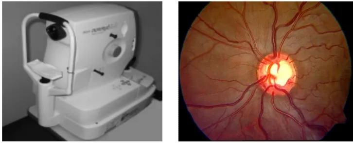
With great improvement in field of medical imaging, Image processing technique helps in early diagnosis of glaucoma and other eye disease. Retinal fundus images assist trained clinicians to diagnose any abnormality and any change in retina. These images are captured by using special devices called ophthalmoscopes. Medical image analysis and processing has great significance in non-invasive treatment and clinical study. The information about the optic disk can be used to examine severity glaucoma. The location of the optic disk is an important issue in retinal image analysis as it is a significant landmark feature. Fig.1 shows the fundus camera and retinal fundus image.
Fig. 1 Digital fundus camera and acquired retinal fundus image
II. IMAGE PROCESSING TECHNIQUE
Various image processing techniques used in automated early diagnosis and analysis of various eye disease are Enhancement, Registration, Fusion, Segmentation, Feature extraction, Pattern matching, Classification, Morphology, Statistical measurements and Analysis [2][3].
Image Enhancement- Image enhancement includes varying brightness and contrast of image. It also includes filtering and histogram equalization. It comes under preprocessing step to enhance various features of image.
Image Registration- Image Registration is an important technique for change detection in retinal image diagnosis. In this process, two images are aligned onto a common coordinate system. Images may be taken at different times and with imaging devices In medical diagnosis, it is essential to combine data from different images and for better analysis and measurements images are aligned geometrically.[4]
Image Fusion- Image fusion is a process of combining information acquired from number of imaging devices. Its goal is to integrate contemporary, multisensor, multi-temporal or multi-view information into a single image, containing all the information so as to reduce the amount of information.
Feature Extraction- It is the process of identifying and extracting region of interest from the image.
© 2013, IJCSMC All Rights Reserved 101
to build object of interest on which analysis and interpretation can be performed. It includes clustering, thresholding etc.
Morphology- Morphology is the science of appearance, shape and organization. Mathematical morphology is a collection of non-linear processes which can be applied to an image to remove details smaller than a certain reference shape. Various morphological operation are erosion, dilation, opening and closing.
Classification- Classification is an important technique of image analysis for estimation of statistical parameter according to the gray level intensities of pixels. It includes labeling of a pixel or group of pixels based on the grey values and other statistical parameters. For understanding the contents of an image, image analysis functions are used [5].
III. LITERATURE REVIEW
Several studies are reported in literature for detection of optic disk and detection and classification of glaucoma. The work is as follows:
In Year 2006, Kevin Noronha performed a work, "Enhancement of retinal fundus Image to highlight the features for detection of abnormal eyes"[6]. This work specifies the methods used to detect main features of retinal fundus images such as optic disk, fovea, and exudates and blood vessels using different techniques. To determine the optic Disk and its centre Author find the brightest part of the fundus and apply Hough transform.
In Year 2007, Sangyeol Lee performed a work, "Validation of Retinal Image Registration Algorithms by a Projective Imaging Distortion Model"[4]. A variety of methods for retinal image registration have been proposed. Authors also present the validation tool for any retinal image registration method by tracing back the distortion path and accessing the geometric misalignment from the coordinate system of reference standard.
In Year 2008, S. Sekhar performed a work," Automated localization of retinal optic disk using hough transform"[7]. The retinal fundus image is widely used in the diagnosis and treatment of various eye diseases such as diabetic retinopathy and glaucoma. The proposed methodology consists of two steps: in the first step, region of interest (ROI) is found by image by means of morphological processing, and in the second step, optic disk is detected using the Hough transform.
In Year 2010, Zhuo Zhang performed a work," ORIGA-light : An Online Retinal Fundus Image Database for Glaucoma Analysis and Research"[8]. Author present an online dataset, ORIGA-light, which aims to share clinical retinal images with the public. Author had updated the system continuously with more clinical ground-truth images. The proposed method focuses on optic disk and cup segmentation.
In Year 2010, Vahabi Z proposed," The new approach to Automatic detection of Optic Disc from non-dilated retinal images"[9]. Author describes a new filtering approach like Sobel edge detection, Texture Analysis, Intensity and Template matching to detect Optic Disc. The proposed algorithm is applied in wavelet domain on 150 images of Messidor dataset.
In Year 2011, Zafer Yavuz performed a work," Retinal Blood Vessel Segmentation Using Gabor Filter And Tophat Transform"[10]. In this, Author gave a method for retinal blood vessels segmentation by applying firstly Gabor filter to enhance blood vessels and then applying top-hat transform. Later on, the output is converted to binary image with p-tile thresholding.
© 2013, IJCSMC All Rights Reserved 102
In Year 2012, R. Geetha Ramani performed a work," Automatic Prediction of Diabetic Retinopathy and Glaucoma through Retinal Image Analysis and Data Mining Techniques"[5]. This paper proposed a novel approach for automatic disease detection. Retinal image analysis and data mining techniques are used to accurately categorize the retinal images as either Normal, Diabetic Retinopathy and Glaucoma affected.
In Year 2012, ManjulaSri Rayudu proposed," Review of Image Processing Techniques for Automatic Detection of Eye Diseases"[14]. The review paper provides information about the application of image processing techniques for automatic detection of eye diseases. The key image processing techniques to detect eye diseases include image registration, fusion, segmentation, feature extraction, enhancement, pattern matching, image classification, analysis and statistical measurements.
IV. GLAUCOMA DETECTION ALGORITHM
For assessment of glaucoma, cup-to-disc ratio is most widely accepted index. Early research was done for detection and localization of optic disk. The various algorithms used in this direction are vessel’s direction matched filter, curvelet transform, active contour model, fuzzy c-mean clustering, artificial neural networks, k-NN regressor, pyramidal decomposition, edge detection, entropy filter and feature vector[15-21].
Other techniques include averaging filter, template matching technique and canny edge detector. S.Sekhar et al. [22] applied Hough transform to detect Optic Disk. After preprocessing a binary image is obtained which can be used to find the contours of OD. Morphological closing is performed on ROI to calculate the magnitude gradient of edge detection and fill the vessels according to (1).
f • B = ( f ⊕ B) Θ B . (1)
For removing any peaks, morphological opening is applied according to (2).
f B = ( fΘB)⊕ B (2)
Where f is the grayscale image, B is binary structuring element;
⊕ is dilation Θ is erosion operators
Gopal Dat Joshi et al.[23] described cup-to-disc ratio(CDR) calculation by applying morphological operations and hough transformation for detecting glaucoma. Within the optic region, cup is segmented using vessel bends(r-bends for specifying cup boundary) and pollar information. These r-bends are non-uniformly distributed on the OD region. So for detection of cup boundary local interpolating spline is applied.
© 2013, IJCSMC All Rights Reserved 103
V. RESULTS
As a part of survey of various image processing techniques, the author has implemented some of the techniques like preprocessing; histogram equalization morphological operation etc. and result are as follows:
a.) Original Image b.) Gray Scale of original image c.) After Histogram Equalization
d.) Original Histogram e.)Enhanced Contrast f.) Binary image
© 2013, IJCSMC All Rights Reserved 104
VI.CONCLUSION
The author of this paper concluded that for detection and diagnosis of glaucoma, firstly, optic disk need to be segmented. After image acquisition, preprocessing is done by applying thresholding, illumination and histogram equalization. The optic disk and cup is segmented using various techniques like Hough transform, k-means clustering, fuzzy c-means clustering, active contour method, matched filter approach, vessel bends, morphological operations etc. Then CDR is calculated and classification is done for deciding whether condition of eye is normal or glaucomatous.
REFERENCES
[1]Initiativkreis zur Glaukomfrüherkennung, Germering, Germany, www.glaukom.de
[2] R.ManjulaSri,KMM Rao, “Novel Image Processing Techniques to Detect Lesions using Lab View”, IEEE Conference Publications on Annual IEEE India Conference INDICON. Publication Year: 2011 , Page(s): 1-4
[3] Viralkumar Bulsara, Surabhi Bothra, Poonam Sharma, K.M.M.Rao, ”Low Cost Medical Image Processing System for
Rural/Semi Urban Healthcare”, IEEE Conference Publications on Recent Advances in Intelligent Computational Systems
(RAICS),Publication Year: 2011 , Page(s): 724 – 728
[4] Sangyeol Lee, Michael D. Abr`amoff, and Joseph M. Reinhardt.” Validation of Retinal Image Registration Algorithms by a
Projective Imaging Distortion Model” 29th Annual International Conference of the IEEE EMBS Cité Internationale, Lyon,
France August 23-26, 2007.
[5] R. Geetha Ramani," Automatic Prediction of Diabetic Retinopathy and Glaucoma through Retinal Image Analysis and Data Mining Techniques”
[6] Kevin Noronha, Jagadish Nayak, S.N. Bhat, “Enhancement of retinal fundus Image to highlight the features for detection of abnormal eyes”
[7] S. Sekhar," Automated localisation of retinal optic disk using hough transform", Department of Electrical Engineering and Electronics, University of Liverpool, UK.
[8] , Zhuo Zhang," ORIGA-light : An Online Retinal Fundus Image Database for Glaucoma Analysis and Research", 32nd Annual International Conference of the IEEE EMBSBuenos Aires, Argentina, August 31 - September 4, 2010
[9]Vahabi Z,” The new approach to Automatic detection of Optic Disc from non-dilated retinal images” Proceedings of the 17th Iranian Conference of Biomedical Engineering (ICBME2010), 3-4 November 2010
[10] Zafer Yavuz," RETINAL BLOOD VESSEL SEGMENTATION USING GABOR FILTER AND TOPHAT TRANSFORM",
2011 IEEE 19th Signal Processing and Communications Applications Conference (SIU 2011) 978-1-4577-0463-511/11 ©2011 IEEE
[11] Nilan jan Dey," Optical Cup to Disc Ratio Measurement for Glaucoma Diagnosis Using Harris Corner", ICCCNT12
[12]Harris C, Stephens, M. , 1 988, A Combined Corner and Edge Detector, Prooeedings of 4th Alvey Vision Conference
[13] Kon stantino s G Derpanis, 200 4, The Harris Corner Detector
[14] ManjulaSri Rayudu," Review of Image Processing Techniques for Automatic Detection of Eye Diseases", 2012 Sixth International Conference on Sensing Technology (ICST) 978-1-4673-2248-5/12 ©2012 IEEE
[15]Aliaa Abdel-Haleim Abdel-Razik Youssif, Atef Zaki Ghalwas, and AmrAhmed SabryAbdel-Rahman Ghoneim ,”Optic Disc
Detection From Normalized Digital Fundus Images by Means of a Vessels’ Direction Matched Filter-IEEE Transactions On
Medical Imaging, Vol. 27, No. 1, January 2008
© 2013, IJCSMC All Rights Reserved 105
[17] Mahdad Esmaeili, Hossein Rabbanin and Alireza Mehri Dehnavi: “Automatic optic disk boundary extraction by the use of
curvelet transform and deformable variational level set model” in Pattern Recognition,Vol. 45 (2012) , pp. 2832–2842
[18] Chisako Muramatsu et al.,”Automated segmentation of optic disc region on retinal fundus photographs: Comparison of
contour modeling and pixel classification methods” in Computer Methods And Programs In Biomedicine Vol.101 (2011) , pp.
23–32.
[19] Meindert Niemeijer, Michael D. Abràmoff and Bram van Ginneken” Fast detection of the optic disc and fovea in color
fundus photographs” in Medical Image analysis, in press.
[20] Rashid Jalal Qureshi et al. “Combining algorithms for automatic detection of optic disc and macula in fundus images;
Computer Vision and Image Understanding ,Vol. 116 (2012) ,pp.138–145
[21] Chih-Yin Ho,Tun-Wen Pai, Hao-Teng Chang and Hsin-Yi Chen, “ An automatic fundus image analysis system for clinical
diagnosis of glaucoma”,-2011 International Conference on Complex, Intelligent, and Software Intensive Systems.
[22] S.Sekhar,W.Al-Nuaimy and A.K.Nandi,”Automated Localisation Of Retinal Optic Disk Using Hough Transform”, 5th IEEE International Symposium on Biomedical Imaging: From Nano to Macro, 2008, pp.1577 - 1580
[23]Gopal Datt Joshi, Jayanthi Sivaswamy, and S. R. Krishnadas,”Optic Disk and Cup Segmentation From Monocular Color
Retinal Images for Glaucoma Assessment’, IEEE Transactions On Medical Imaging, Vol. 30, No. 6, June 2011,pp.1192-1205.