R E V I E W
Update on terbina
fi
ne with a focus
on dermatophytoses
Jason G Newland1,3
Susan M Abdel-Rahman2,3
Divisions of Infectious Diseases1
and Clinical Pharmacology and Medical Toxicology2, Children’s
Mercy Hospitals and Clinics, Kansas City, MO, USA;
3Department of Pediatrics,
University of Missouri-Kansas City, School of Medicine, Kansas City, MO, USA
Correspondence: Susan M Abdel-Rahman Associate Professor of Pediatrics, Division of Clinical Pharmacology and Medical Toxicology, Children’s Mercy Hospital, 2401 Gillham Road, Suite 0411, Kansas City, MO 64108, USA Tel +1 816 855 1759 Fax +1 816 855 1958 Email [email protected]
Abstract: Since terbinafi ne was introduced on the world market 17 years ago, it has become
the leading antifungal for the treatment of superfi cial fungal infections, aided by unique pharma-cologic and microbiologic profi les. This article reviews mode of action, antimycotic spectrum and disposition profi le of terbinafi ne. It examines the data, accumulated over 15 years, on the comparative effi cacy of terbinafi ne (vs griseofulvin, itraconazole, fl uconazole) in the management of the infections for which it is primarily indicated (eg, dermatophytoses) and provides a brief discussion on its use for the treatment of non-dermatophyte infections. Finally, the available data on the newest topical and systemic formulations are introduced.
Keywords: tinea, Trichophyton, Microsporum, allylamine, antifungal
Introduction
In 2008, oral terbinafi ne reached the 12-year mark in the United States (US) and 17 years on the world market. Since its launch, terbinafi ne has garnered the top slot among topical antifungals and the oral formulation is estimated to have captured nearly 80% of the greater than US$1.5 billion worldwide onychomycosis market (although it makes up only a minority of prescriptions written for children).1,2 Terbinafi ne remains
the only commercially available orally available allylamine and shares the topical allylamine/benzylamine market with naftifi ne, butenafi ne and amorolfi ne. In recent years several new formulations have been added to the portfolio of this antimycotic including a pediatric oral granule approved by the US Food and Drug Administration in September of 2007 and a single-dose, fi lm-forming topical solution that is now available over the counter in a number non-US markets.
This article will review the accumulated data on the mycology and pharmacology of terbinafi ne including its mode of action, antimycotic spectrum, disposition profi le and therapeutic effi cacy. The primary focus will surround dermatophytoses with a brief discussion on the role, to date, of terbinafi ne in non-dermatophyte infections.
Clinical mycology
Discovered in 1983, terbinafi ne is a member of the allylamine class of antifungals. It differs from its parent compound, naftifi ne, by the presence of a tert-butyl acetylene substitution of the phenyl ring on the side chain of the molecule. This substitution confers an increase in oral effi cacy and an additional 10 to 100 times the in vitro activity of naftifi ne.3,4
Terbinafi ne inhibits fungal growth by disrupting sterol biosynthesis. It abrogates the formation of ergosterol by inhibiting squalene epoxidase, the catalytic enzyme responsible for converting squalene to 2,3-oxidosqualene (an ergosterol precursor). The resultant defi ciency in ergosterol compromises cell wall integrity and contrib-utes to impaired growth and/or death of the pathogen.5–7 Notably, the biosynthesis
of cholesterol in higher order eukaryotes similarly relies on the activity of squalene
Clinical, Cosmetic and Investigational Dermatology downloaded from https://www.dovepress.com/ by 118.70.13.36 on 21-Aug-2020
Newland and Abdel-Rahman
epoxidase; however, terbinafi ne demonstrates a markedly lower binding affi nity for the mammalian enzyme. In vitro, the minimum concentration of terbinafi ne required to inhibit 95% of squalene epoxidase activity (IC95) is two to three orders of magnitude greater for the mammalian enzyme (300 μM) than for enzymes isolated from pathogenic yeast (0.6–2.1 μM).7
While the majority of clinical terbinafi ne use is observed with infections caused by dermatophytes, the susceptibility of numerous organisms including pathogenic yeast, dematia-ceae, thermally dimorphic fungi and hyaline hyphomycetes has been evaluated.8–16 Although the nature of the assays
employed precludes direct comparison of minimum inhibi-tory concentrations (MICs) between studies (ie, some were performed prior to standardization of a reference method by the Clinical and Laboratory Standards Institute (CLSI),17
some universal trends are repeated throughout. Namely, terbinafi ne demonstrates the greatest activity against species within the Trichophyton, Microsporum, and Epidermophyton
genera followed by the dematiaceae, the fi lamentous fungi and a few selected pathogenic yeast.
The terbinafi ne MICs observed against the dermatophytes are typically several orders of magnitude lower than those reported for other fungi.18–23 This heightened susceptibility
is refl ected by an MIC that is an order of magnitude lower than IC95 for sterol biosynthesis.6 Given that dermatophyte
growth can be fully inhibited despite only partial inhibi-tion of sterol synthesis, the activity of terbinafi ne is likely accounted for by other processes including the intracellular accumulation of squalene.3 In contrast, the MIC to IC
95 ratio
for several species of fermentative yeast equals or exceeds one.6 Compared with dermatophytes, these organisms have
adapted to survive under anaerobic growth conditions which are characterized by low ergosterol and high squalene concentrations.6 Reasonably, it is expected that such
organ-isms would be less susceptible to the effects of a squalene epoxidase inhibitor.
The MICs reported for terbinafi ne against various derma-tophytes are typically comparable to or lower than those of other antifungals active against these organisms, namely the triazoles, imidazioles and griseofulvin.24 However, a direct
comparison of MICs between therapeutic agents needs to be considered in the context of achievable concentrations at the site of infection. No signifi cant differences in terbinafi ne sus-ceptibilities exist between US and non-US isolates of selected
Trichophyton and Microsporum species.25 Further, the
putative increase in resistance to azole antifungals observed with the “heartier” arthroconidia of the dermatophytes has
not been observed with terbinafi ne. Both arthroconidia and microconidia of selected Trichophyton species demonstrate the same susceptibility profi le to terbinafi ne in vitro.26
In addition to diminished susceptibility, non-dermatophyte species of fungi demonstrate resistance mechanisms not observed in the dermatophytes. Under the selective pressure of terbinafi ne exposure, an increase in the expression of energy-dependent effl ux transporters can be observed in yeast;27 however, the expression level of orthologous
trans-porters remains unchanged in dermatophytes.28 Similarly,
fi lamentous fungi can upregulate the expression of an enzyme that catalyzes the breakdown of terbinafi ne29 but this has not
been reported for dermatophytes.
Overall, an extremely low rate of spontaneous mutation conferring resistance of dermatophytes to terbinafi ne exists in vitro.30 Nonetheless, reduced susceptibility to terbinafi ne
has been observed in clinical dermatophyte isolates. These appear to arise from clones harboring one of two described sequence variations in the squalene epoxidase gene.31,32 As
the reported mutations do not appear to impact fungal growth in the absence of terbinafi ne,33 they likely signal changes to
the terbinafi ne binding domain and ultimately binding affi nity of the drug for the protein.34
While spontaneous resistance is rare, increasing reports describe cross-resistance developing between other anti-fungals and terbinafine. Under the selective pressure of echinocandin exposure in vitro, the upregulation of effl ux transporters in yeast also reduced susceptibility to terbinafi ne.35 Similarly, azole “pre-exposure” in yeast can
diminish susceptibility of the organisms to terbinafi ne.36–38
Notably, the aforementioned mechanisms of cross-resistance have not been reported for dermatophytes, in vitro or in vivo, after protracted imidazole treatment.36–38
Of potentially signifi cant clinical relevance is the activity of terbinafi ne when used in combination with other anti-fungals for the management of invasive mycoses. Against
Aspergillus fumigatus, indifference was primarily observed
when terbinafi ne was combined with amphotericin B. Simi-larly, terbinafi ne did not improve the activity of fl uconazole or itrazonazole against A. fumigatus; however, the triazoles demonstrated synergism when added to terbinafi ne.39 Against
fl uconazole-resistant yeast, synergy with fl uconazole and itraconazole was observed in a fraction of isolates, Candida
glabrata⬎ Candida tropicalis, ⬎ Candida kreusi.40,41 In
these pathogenic yeast antagonism has also been observed among these antifungals.41 Against ocular isolates of
Fusarium, the combination of amphotericin B and terbinafi ne
was synergistic, while the combination of terbinafi ne and
Clinical, Cosmetic and Investigational Dermatology downloaded from https://www.dovepress.com/ by 118.70.13.36 on 21-Aug-2020
Terbinafi ne in dermatophytoses
triazoles demonstrated indifference.42 The ultimate utility
of terbinafi ne in the management of invasive infections will be determined as more experience is obtained utilizing this agent as adjunct therapy.
Clinical pharmacology
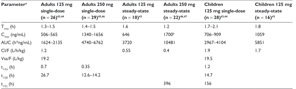
(Tables 1 and 2)
Terbinafi ne is effi ciently absorbed following oral administration (bioavailability approx. 70%) and this does not appear to be affected by feeding status.51,52 Over the range of clinically
relevant doses (125–750 mg) terbinafi ne demonstrates a linear absorption profi le with total body exposure increasing in direct proportion to dose.53 The rate of absorption does not appear
to differ substantially between children and adults. However, the extent of absorption as refl ected by maximum plasma concentrations is markedly lower in children when doses are normalized per kilogram of body weight.43
Following topical administration to normal skin, cream- and gel-based terbinafi ne formulations attain concentrations rang-ing from 746 to 949 ng/cm2.48,54 Maximum stratum corneum
concentrations increase by 15% with 7 days of application; however, the area under the plasma concentration vs time curve (AUC) can increase by as much as 40% over 1 week.54 Notably,
concentrations obtained in the horny layer of patients with an active infection can be up to an order of magnitude lower than that observed in healthy individuals.55 While topical
prepara-tions are well absorbed into the stratum corneum, the resultant systemic exposure is several orders of magnitude lower than observed after oral terbinafi ne administration (Tables 1 and 2).
Terbinafine is extensively distributed with estimates of apparent distribution volume approaching 20 L/kg.53,56
This relatively large volume of distribution results from the drugs
high degree of lipophilicity, extensive protein binding profi le and ability to concentrate in adipose and keratin rich tissue.56,57
At steady-state, concentrations observed in sebum, stratum corneum and hair exceed those observed in the plasma by as much as an order of magnitude. Although lower in the stratum corneum of hyperkeratotic tissue, terbinafi ne concentrations remain elevated following the discontinuation of oral therapy and persist in excess of 1 month after stopping treatment.58
At least seven cytochromes P450 (CYP) appear to be responsible for metabolizing terbinafi ne into more than 15 metabolites.59 In adults, the N-demethyl and carboxybutyl
metabolites constitute the largest fraction of the metabolites observed. Maximum circulating concentrations and total body exposure are comparable or in excess of those observed with the parent compound. Notably, the circulating half-life for the carboxy metabolites were twice as long as that of terbinafi ne.46
Although the metabolites lack an appreciable antifungal activity, they may contribute to the drug interactions and/or side effects observed following administration.51,60
Given the polyfunctional nature of terbinafine as a substrate for the CYP450, the magnitude of potential drug interactions would be predicted to be low as compared with other drugs.61–63 However, this is not the case for interactions
mediated by CYP2D6. Terbinafi ne exhibits potent inhibition of this enzyme in vitro (apparent inhibitor rate constant (ki) approx. 30 nM) and correspondingly marked reduction in the metabolism of the CYP2D6 substrate dextromethorphan in vivo.60,64 Importantly, the activity of CYP2D6 may not return
to normal for months after the completion of a prolonged course of therapy.64 Clinically, terbinafi ne is demonstrated to
interact with concurrently administered CYP2D6 substrates including amitriptyline, nortriptyline, desipramine, and
Table 1 Pharmacokinetic parameter estimates of terbinafi ne following oral administration
Parametera Adults 125 mg single-dose (n = 26)43,44
Adults 250 mg single-dose (n = 29)45,46
Adults 125 mg steady-state (n = 10)43
Adults 250 mg steady-state (n = 22)46,47
Children
125 mg single-dose (n = 28)43,44
Children 125 mg steady-state (n = 16)43
Tmax (h) 1.3–1.5 1.4–1.5 1.6 1.2 1.7–2.1 1.8
Cmax (ng/mL) 506–565 1340–1656 646 1700b 706–909 1059
AUC (h*ng/mL) 1624–2135 4740–6762 3720 10481 2967–4104 5851
Cl/F (L/h/kg) 1.2 0.55 0.4 1.9 1.7
Vss/F (L/kg) 19.2 19.5
t1/2α(h) 0.7 0.35 1.2
t1/2β(h) 26.7 12.6–14.2 14.7
t1/2γ(h) 396 156
Notes: aValues represent the mean reported values from the referenced studies (when more than one study is referenced, values represent the range of reported mean values). bCorresponding peak tissue concentrations: hair, 2.4 μg/g; stratum corneum, 14.4 μg/g; sebum, 56.1 μg/g.
Abbreviations: Tmax, time at maximum plasma concentration; Cmax, maximum plasma concentration; AUC, area under the plasma concentration vs time curve; Cl/F, apparent oral clearance; Vss/F, steady-state volume of distribution; t1/2α, alpha-phase half-life; t1/2β, beta-phase half-life, t1/2γ, gamma-phase or terminal half-life.
Clinical, Cosmetic and Investigational Dermatology downloaded from https://www.dovepress.com/ by 118.70.13.36 on 21-Aug-2020
Newland and Abdel-Rahman
venlafaxine.65–68 Other drugs harboring the potential to interact
with terbinafi ne include perphenazine, metoprolol, encainide and propafenone.69
For drugs that are not substrates of CYP2D6 (eg, anti-coagulants, corticosteroids, oral contraceptives, tolbuta-mide, cyclosporine, midazolam, digoxin and terfenadine) terbinafi ne has only a modest or minimal affect on their metabolism.70–74 However, as a substrate of the cytochromes
P450, the pharmacokinetics of terbinafi ne are altered with the concurrent administration of several agents (eg, cimetidine, terfenadine, rifampin).70,75
The clearance of terbinafi ne is triphasic with the terminal elimination half-life approximating 100 hours after a single dose and 22 days with durations of therapy spanning several months.44,76 Approximately 80% of terbinafi ne’s
metabo-lites are excreted by the kidney with the remaining fraction eliminated in the feces.44 This protracted rate of elimination
accounts for the magnitude of accumulation observed with terbinafi ne after repeated dosing and the persistence in plasma and tissues long after discontinuation of the drug.47 While
this confers a distinct advantage to the allylamine permitting shorter courses of therapy, it poses a unique disadvantage for patients experiencing drug-related adverse events.
Therapeutic use
Terbinafine is indicated for use in the management of cutaneous dermatophytoses (eg, tinea corporis, tinea cruris and tinea pedis), onychomycosis and most recently tinea capitis. In addition, terbinafi ne use has been explored in a number of superfi cial and systemic mycoses involving pathogens other than the dermatophytes. The results of open-label and randomized trials exploring the utility of terbinafi ne in various infections is detailed in the following sections.
Cutaneous dermatophytoses
Dermatophyte infections of the glabrous skin, groin and feet can be caused by any of a number of dermatophyte species.77
While these infections typically respond to topical antifungals, oral therapy is often indicated when the lesions are widespread or chronic in nature.78
Terbinafi ne applied topically as a 1% cream, gel or solu-tion demonstrates utility in the management of both tinea corporis and tinea cruris.79 The application of terbinafi ne
once-daily for 7 to 14 days resulted in mycological cure rates ranging from 84% to 94%, clinical cure rates ranging from 75% to 84% and overall effi cacy rates ranging from 65% to 83%;80–86 The observed treatment response with topical
terbinafi ne is signifi cantly greater than reported for placebo wherein clinical, mycological and complete cure rates range from 8% to 22%.81,83 Topically applied terbinafi ne also
dem-onstrated statistically greater mycological response rates than a 2-week course of 2% ketoconazole cream,80 whereas clinical
response rates were comparable to a 0.6% gel containing the garlic-derived, sulfuric compound ajoene.84
Following oral administration for the treatment of tinea corporis and tinea cruris, clinical and mycological cure rates observed with terbinafi ne have ranged from 71% to 100% and 78% to 100%, respectively.78,87–89 No signifi cant differences
in mycological or clinical cure rates were observed between terbinafi ne and griseofulvin in these studies;87–89 however,
higher relapse rates were observed with griseofulvin.88,89
Tinea imbricata, a variant of tinea corporis found predomi-nantly in tropical countries, manifests as concentric rings of papular plaques that are chronic in nature and relatively recalcitrant to antifungal therapy. In a single random-ized, controlled trial 4 weeks of treatment with terbinafi ne demonstrated higher overall effi cacy rates (100%) and lower relapse rates (16%) as compared with itraconazole (89% effi cacy, 75% relapse).90
Tinea pedis is a dermatophyte infection of the foot that generally takes one of three forms: 1) interdigital infections characterized by cracking and fi ssuring of the skin in the webbing between the toes, 2) acute vesicular infections which present with erythema, bullous eruptions and bacterial
Table 2 Local and systemic estimates of exposure following topical terbinafi ne application
Parametera 1% gel × 7 days
healthy skin48,49
1% cream 7 days48,50,54
1% FFS 1 application50
Stratum corneum Cmax (μg/cm2) 0.91 0.94–2 5
Stratum corneum AUC (h*μg/cm2) 12.7 11.7–13.5 104.2
Tissue t1/2 (h) 1.2 68 162
Plasma Cmax (ng/mL) 3.82
Plasma AUC (h*ng/mL) 63
Notes: aValues represent the mean reported values from the referenced studies (when more than one study is referenced, values represent the range of reported mean values).
Abbreviations: Cmax, maximum observed concentration, AUC, area under the concentration vs time curve; t1/2, half-life.
Clinical, Cosmetic and Investigational Dermatology downloaded from https://www.dovepress.com/ by 118.70.13.36 on 21-Aug-2020
Terbinafi ne in dermatophytoses
super-infections and 3) a chronic form of infection with scaling that covers the entire foot typically referred to as “dry” or “moccasin” type tinea pedis.91 The nature and
extent of infection often determines whether oral therapy is required or whether symptomatic relief can be obtained with topical therapy.92
Although topical terbinafi ne formulations in excess of 1% have been investigated, superfi cial treatment of tinea pedis typically relies on 5 to 7 days of application with the 1% cream, gel or solution (as described above for infections of the skin and groin). Mycologic cure rates are comparable irrespective of formulation, ranging from 82% to 97% with overall effi cacy rates of 64% to 86%. Response rates are signifi cantly greater than observed with the vehicle alone wherein mycologic cure and overall effi cacy range from 22% to 37% and 4% to 26%, respectively.83,93–96 Mycological cure
rates, after 1 week of treatment, are also comparable to or greater than those observed after 4 weeks of treatment with topical azole preparations.97–99
Despite the advantage of a strong effi cacy profi le with the application of terbinafi ne for one week, efforts to sim-plify topical administration, decrease treatment duration and improve compliance has lead to the development of a poly-meric fi lm-forming solution (FFS) designed as a “one-time” dose. The acrylate/cellulose/triglyceride based formulation leaves a nearly invisible, highly-concentrated fi lm on the skin after the carrier solvent (ethanol) has evaporated. This fi lm remains on the site of infection nearly 6 times longer than other topical preparations and results in stratum cor-neum concentrations that are sustained above the MIC in excess of 2 weeks after application.50,100 FFS concentrations
as high as 10% have been evaluated; however, mycologi-cal cure rates (80%–84%) and overall effi cacy (61%–70%) 6 weeks after application did not appear to demonstrate dose-dependence.100 Consequently, the currently marketed
formulation contains 1% terbinafi ne. In a study examining slightly longer-term follow-up, mycological cure rates were observed in 72% of those treated. Notably, the duration of infection prior to treatment did not appear to infl uence the likelihood of mycological cure. When participants were re-evaluated at 3 months, 12.5% of those individuals consid-ered mycologically cured at 6 weeks demonstrated positive cultures, comparable to the rates observed with other topical terbinafi ne formulations.101
For more refractory (eg, hyperkeratotic) infections of the foot, effi cacy rates comparable to those described above can be observed with topical treatment; however, this may require protracted treatment durations (ie, several months).55 In these
cases, oral therapy has emerged as a reliable alternative. Orally administered terbinafi ne can be highly effective in the management of tinea pedis with overall effi cacy rates exceeding 90% depending on the nature of infection and the regimen used. The majority of investigations have examined the effi cacy of orally administered terbinafi ne at daily doses of 250 mg (either divided or once daily). Effi cacy rates after 6 weeks of treatment ranged from 59% to 75%, increasing to 65% to 88% 12 weeks after the end of therapy.102,103 This is
compared with placebo and griseofulvin where effi cacy rates were 0% and 27%, respectively at the end of treatment and 0% and 45%, respectively, 2 weeks post treatment.102,103 With
shorter courses of oral treatment (250 mg daily × 2 weeks), mycological and clinical cure rates are markedly less impres-sive (23%–28% and 8%–43%, respectively). However, when followed out for 6 to 16 weeks, mycological cure rates (78%–86%) and clinical effi cacy (71%–94%) improve dramatically suggesting utility with shorter treatment dura-tions of terbinafi ne.104,105 Cure rates observed with
terbin-afi ne were comparable to those observed with itraconazole (100 mg/day); however, long-term follow-up suggested that terbinafi ne was slightly superior to 4 weeks of itraconazole treatment and signifi cantly superior to 2 weeks of treatment with the triazole.104,105 Data from a single study each suggest
that reducing (125 mg/day) or increasing (500 mg/day) the dose of terbinafi ne may not substantially alter cure rates compared traditional dosing with 250 mg/day.51,58
Onychomycosis
Approximately one-half of all nail problems are accounted for by onychomycosis, a fungal infection wherein the nails become discolored, thickened and prone to peeling or splitting.106 Dermatophytes are principally responsible for
infections of the toenail, whereas over 50% of fi ngernail infections can be caused by non-dermatophyte species.107,108
Of the commercially available oral antifungals, griseofulvin, itraconazole and terbinafine are most commonly used for the management of onychomycosis. Efficacy rates and treatment durations vary although treatment with the latter agents typically requires shorter courses than does the former. Irrespective, the majority of studies described below document cure rates at 12 months or beyond (earlier for fi ngernails) to account for the protracted growth rate of nails and the extended time-frames over which drug remains in the affected nails.
Since its approval, scores of studies have evaluated numerous terbinafi ne dosing regimens for the treatment of onychomycosis. With the administration of daily 250 mg
Clinical, Cosmetic and Investigational Dermatology downloaded from https://www.dovepress.com/ by 118.70.13.36 on 21-Aug-2020
Newland and Abdel-Rahman
doses, mycologic and clinical response rates observed in toenails ranged from 72% to 92% and 45% to 77%, respectively.109–113 Notably, there was little difference in both
clinical and mycological response rates whether patients were treated for 12, 18 or 24 weeks.109,111–113 Infections of the
fi ngernail demonstrated comparable response rates ranging from 71% to 100%.109,111,114 When non-dermatophyte
patho-gens are considered in subgroup analyses, response rates approximate 40% for infections with Candida and greater than 90% when treating Scopulariopsis brevicaulis.115–117
The combination of daily terbinafi ne doses (250 mg) with chemical or mechanical removal of the nail offers little to no additional increase in effi cacy over terbinafi ne alone.118,119
Similarly, only limited increases in effi cacy are observed when daily oral therapy is combined with adjunctive therapy including once-weekly topical amorolfi ne or once-daily topi-cal ciclopirox.120–123 In each study topical therapy appeared
to confer slight improvements in response; however, limited sample sizes restrict their signifi cance and superiority over terbinafi ne alone could not be determined.
Intermittent terbinafi ne dosing has also been explored for the management of onychomycosis. When the standard regimen of 250 mg once daily was compared to intermittent dosing administered at 350 mg daily for 2 weeks followed by 2 weeks “off ” for the management of subungual onychomycosis, both mycological (56%–58% vs 43%–50%) and complete cure (26%–30% vs 20%–24%) favored traditional dosing; however, only the latter was statistically signifi cant.124 When compared with 500 mg administered
daily for 1 week (followed by 3 weeks off) for the treatment of distal subungual onychomycosis, traditional dosing again proved superior to intermittent dosing. Mycological cure of the target toenail (71% vs 59%); clinical cure of the target toenail (45% vs 29%); complete cure of the target toenail (40% vs 28%); and complete cure of all 10 toenails (25% vs 15%) were all statistically greater with standard dosing.125 No
signifi cant differences in complete cure have been observed based on the number of pulses administered; however, a clear trend is noted with response rates increasing steadily from one to four pulses.126 As noted with traditional dosing, higher
cure rates were observed for fi ngernails treated with pulse-dosing as compared with toe nails. Mycological and clinical cure rates were 89% and 72% for dermatophytes, albeit lower (67%) for infections caused by yeast.127 As expected based
on the comparative data generated from traditional dosing trials, the combination of pulse therapy with ancillary topical therapy does not substantially improve outcome over treat-ment with terbinafi ne alone.126–128
In two comparative trials, terbinafine (250 mg/day) was significantly superior to griseofulvin (500 mg/day). Mycological cure rates were significantly higher with terbinafi ne (84%–92% vs 45%–63%), time to mycological cure significantly shorter (73 vs 93 days) and clinical cure significantly higher (76% vs 39%) as compared with griseofulvin.129,130 However, the exclusion of
non-dermatophyte pathogens in one study and the administration of sub-clinical griseofulvin doses in both studies likely resulted in an overestimate of terbinafi ne’s superiority. In a single trial employing the recommended dose of griseofulvin (1000 mg daily) mycological response was similar between terbinafi ne and griseofulvin (88% vs 82%, respectively); however, clinical response at the end of study was greater with terbinafi ne (81% vs 62%). Not surprisingly time to negative mycologic cultures was shorter for terbinafi ne (130 days vs 172 days).131
In three studies, traditional dose itraconazole (200 mg/day) was compared with traditional dose terbinafi ne (250 mg/day). In only one investigation where infections were solely restricted to those caused by dermatophytes did mycological cure rates favor terbinafi ne (81%–92% vs 63%–67%). In the remaining investigations no difference in mycological cure rates were observed between the allylamine and the triazole.132–134 The
remainder of studies comparing the two agents examined pulse-dosing of itraconazole. In the treatment of distal and lateral subungual onychomycosis restricted to dermatophytes, no appreciable difference was observed between itracon-azole (400 mg/day one week “on” three weeks “off ”) and terbinafi ne (mycological cure rates: 75%–90% vs 76%–87%, clinical cure rates 53%–82% vs 50%–79%).135–138 Not
surpris-ingly, itraconazole demonstrated superior cure rates among the non-dermatophyte moulds (62% vs 44%) and Candida
species (92% vs 40%).138 These studies are contradicted by
a large multi-national trial wherein mycological cure rates at 18 months were significantly greater with terbinafine (76%–80%) than observed with pulse-dose itraconazole (38%–49%).139 In selected subpopulations, complete cure rates
observed after 4 years remained superior in the terbinafi ne arm (24%–78%) compared with those receiving itraconazole (24%–28%) as did mycological cure (46% vs 13%).140,141
Finally, two investigations compare daily terbinafi ne with once weekly fluconazole (150 mg). Clinical cure rates (21%–38% vs 67%–81%), mycological cure rates (31%–51% vs 75–89%) and overall effi cacy (31% vs 62%) were inferior in fl uconazole treated patients.135,142
Despite reasonable effi cacy rates, the results of numerous trials indicate that a substantial fraction of onychomycosis patients treated with terbinafi ne remain uncured at the end
Clinical, Cosmetic and Investigational Dermatology downloaded from https://www.dovepress.com/ by 118.70.13.36 on 21-Aug-2020
Terbinafi ne in dermatophytoses
of treatment. Consequently several investigations have attempted to address whether additional intrinsic or extrinsic factors infl uence treatment response. Although the majority of these investigations were not adequately powered to defi ni-tively determine an association between selected covariates and treatment failure, several trends could be observed. The studies suggested that a higher fraction of patients 1) receiv-ing lower terbinafi ne doses (125 mg vs 250 mg), 2) with distal and lateral subungual onychomycosis, 3) with big toenail involvement, or 4) with positive mycology 3 months into treatment remain uncured at the time of follow-up.143–145 The
link between other factors such as demographic characteris-tics, the rate of nail growth, the species of infecting pathogen, the extent and duration of disease at the time of presentation and the number of nails involved remain unclear.144–146
Tinea capitis
Ringworm of the scalp is a dermatophyte infection commonly seen among preschool and school-aged children. It is one of the few dermatophyte infections that does not adequately respond to topical therapy, often requiring 6 to 8 weeks of oral antifungal treatment.147 Griseofulvin remains the gold
standard in the management of this infection, but treatment failures are common and it is not unusual for children to remain on therapy for extended durations.148
Several studies have examined the role of terbinafi ne in the treatment of tinea capitis; however, results need to be considered in the context of the geographic origin of the study participants given that the primary causative organ-isms and, thus response rates, are expected to vary between countries.149–151 The majority of these studies examine a
weight-based dosing scheme with 62.5 mg of terbinafi ne administered to children under 20 kg, 125 mg administered to children between 20 and 40 kg and an adult dose of 250 mg administered to children over 40 kg.
With the administration of terbinafi ne for one week,
Trichophyton infections are effectively treated in 56%
of children.152,153 Effi cacy rates ranged from 69% to 86%
after 2 weeks of treatment, averaged 65% with 4 weeks of treatment and ranged from 80% to 100% after 6 weeks of therapy.44,152–155 Corresponding mycological cure rates
observed after 1, 2, 4 and 6 weeks of treatment were 60%, 76%, 72% and ⬎90%, respectively.44,152,154 By contrast,
infections with Microsporum are less responsive to terbinafi ne. Response rates as low as 15% are observed with 1 to 2 weeks of therapy.153 However, extended durations of
treatment and/or doubling of the dose appeared to improve the likelihood of effi cacy in children harboring Microsporum
and clinical and mycological response rates begin to approxi-mate those observed for Trichophyton.153,156–158
In comparative trials, terbinafi ne, when used for dura-tions of 4 weeks, appeared to be as effective as griseofulvin administered for slightly longer intervals (8 weeks). Clinical and mycological cure rates ranged from 64% to 93% and 70% to 93%, respectively for terbinafi ne and 67% to 80% and 72% to 88%, respectively for griseofulvin.159–161 As above, the
exception lies with infections caused by Microsporum where griseofulvin appears to be superior to terbinafi ne even when the allylamine is administered for 12 weeks.160–162 Pulse
regi-mens wherein the standard dose or double the standard dose of terbinafi ne is administered daily for 1 week (followed by 3 weeks off) do not appear to confer any advantage or dis-advantage in the treatment of Microsporum infections over standard regimens.163 By contrast, terbinafi ne appeared to be
slightly more effective than both itraconazole and fl uconazole in treating tinea capitis.164 It should be noted that in a number
of the aforementioned studies, the griseofulvin doses employed (as low as 6 mg/kg) were markedly lower than those recom-mended for use in clinical practice. Consequently, the studies may have underestimated the effi cacy of griseofulvin.
A single publication is available detailing the results of two randomized-controlled trials that compared 6 weeks of treat-ment with the recently marketed terbinafi ne oral granule formu-lation (5–8 mg/kg daily) with griseofulvin (10–20 mg/kg daily) for the management of tinea capitis. The new formulation is designed to be sprinkled on foods thereby improving the reliability with which the drug can be administered to young children (⬎4 years of age). In these investigations, the complete cure rate observed with terbinafi ne was superior to griseofulvin in one trial (46% vs 34%) but not the other (44% vs 43%). Similarly mycological cure rates were signifi cantly higher in trial 1 (62% vs 50%) but not in trial 2 (61% vs 60%). In affi rmation of the results conferred in previously conducted trials, subgroup analyses in these trials revealed that terbinafi ne was signifi cantly better than griseofulvin when children were infected with Trichophyton tonsurans whereas griseofulvin proved superior for the treatment of infections with Micros-porum canis.165 This multi-national study also reported greater
effi cacy among US vs non-US infections which was likely accounted for by the higher fraction of children in the US that are infected with T. tonsurans.
Non-dermatophyte infections
Although not within the scope of this review, we would be remiss not to point out the increasing utilization of terbi-nafi ne, alone and in combination, for the management of
Clinical, Cosmetic and Investigational Dermatology downloaded from https://www.dovepress.com/ by 118.70.13.36 on 21-Aug-2020
Newland and Abdel-Rahman
non-dermatophyte infections. Despite its higher MIC to pathogenic yeast, topically administered terbinafi ne appeared effective for the management of tinea versicolor,166 and orally
administered terbinafi ne (250 mg twice daily) has been used successfully for the treatment of cutaneous candidiasis.167
Given, however, that the majority of superfi cial Candida
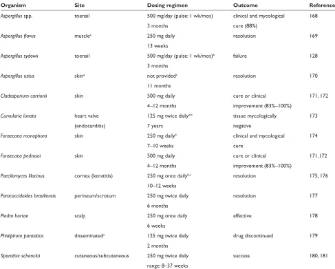
infections will respond to topical antifungals, the role of terbinafi ne in these infections remains unclear. A brief sum-mary of additional non-dermatophyte infections wherein terbinafi ne has been utilized is provided in Table 3.
Adverse effects
Terbinafine has been extensively used with a relatively low incidence of reported adverse drug reactions. Clini-cal trials evaluating the efficacy of orally administered
terbinafine in both children and adults have noted adverse event rates in as high as 52%; however, less than 10% were attributed to the drug.165,182–184 Most terbinafine related
adverse events are mild or moderate and include gastroin-testinal complaints (eg, nausea, abdominal pain, vomiting, diarrhea), cutaneous eruptions, weight gain, appetite changes, headaches, and vertigo.43,152,165,185,186 Those adverse
reactions involving the gastrointestinal system and skin are most commonly associated with discontinuation of therapy,165,186 with the risk of discontinuation estimated
at 3.4%.187
Serious adverse drug reactions, most commonly involving the liver and the hematologic system, are only rarely reported with terbinafi ne use (0.04%).188 Hepatotoxicity ranging
from mild transaminitis to fulminant liver failure has been
Table 3 Case-reports and open-label studies examining terbinafi ne use in non-dermatophyte infections
Organism Site Dosing regimen Outcome Reference
Aspergillus spp. toenail 500 mg/day (pulse: 1 wk/mos) clinical and mycological 168
3 months cure (88%)
Aspergillus fl avus musclea 250 mg daily resolution 169
13 weeks
Aspergillus sydowii toenail 500 mg/day (pulse: 1 wk/mos)b failure 128
3 months
Aspergillus ustus skina not providedc resolution 170
11 months
Cladosporium carrionii skin 500 mg daily cure or clinical 171, 172
4–12 months improvement (83%–100%)
Curvularia lunata heart valve 125 mg twice dailyd,e tissue mycologically 173
(endocarditis) 7 years negative
Fonsecaea monophora skin 250 mg dailyb clinical and mycological 174
7–10 weeks cure
Fonsecaea pedrosoi skin 500 mg daily cure or clinical 171,172
4–12 months improvement (83%–100%)
Paecilomyces lilacinus cornea (keratitis) 250 mg once dailyb,c resolution 175, 176
10–12 weeks
Paracoccidoides brasiliensis perineum/scrotum 250 mg twice daily resolution 177
6 months
Piedra hortae scalp 250 mg once daily effective 178
6 weeks
Phialphora parasitica disseminateda 125 mg twice daily drug discontinued 179
2 months
Sporothix schenckii cutaneous/subcutaneous 250 mg twice daily success 180, 181
range: 8–37 weeks
Notes:aPatient immunocompromised or immunosuppressed; bConcurrent treatment with itraconazole; cConcurrent treatment with voriconazole; dConcurrent treatment with amphotericin B; eConcurrent treatment with ketoconazole.
Clinical, Cosmetic and Investigational Dermatology downloaded from https://www.dovepress.com/ by 118.70.13.36 on 21-Aug-2020
Terbinafi ne in dermatophytoses
reported as a consequence of oral terbinafi ne use. It is estimated that 2.2% of patients treated with terbinafi ne will experience changes in their liver function tests.187 The onset
typically occurs after 3 weeks of therapy and resolution can take as long as 3 months after discontinuation of the drug. While the majority of cases resolve following discontinu-ation of terbinafi ne, reports are available detailing patients that have progressed to liver transplantation and death.189,190
Notably, hepatic fi ndings are not restricted to adults. An FDA review of the recently marketed terbinafine oral granule identifi ed two cases, among 1042 children treated, of elevated transaminases leading to discontinuation of the drug.191 Terbinafi ne-induced acute autoimmune hepatitis
has also been reported in a patient infected with hepatitis B virus.192,193
Blood dyscrasias including leucopenia, agranulocytosis, neutropenia and pancytopenia represent the other primary group of severe adverse drug reactions reported with terbinafi ne use. Most cases occur between weeks 4 to 5 of therapy and resolve within a week after stopping the medication. One fatality has been reported in a 79-year-old female who developed septic shock while being treated for terbinafi ne induced agranulocytosis.194 Among children
receiving the new granule formulation, leucopenia and/or neutropenia was observed in 1.8% (19/1042) of the children treated.191
Severe dermatologic eruptions including toxic epider-molysis, acute generalized exanthematous pustulosis, and Steven’s Johnson syndrome have also been associated with terbinafi ne use.195 Recently, reports have also linked
dermatomyositis and subacute cutaneous systemic lupus erythematosus to terbinafi ne.196,197 In one patient, a skin
eruption presenting 4 weeks after the onset of therapy and occurring with the triad of fever, hepatic dysfunction and lymphadenopathy led investigators to conclude the presence of hypersensitivity syndrome. Resolution was experienced within 6 weeks of discontinuing the drug.198–203
An adverse effect unique to terbinafine is altered taste perception. Although numerous case reports have been published, there exist no reliable estimates of this side effect in patients taking terbinafi ne.204–210 Ageusia
has taken up to 6 weeks to resolve, and was reported in association with loss of smell and discoloration of the tongue, in one patient each.208,210 Possible risk factors for
developing terbinafi ne associated taste loss include an age greater than 65 years and a body mass index less than 21 kg/m2.211 Notably, a singular trial comparing terbinafi ne
with griseofulvin for the treatment of tinea capitis, reported
a change of eating habits in 4.7% and 5.5% of children, respectively. Whether, however, this was due to changes in taste perception is unknown.165
Of note, fewer patients receiving terbinafi ne pulse therapy as compared with traditional dosing experience elevations in liver enzymes or taste disturbances; however, the overall percentage of patients discontinuing therapy for adverse events was comparable between dosing strategies.124
While only a few reports exist, ocular side effects have been observed with oral terbinafi ne use.212–214 Bilateral
anterior optic neuropathy with decreased vision and optic disc edema was reported in a patient 2 weeks after starting terbinafi ne (500 mg/day).214 After discontinuing
the medication his vision improved. Anterior uveitis was reported in a second patient with acquired immune defi ciency syndrome after 12 days of therapy. As in the previous case, symptoms resolved with discontinuation of terbinafi ne.213
Among patients treated with topical terbinafi ne prepa-rations, adverse events are primarily restricted to mild to moderate local skin reaction which may occur in as many as 6% of patients.99
Conclusions
Terbinafi ne is among the most commonly used antifungal agents for the treatment of dermatophyte infections of the skin and nails. The success experienced by this drug can, in large part, be attributed to its favorable mycologic and pharmacokinetic profi les. Terbinafi ne possesses many of the characteristics required of a drug used for infections where clinical resolution is largely dependent on the slow turnover of infected tissue, namely, excellent penetration at the site of infection and sustained fungicidal activity for extended durations after the discontinuation of therapy. Numerous clinical trials corroborate the suitability of terbinafi ne for the treatment of dermatophytoses with effi cacy rates comparable to or greater than existing antifungals despite shorter treatment durations. While the role of terbinafi ne use in systemic mycoses has been limited to date, the agent may eventually fi nd utility as an adjunctive agent in the management of recalcitrant infections. Although terbinafi ne is likely to remain the fi rst-line agent for a number of der-matophyte infections, prescribers should remain cognizant of the infrequent but severe adverse reactions that have been observed (eg, agranulocytosis, hepatotoxicity) and the potential for signifi cant drug–drug interactions with medications that rely on CYP2D6 as a primary route of metabolism.
Clinical, Cosmetic and Investigational Dermatology downloaded from https://www.dovepress.com/ by 118.70.13.36 on 21-Aug-2020
Newland and Abdel-Rahman
Disclosures
The authors disclose no confl icts of interest.
References
1. Brown M, Traynor M. Treatment of fungal nail infections. Drug Delivery Report. 2007(Spring/Summer):17–19.
2. Suh DC, Shin H, Raut M, Tavakkol A. Usage patterns of medical services and prescription drugs in patients with tinea capitis. J Am Acad Dermatol. 2004;50:86.
3. Birnbaum JE. Pharmacology of the allylamines. J Am Acad Dermatol.
1990;23(4 Pt 2):782–785.
4. Ganzinger U, Stutz A, Petranyi G, Stephen A. Allylamines: topical and oral treatment of dermatomycoses with a new class of antifungal
agents. Acta Derm Venereol Suppl (Stockh). 1986;121:155–160.
5. Ryder NS. Specifi c inhibition of fungal sterol biosynthesis by SF
86–327, a new allylamine antimycotic agent. Antimicrob Agents
Chemother. 1985;27(2):252–256.
6. Ryder NS. Terbinafi ne: mode of action and properties of the squalene
epoxidase inhibition. Br J Dermatol. 1992;126 Suppl 39:2–7.
7. Ryder NS, Dupont MC. Inhibition of squalene epoxidase by allylamine antimycotic compounds. A comparative study of the fungal and
mammalian enzymes. Biochem J. 1985;230(3):765–770.
8. Clayton YM. Relevance of broad-spectrum and fungicidal activity
of antifungals in the treatment of dermatomycoses. Br J Dermatol.
1994;130 Suppl 43:7–8.
9. Dixon DM, Polak A. In vitro and in vivo drug studies with three
agents of central nervous system phaeohyphomycosis. Chemotherapy.
1987;33(2):129–140.
10. Odds FC. Effects of temperature on anti-Candida activities of antifungal
antibiotics. Antimicrob Agents Chemother. 1993;37(4):685–691.
11. Petranyi G, Meingassner JG, Mieth H. Activity of terbinafi ne in experimental fungal infections of laboratory animals. Antimicrob Agents Chemother. 1987;31(10):1558–1561.
12. Petranyi G, Meingassner JG, Mieth H. Antifungal activity of the allylamine derivative terbinafi ne in vitro. Antimicrob Agents Chemother. 1987;31(9):1365–1368.
13. Petranyi G, Ryder NS, Stutz A. Allylamine derivatives: new class of synthetic antifungal agents inhibiting fungal squalene epoxidase.
Science. 1984;224(4654):1239–1241.
14. Schmitt HJ, Bernard EM, Andrade J, Edwards F, Schmitt B, Armstrong D. MIC and fungicidal activity of terbinafi ne against clinical isolates of
Aspergillus spp. Antimicrob Agents Chemother. 1988;32(5):780–781.
15. Shadomy S, Espinel-Ingroff A, Gebhart RJ. In-vitro studies with
SF 86–327, a new orally active allylamine derivative. Sabouraudia.
1985;23(2):125–132.
16. Venugopal PV, Venugopal TV. Antidermatophytic activity of
allyl-amine derivatives. Indian J Pathol Microbiol. 1994;37(4):381–388.
17. Standards NCfCL. Reference method for broth dilution antifungal suscep-tibility testing of fi lamentous fungi. Approved Standard. NCCLS document M38-A2. Wayne, PA: Clinical and Laboratory Standards Institute; 2008. 18. Goh CL, Tay YK, Ali KB, Koh MT, Seow CS. In vitro evaluation of
griseofulvin, ketoconazole, and itraconazole against various
dermato-phytes in Singapore. Int J Dermatol. 1994;33(10):733–737.
19. Grant SM, Clissold SP. Itraconazole. A review of its pharmacodynamic and pharmacokinetic properties, and therapeutic use in superfi cial and
systemic mycoses. Drugs. 1989;37(3):310–344.
20. Heel RC, Brogden RN, Carmine A, Morley PA, Speight TM, Avery GS. Ketoconazole: a review of its therapeutic effi cacy in superfi cial and
systemic fungal infections. Drugs. 1982;23(1–2):1–36.
21. Howard R, Aly R, Frieden I. Analysis of trychophyton tonsurans sensitivity to griseofulvin in vitro (abstract). Clin Res. 1993;41:420A. 22. Venugopal PV, Venugopal TV. Disk diffusion susceptibility testing of
dermatophytes with allylamines. Int J Dermatol. 1994;33(10):730–732.
23. Venugopal PV, Venugopal TV. Antidermatophytic activity of garlic
(Allium sativum) in vitro. Int J Dermatol. 1995;34(4):278–279.
24. da Silva Barros ME, de Assis Santos D, Hamdan JS. Evaluation of susceptibility of Trichophyton mentagrophytes and Trichophyton rubrum clinical isolates to antifungal drugs using a modifi ed CLSI microdilution
method (M38-A). J Med Microbiol. 2007;56(Pt 4):514–518.
25. Ghannoum MA, Wraith LA, Cai B, Nyirady J, Isham N. Susceptibility of dermatophyte isolates obtained from a large worldwide terbinafi ne tinea capitis clinical trial. Br J Dermatol. 2008;159(3):711–713. 26. Coelho LM, Aquino-Ferreira R, Maffei CM, Martinez-Rossi NM.
In vitro antifungal drug susceptibilities of dermatophytes microconidia
and arthroconidia. J Antimicrob Chemother. 2008;62(4):758–761.
27. Zeng YB, Qian YS, Ma L, Gu HN. Genome-wide expression profi l-ing of the response to terbinafi ne in Candida albicans usl-ing a cDNA
microarray analysis. Chin Med J (Engl). 2007;120(9):807–813.
28. Cervelatti EP, Fachin AL, Ferreira-Nozawa MS, Martinez-Rossi NM. Molecular cloning and characterization of a novel ABC transporter
gene in the human pathogen Trichophyton rubrum. Med Mycol.
2006;44(2):141–147.
29. Graminha MA, Rocha EM, Prade RA, Martinez-Rossi NM. Terbinafi ne resistance mediated by salicylate 1-monooxygenase in Aspergillus
nidulans. Antimicrob Agents Chemother. 2004;48(9):3530–3535.
30. Osborne CS, Hofbauer B, Favre B, Ryder NS. In vitro analysis of the ability of Trichophyton rubrum to become resistant to terbinafi ne.
Antimicrob Agents Chemother. 2003;47(11):3634–3636.
31. Osborne CS, Leitner I, Favre B, Ryder NS. Amino acid substitution in Trichophyton rubrum squalene epoxidase associated with resistance to
terbinafi ne. Antimicrob Agents Chemother. 2005;49(7):2840–2844.
32. Osborne CS, Leitner I, Hofbauer B, Fielding CA, Favre B, Ryder NS. Biological, biochemical, and molecular characterization of a new
clini-cal Trichophyton rubrum isolate resistant to terbinafi ne. Antimicrob
Agents Chemother. 2006;50(6):2234–2236.
33. Rocha EM, Almeida CB, Martinez-Rossi NM. Identifi cation of genes
involved in terbinafi ne resistance in Aspergillus nidulans. Lett Appl
Microbiol. 2002;35(3):228–232.
34. Martinez-Rossi NM, Peres NT, Rossi A. Antifungal resistance
mecha-nisms in dermatophytes. Mycopathologia. 2008;166(5–6):369–383.
35. Schuetzer-Muehlbauer M, Willinger B, Krapf G, Enzinger S, Presterl E, Kuchler K. The Candida albicans Cdr2p ATP-binding cassette (ABC)
transporter confers resistance to caspofungin. Mol Microbiol.
2003;48(1):225–235.
36. Kohli A, Smriti, Mukhopadhyay K, Rattan A, Prasad R. In vitro low-level resistance to azoles in Candida albicans is associated with
changes in membrane lipid fl uidity and asymmetry. Antimicrob Agents
Chemother. 2002;46(4):1046–1052.
37. Magaldi S, Mata S, Hartung C, Verde G, Deibis L, Roldan Y, et al. In vitro susceptibility of 137 Candida sp isolates from HIV positive patients to
several antifungal drugs. Mycopathologia. 2001;149(2):63–68.
38. vanden Bossche H, Marichal P, Odds FC, Le Jeune L, Coene MC. Char-acterization of an azole-resistant Candida glabrata isolate. Antimicrob Agents Chemother. 1992;36(12):2602–2610.
39. Ryder NS, Leitner I. Activity of terbinafi ne against Aspergillus in vitro, in combination with amphotericin B or triazoles (Abstract E54). 36th Interscience Conference on Antimicrobial Agents and Chemotherapy; 1996 Sept 15–18; New Orleans, LA. 1996.
40. Fothergill AW, Leitner I, Meingassner JG, Ryder NS, Rinaldi MG. Combination antifungal susceptibility testing of terbinafi ne and the triazoles fl uconazole and itraconazole (Abstract E53). 36th Inter-science Conference on Antimicrobial Agents and Chemotherapy; 1996 September 15–18; New Orleans, LA. 1996.
41. Rodero L, Vitale R, Hochenfellner F, Canteos C, Davel G. In vitro activ-ity of terbinafi ne in combination with triazole drugs against fl uconazole resistant candida species (Abstract E56). . 36th Interscience Conference on Antimicrobial Agents and Chemotherapy; 1996 September 15–18; New Orleans, LA. 1996.
42. Li L, Wang Z, Li R, Luo S, Sun X. In vitro evaluation of combina-tion antifungal activity against Fusarium species isolated from ocular
tissues of keratomycosis patients. Am J Ophthalmol. 2008;146(5):
724–728.
Clinical, Cosmetic and Investigational Dermatology downloaded from https://www.dovepress.com/ by 118.70.13.36 on 21-Aug-2020
Terbinafi ne in dermatophytoses
43. Abdel-Rahman SM, Herron J, Fallon-Friedlander S, Hauffe S, Horowitz A, Riviere GJ. Pharmacokinetics of terbinafi ne in young children treated for tinea capitis. Pediatr Infect Dis J. 2005;24(10):886–891.
44. Nejjam F, Zagula M, Cabiac MD, Guessous N, Humbert H, Lakhdar H. Pilot study of terbinafi ne in children suffering from tinea capitis:
evaluation of effi cacy, safety and pharmacokinetics. Br J Dermatol.
1995;132(1):98–105.
45. Jiang X, Wang N, Zhang ZJ, Tian Y, Chen Y. Pharmacokinetics and comparative bioavailability of two terbinafi ne hydrochloride formula-tions after single-dose administration in Chinese healthy subjects.
Arzneimittelforschung. 2008;58(7):363–366.
46. Kovarik JM, Mueller EA, Zehender H, Denouel J, Caplain H, Millerioux L. Multiple-dose pharmacokinetics and distribution in
tissue of terbinafi ne and metabolites. Antimicrob Agents Chemother.
1995;39(12):2738–2741.
47. Faergemann J, Zehender H, Denouel J, Millerioux L. Levels of terbinafi ne in plasma, stratum corneum, dermis-epidermis (without stratum corneum), sebum, hair and nails during and after 250 mg
terbinafi ne orally once per day for four weeks. Acta Derm Venereol.
1993;73(4):305–309.
48. Cramer J, Kshatriya B, Snoddy A. Skin pharmacokinetics of terbinafi ne in healthy subjects following once-daily application of 1% emulsion gel or 1% cream for 1, 5, or 7 days. American College of Clinical Pharmacy Annual Meeting; 2006 October 26; St. Louis, MO. 2006.
49. Denouell J, Burtin P, Kshatriya B, Snoddy A. Pharmacokinetics of terbinafi ne 1% emulsion gel in healthy volunteers and in patients with tinea cruris/corporis. American College of Clinical Pharmacy Annual Meeting; 2006; St. Louis, MO. 2006. p. October 26.
50. Kienzler JL, Queille-Roussel C, Mugglestone C, Ortonne JP, Larnier C. Stratum corneum pharmacokinetics of the anti-fungal drug, terbinafi ne, in a novel topical formulation, for single-dose application in
dermato-phytoses. Curr Med Res Opin. 2007;23(6):1293–1302.
51. Gupta AK, Sauder DN, Shear NH. Antifungal agents: an overview.
Part II. J Am Acad Dermatol. 1994;30(6):911–33; quiz 34–6.
52. Jensen JC. Clinical pharmacokinetics of terbinafi ne (Lamisil). Clin Exp Dermatol. 1989;14(2):110–113.
53. Kovarik JM, Kirkesseli S, Humbert H, Grass P, Kutz K. Dose-proportional pharmacokinetics of terbinafi ne and its N-demethylated metabolite in
healthy volunteers. Br J Dermatol. 1992;126 Suppl 39:8–13.
54. Hill S, Thomas R, Smith SG, Finlay AY. An investigation of the phar-macokinetics of topical terbinafi ne (Lamisil) 1% cream. Br J Dermatol. 1992;127(4):396–400.
55. Tanuma H, Doi M, Ohta Y, Nishiyama S, Katsuoka K, Kaneko S, et al. Usefulness of 1% terbinafi ne HCl (Lamisil) cream for hyperkera-totic-type tinea pedis and its transfer into the horny layer. Mycoses. 2000;43(11–12):417–432.
56. Jensen JC. Pharmacokinetics of Lamisil in humans. J Dermatol Treat.
1990;1 Suppl 2:15–18.
57. Ryder NS, Frank I. Interaction of terbinafi ne with human serum and
serum proteins. J Med Vet Mycol. 1992;30(6):451–460.
58. Kikuchi I, Tanuma H, Morimoto K, Kawana S. Usefulness and pharma-cokinetic study of oral terbinafi ne for hyperkeratotic type tinea pedis.
Mycoses. 2008;51(1):7–13.
59. Humbert H, Cabiac MD, Denouel J, Kirkesseli S. Pharmacokinetics of terbinafi ne and of its fi ve main metabolites in plasma and urine,
fol-lowing a single oral dose in healthy subjects. Biopharm Drug Dispos.
1995;16(8):685–694.
60. Abdel-Rahman SM, Marcucci K, Boge T, Gotschall RR, Kearns GL, Leeder JS. Potent inhibition of cytochrome P-450 2D6-mediated
dex-tromethorphan O-demethylation by terbinafi ne. Drug Metab Dispos.
1999;27(7):770–775.
61. Back DJ, Tjia JF, Abel SM. Azoles, allylamines and drug metabolism.
Br J Dermatol. 1992;126 Suppl 39:14–18.
62. Schuster I. The interaction of representative members from two classes of antimycotics--the azoles and the allylamines--with cytochromes
P-450 in steroidogenic tissues and liver. Xenobiotica. 1985;15(6):
529–546.
63. Vickers AE, Sinclair JR, Zollinger M, Heitz F, Glanzel U, Johanson L, et al. Multiple cytochrome P-450s involved in the metabolism of
ter-binafi ne suggest a limited potential for drug-drug interactions. Drug
Metab Dispos. 1999;27(9):1029–1038.
64. Abdel-Rahman SM, Gotschall RR, Kauffman RE, Leeder JS, Kearns GL. Investigation of terbinafi ne as a CYP2D6 inhibitor in vivo.
Clin Pharmacol Ther. 1999;65(5):465–472.
65. Castberg I, Helle J, Aamo TO. Prolonged pharmacokinetic drug
interaction between terbinafi ne and amitriptyline. Ther Drug Monit.
2005;27(5):680–682.
66. Hynninen VV, Olkkola KT, Bertilsson L, Kurkinen K, Neuvonen PJ, Laine K. Effect of terbinafi ne and voriconazole on the pharmacokinet-ics of the antidepressant venlafaxine. Clin Pharmacol Ther. 2008;83(2): 342–348.
67. Madani S, Barilla D, Cramer J, Wang Y, Paul C. Effect of terbinafi ne on the pharmacokinetics and pharmacodynamics of desipramine in healthy volunteers identifi ed as cytochrome P450 2D6 (CYP2D6) extensive
metabolizers. J Clin Pharmacol. 2002;42(11):1211–1218.
68. van der Kuy PH, Hooymans PM. Nortriptyline intoxication induced by
terbinafi ne. BMJ. 1998;316(7129):441.
69. Venkatakrishnan K, von Moltke LL, Greenblatt DJ. Effects of the antifungal agents on oxidative drug metabolism: clinical relevance.
Clin Pharmacokinet. 2000;38(2):111–180.
70. Lamisil Package Insert. Sandoz Pharmaceutical Corporation. 1996.
71. Ahonen J, Olkkola KT, Neuvonen PJ. Effect of itraconazole and terbinafi ne on the pharmacokinetics and pharmacodynamics of
midazolam in healthy volunteers. Br J Clin Pharmacol. 1995;40(3):
270–272.
72. Back DJ, Stevenson P, Tjia JF. Comparative effects of two antimycotic agents, ketoconazole and terbinafi ne on the metabolism of tolbutamide, ethinyloestradiol, cyclosporin and ethoxycoumarin by human liver
microsomes in vitro. Br J Clin Pharmacol. 1989;28(2):166–170.
73. Seyffer R, Eichelbaum M, Jensen JC, Klotz U. Antipyrine metabolism
is not affected by terbinafi ne, a new antifungal agent. Eur J Clin
Pharmacol. 1989;37(3):231–233.
74. Shah IA, Whiting PH, Omar G, Ormerod AD, Burke MD. The effects of retinoids and terbinafi ne on the human hepatic microsomal metabolism
of cyclosporin. Br J Dermatol. 1993;129(4):395–398.
75. Breckenridge A. Clinical signifi cance of interactions with antifungal
agents. Br J Dermatol. 1992;126 Suppl 39:19–22.
76. Zehender H, Cabiac MD, Denouel J, Faergemann J, Donatsch P, Kutz K, et al. Elimination kinetics of terbinafi ne from human plasma and tissue following multiple-dose administration, and comparison with 3 main
metabolites. Drug Invest. 1994;8:203–210.
77. Odom R. Pathophysiology of dermatophyte infections. J Am Acad
Dermatol. 1993;28(5 Pt 1):S2–S7.
78. Farag A, Taha M, Halim S. One-week therapy with oral terbinafi ne in
cases of tinea cruris/corporis. Br J Dermatol. 1994;131(5):684–686.
79. Evans EG, Seaman RA, James IG. Short-duration therapy with
terbinafi ne 1% cream in dermatophyte skin infections. Br J Dermatol.
1994;130(1):83–87.
80. Bonifaz A, Saul A. Comparative study between terbinafine 1% emulsion-gel versus ketoconazole 2% cream in tinea cruris and tinea
corporis. Eur J Dermatol. 2000;10(2):107–109.
81. Budimulja U, Bramono K, Urip KS, Basuki S, Widodo G, Rapatz G, et al. Once daily treatment with terbinafi ne 1% cream (Lamisil) for one week is effective in the treatment of tinea corporis and cruris.
A placebo-controlled study. Mycoses. 2001;44(7–8):300–306.
82. Greer DL, Jolly HW Jr. Treatment of tinea cruris with topical terbinafi ne.
J Am Acad Dermatol. 1990;23(4 Pt 2):800–804.
83. Lebwohl M, Elewski B, Eisen D, Savin RC. Effi cacy and safety of terbinafi ne 1% solution in the treatment of interdigital tinea pedis and tinea corporis or tinea cruris. Cutis. 2001;67(3):261–266.
84. Ledezma E, Lopez JC, Marin P, Romero H, Ferrara G, De Sousa L, et al. Ajoene in the topical short-term treatment of tinea cruris and tinea corporis in humans. Randomized comparative study with terbinafi ne.
Arzneimittelforschung. 1999;49(6):544–547.
Clinical, Cosmetic and Investigational Dermatology downloaded from https://www.dovepress.com/ by 118.70.13.36 on 21-Aug-2020