Reprogramming of melanoma cells
by embryonic microenvironments
ALEJANDRO DÍEZ-TORRE, RICARDO ANDRADE, CRISTINA EGUIZÁBAL, ELIXABETE LÓPEZ,
JON ARLUZEA, MARGARITA SILIÓ and JUAN ARÉCHAGA*
Laboratory of Stem Cells, Development and Cancer. Department of Cell Biology and Histology and Analytical and High Resolution Biomedical Microscopy Core Facility, University of the Basque Country,
Leioa, Vizcaya, Spain
ABSTRACT In recent years, the reversion of the cancer phenotype of human melanoma cells in developing zebrafish and chick embryos has been reported. The aim of this review is to revise these and other related contributions regarding the regulation of embryonic cancer and to provide a framework with which to understand results from our laboratory on the interactions of human melanoma cells with post-implanted mouse embryos cultured in vitro. To this end, we used the A375 human melanoma cell line transfected with the green fluorescent protein (GFP) gene. Labeled cells were transplanted onto the surface of the developing visceral endoderm of 7.5 dpc mouse embryos. Subsequently, we cultured the transplanted embryos for three days and monitored the movements of GFP labeled human melanoma cells by confocal microscopy. Our results show that ectopic melanoma cells internalize and migrate inside the embryo body in a way reminiscent of neural crest cells. The absence of localized tumor growth after 72 hours of in vitro embryo co-culture suggests that malignant phenotype inhibiting factors are active at the gastrulating stage and during early organogenesis. These results complement previous reports of growth regulation of B16 mouse melanoma cells by 10 dpc mouse embryonic skin (Gerschenson et al., 1986). Further research is required to elucidate the final fate of melanoma cells in mammalian embryos and the details of the signaling pathways underlying tumor growth regulation. Under-standing the regulation of melanoma cells by young embryos could represent a starting point for a developmental theory of the pathogenesis of melanoma, and for future developments of more physiologically-based anticancer therapies for this and indeed, other types of aggressive tumor.
KEY WORDS:
cancer stem cell, cancer microenvironment, melanoma reprogramming, melanoma regulation,
embryonic control of cancer, stem cell reprogramming
In the last decades, our knowledge about tumor pathogenesis has been growing in a constant manner. Even though many oncogenes and tumor suppressor genes have been identified, it is broadly accepted that they are not enough for governing tumor behavior and that the crosstalk between tumor cells and their microenviron-ment plays a critical role in cancer progression. Indeed, many studies have demonstrated that the malignant phenotype can be reverted by changes in the environmental conditions without altering the tumor cell genotype (Brinster, 1974; Postovit et al., 2007). The epigenetic reprogramming of malignant cells by em-bryonic environments has been suggested to be due to common regulatory signals shared by embryonic and tumor stem cells (Abbott et al., 2008). Supporting this proposal, several factors,
BIOLOGY
www.intjdevbiol.com*Address correspondence to: Juan Aréchaga. Department of Cell Biology and Histology, Faculty of Medicine, University of the Basque Country, E-48940 Leioa, Vizcaya, Spain. Fax: +34-946-013-266. e-mail: [email protected]
Final author-corrected PDF published online: 5 November 2009.
ISSN: Online 1696-3547, Print 0214-6282
© 2009 UBC Press Printed in Spain
including members of the Wingless (Wnt), Notch and Transform-ing Growth Factor Beta (TGF-beta) superfamilies, have been recently identified as common molecular messengers involved in the interaction of both malignant tumor cells and embryonic stem cells with their respective microenvironment, suggesting a con-vergence of embryonic and cancer stem cell signaling pathways (Topczewska et al., 2006; Balint et al., 2005; Dissanayake et al., 2007).
observations, Pierce and co-workers (Gerschenson et al., 1986) specifically showed that the incidence of tumors derived from B16 Murine melanoma cells transplanted under the skin of 10 dpc mouse embryos (at the time of the arrival of migrating premelanocytes) was significantly reduced. These in vivo obser-vations were further confirmed in vitro using embryonic skin conditioned medium to see its effect on B16 melanoma cell colony formation efficiency. Moreover, after the exposure to embryonic skin derived factors, the melanoma B16 cell line reduced consid-erably its ability to form tumors in vivo.
Human melanoma regulation by the zebrafish embryo
Zebrafish embryo model has been used to study the crosstalk between tumor cells and the embryonic environment in order to clarify which signaling pathways involved in embryonic devel-opment also participates in the maintenance of tumor cell plasticity. With this aim, Lee et al. (2005) transplanted human melanocytes and metastatic melanoma cells into zebrafish blastula-stage embryos and monitored their behavior after transplantation. Both melanocytes and melanoma cells were able to survive, divide and migrate into the developing embryo without forming tumors. The maintenance of the dedifferenti-ated state of melanoma cells was reflected by the random and scattered distribution of the tumor cells into the interstitial spaces of the embryo whereas the melanocytes were prone to localize in the skin, suggesting that they were responding to homing signals that direct them to their normal fate in the embryo. The melanoma cells injected into the blastula-stage embryos lost their tumorigenic phenotype and could be ob-served in the adult animals for a period of at least 3 months without forming tumors. Furthermore, cells from other kind of tumors, like human colon cancer, have been shown to undergo proliferation rate reduction and apoptosis when treated with zebrafish embryo-derived extracts (Cucina et al., 2006). Inter-estingly enough, it has been communicated that melanoma cells transplanted into the zebrafish embryo when the
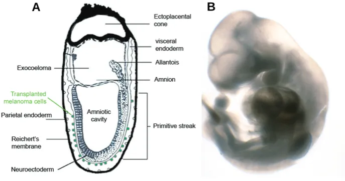
organo-Fig. 1. Initial and final embryonic stage of our observations. (A)Schematic drawing of the initial position of labeled human melanoma cells (in green) on the surface of the visceral endoderm of a ~7.5 dpc mouse embryo. Reichert’s membrane had to be removed in order to allow the embryo to grow in culture. (B)Mouse embryo of ~10.5 dpc after 3 days of in vitro culture.
pluripotency of single undifferentiated embryonal carcinoma cells (EC cells) engrafted in an adult syngenic mouse. Such cells are able to produce a teratocarcinoma containing different cell-lineages derived from the three germ layers (Kleinsmith and Pierce 1964). This was really the first experiment that showed a similarity between the biological behavior of cancer and embry-onic stem cells (ES cells). The next step up in establishing the convergence in signaling pathways regulating tumor and embry-onic stem cell plasticity was taken by Ralph Brinster when he demonstrated that murine blastocysts injected with EC cells can give rise to healthy chimeric mice (Aréchaga, 1998). The absence of tumors in some of the resulting adult chimeras proved the ability of the embryonic microenvironment to abrogate the meta-static phenotype of malignant tumor cells. Brinster evidenced the presence of EC cell derived tissues in the chimeric mice by using mouse strains with different skin pigmentation. Mintz and Illmensee (1975) went further in their observations and demonstrated that EC cells injected into the blastocyst take part in the development of tissues derived from the three germ layers and even colonized the germ line, in agreement with the EC cell plasticity previously described by Pierce's group (Aréchaga, 1993). A decade after these great pioneer assays, supplemental experiments added new evidences supporting the tumor-suppressing ability of the embryonic microenvironment, not only in the mouse but also in other animal models, such as the chick embryo. Dolberg and Bissell (1984), for example, showed that the Rous sarcoma virus (which rapidly transform chick adult cells in vivo and chick embryo derived fibroblast in vitro) is non-tumorigenic when 4 days chicken embryos are infected, even though the virus does replicate in the embryo cells. Moreover, when infected embryo tissues are dis-sected and their cells are maintained in vitro, they manifest a transformed phenotype after just 24 hours of culture. Pierce and colleagues (1982) threw new light on this issue when observed that only those carcinomas with a normal embryonic counterpart are regulated by the blastocyst and that the malignant phenotype abrogation of carcinoma cells takes place just in their closely related embryonic fields (Aréchaga 1993). Supporting these
g e n e s i s i s c o m p l e t e a r e n o t reprogrammable, give rise tumors and induce angiogenesis (Haldi et al., 2006). These results show that the ability of the embryo microenvironment to reprogram-ming malignant tumor cells is signifi-cantly reduced after organogenesis.
A different set of experiments with zebrafish embryos and melanoma cells demonstrated that the ability of tumor cells to interact with the developing em-bryo is linked to their aggressive and plastic phenotype. Invasive melanoma cells, but not the poorly aggressive ones, lead to the formation of an abnormal anterior appendage when transplanted into the animal pole and produce a dupli-cation of the body axis when placed near the yolk margin. In all these processes, melanoma cell activity is limited to the organization of the host zebrafish cells without directly inducing abnormal
out-B
growths of the embryo (Topczewska et al., 2006). Subsequent experiments demonstrated that the ability of melanoma stem cells to direct the embryonic development is due to the secre-tion of Nodal, a potent morphogen of the TGF-beta superfamily (Schier, 2003). Knowing that Nodal is highly secreted by ag-gressive melanoma cells but it is not detected in normal skin or non-invasive melanoma cells, and having seen its effects on embryonic developments, it seems to be involved in both embryonic and tumorigenic signaling pathway. Additional ex-periments have confirmed this observation. The expression of Goosecoid, a Nodal-responsive gene, was shown to be up-regulated in zebrafish cells surrounding the implanted mela-noma cells one hour after transplantation. On the other hand, it has been proven that the formation of melanoma cells mediated ectopic outgrowths in the zebrafish embryo is totally disrupted by the inhibition of Nodal expression with specific morpholinos or by the abrogation of its activity by inhibiting its downstream mediator activin-like kinase (Topczewska et al., 2006).
Reprogramming of human melanoma cells by the
chick embryo
Due to the common migratory and invasive properties shared by neural crest cells and malignant melanoma cells, the chick embryo model has been especially useful in the study of the neural crest regions to reprogramming human metastatic mela-noma phenotype. Melanocytes differentiate from the neural crest (NC), which is constituted by a multipotent cell population
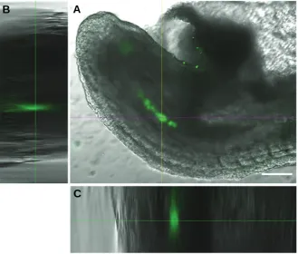
Fig. 2.A375-GFP-Np human melanoma cells internalized into the central axis of the caudal region of the mouse embryo.(A)Merge of green fluorescence and transmitted light channels obtained by confocal microscopy. (B,C) Orthogonal views of a 200 μm Z-stack showing the internal location of the fluorescent labeled melanoma cells. Bar, 100 μm.
MLANA and the neuronal marker TUJ1, suggesting a transition from malignant melanoma to benign melanocytic phenotype.
The mammalian embryo approach to study the
embry-onic control of cancer cells
As mentioned before, the pioneer experiments demonstrating the ability of embryonic microenvironment to revert tumor malig-nant phenotype were carried out by Brinster and Pierce groups, using the mouse blastocysts as a model system (Aréchaga 1993, 1998). Pierce and co-workers also explored cancer growth regu-lation by their related mouse embryonic fields during organogen-esis after several in vitro approaches (Pierce et al., 1986). Nevertheless, as we have seen, recent progress in this issue have substituted the mouse embryos for alternative animal mod-els whose whole development can be easily followed in vitro, such as zebrafish or chick embryos. But, even though these models offer many advantages for longer observations of tumor cell behavior into the embryo and adult animals, it is advisable to compare the results obtained in such models with those obtained in mammalian embryos, which are presumably more closely related to the environment found in the human embryos. Follow-ing this approach, HochedlFollow-inger et al. (2004) have been able to reprogram RAS-inducible melanoma cells by activated mouse oocytes after nuclear transfer, obtaining normally developed embryos that reach the blastocyst stage and derived pluripotent ES cells lines. Later, Astigiano and colleagues (2005) have recently recovered the use of the mouse model in order to clarify
B
C
A
that migrates from the neural tube and in-vades throughout the embryo body during vertebrate development. Neural crest cell to melanoblast transition involves the restriction of pluripotent cell plasticity in order to deter-mine the melanogenic lineage and finally the melanocyte phenotype. Wnt and BMP signal-ing pathways mainly govern this specification process. The melanocyte fate determination is also directed by the transcriptional repres-sor FoxD3, represrepres-sor of melanogenesis in premigratory NC cells and in other NC lin-eages (Thomas and Erickson 2009). The fate of each migratory neural crest cell seems to be determined by a combination of signals produced by the neural tube itself and the microenvironment along the migratory route (Lumsden et al., 1991; Trainor and Krumlauf 2000).for how long the embryonic environment shows the ability to abolish the malignant phenotype. With this purpose, several EC cell lines were injected in intrauterine post-implantation mouse embryos at different developmental stages, from E8.5 embryos to newborn and adult mice. The transplanted cell viability, migration and tumor formation ability were then monitored and it was observed that the percentage of animals that develop tumors increases with the age of the embryo at the moment of the carcinoma cell transplantation. These assays confirm that the ability to abrogate malignancy by the embryonic environment is gradually reduced as the development progresses and, eventu-ally, it is completely lost after the organogenesis stage.
However, the previously mentioned experiments on the rever-sion of the cancer phenotype by mammalian embryos present some disadvantages. Thus, observations are not currently pos-sible beyond the blastocyst stage; however, live chimeras with derived ES cell lines have been obtained. These lines were previously cultured for some passages in vitro, and the resulting animals do develop tumors (Hochedlinger et al., 2004). Other inconveniences include the difficulty of injecting cells in utero into specific places (Gerschenson et al., 1986; Astigiano et al., 2005). Here we present a new approach using post-implanted mouse embryos cultured in vitro to analyze the ability of the embryonic microenvironment to regulate melanoma cell malignant behavior during organogenesis.
Our experiments are based on the culture system developed by Denis New (Aréchaga, 1997) that allows the culture of post-implantation mouse embryos for some days. Briefly, A375 human melanoma cells expressing the fusion protein Green Fluorescent Protein-Nucleoplasmin (GFP-Np) into their nucleus were attach to the surface of the visceral endoderm of gastrulating mouse embryos in order to observe their interactions and to localize their position after three days of culture (Fig. 1). The observations by confocal microscopy showed that melanoma cells were located mainly in the caudal region of the embryo, and in most of the cases they have been able to migrate and appear internalized in the embryo tissues. The distribution of the GFP labeled melanoma
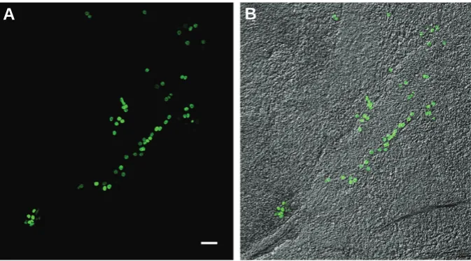
cells forming a row along the midline of the embryo body also suggests that they migrate in an orderly manner in response to some migratory cues (Figs. 2 and 3). Tumor formation was not detected in any of the analyzed embryos.
These observations with human melanoma cells support pre-vious works that suggested the reprogramming of mouse mela-noma genome by the embryonic microenvironments during early embryogenesis and organogenesis (Gerschenson et al., 1986; Hochedlinger et al., 2004). Our results with cultured post-im-planted mouse embryos confirm also those obtained recently with zebrafish and chick embryos (Lee et al., 2005; Kulesa et al., 2006), what points out the universality of the mechanisms in-volved in cancer cell reprogramming by embryonic microenviron-ment.
Discussion and perspectives
An increasing number of studies support the idea that some characteristics shared by embryonic and cancer stem cells, such as plasticity and undifferentiated phenotype, are regulated, at least partially, by common mechanisms. Similarities between both cell types are corroborated by the current cancer stem cell theory, which postulates that the origin of several types of cancer lay on tumor stem cell niches (Pierce, 1983). Over the past few years, several assays have demonstrated the reversion of the metastatic phenotype of human melanoma cells by the embryonic microenvironment using zebrafish and chick embryos as models (Lee et al., 2005; Kulesa et al., 2006) but the use of a post-implanted mammalian experimental model could provide better conditions and cues closer to those found in the human microen-vironments. In our experiments, the absence of localized tumoral growth after 72 hours of embryo co-culture with metastatic mela-noma cells confirm that malignant phenotype inhibiting factors are active at the gastrula stage and early organogenesis. Although further research is needed to elucidate the specific fate of trans-planted melanoma cells and the signaling pathways involved in cancer inhibitory mechanisms of the mammalian embryo, the
Fig. 3.Squash preparation of a mouse post-implanted embryo colonized by A375-GFP-Np human melanoma cellsafter 3 days of co-culture. Human melanoma cells seem to migrate along the body axis. (A)Green fluorescence channel. (B)Merge of green fluorescence and transmitted light channels. Bar, 50 μm.
present approach confirm the presence of cancer regulatory mechanisms in the mam-malian embryos cultured in vitro. A good starting point to understand the main molecu-lar changes underlying the pathogenesis of this aggressive tumor (Palmieri et al., 2009) from the development point of view and for future progresses to the development of bio-logical based anticancer therapies.
Materials and Methods
Mouse embryos
7.5 dpc mouse embryos were obtained from OF1 random bread albino Swiss strain (Crifa, France) following the method developed by New (1978). According to it, the uterine horns contain-ing embryos were extracted into Petri dishes with PBS solution and uterine walls were ripped and discarded to uncover the decidua mass surround-ing the egg cylinder stage embryos. The decidua swelling was transfer to a new dish with fresh PBS solution and then dissected carefully into two halves
to obtain the embryos. After, the Reichert’s membrane of collected embryos was tore and they were transferred to 2 ml of pre-equilibrated culture medium in 30ml tubes (two embryos per tube).
A375 human melanoma cells expressing GFP-Np fusion protein
A375 human melanoma cells were obtained from American Type Culture Collection (ATCC, Ref. CRL-1619). Cells were cultured in Dulbecco’s modified Eagle’s Medium (DMEM) supplemented with 10% fetal calf serum (FCS) and 1% penicillin/streptomycin at 37°C in 5% CO2. To yield the vector pGFP-Np, we employed a full length nucleoplasmin cDNA sequence was obtained by PCR from pET-11b using N-term (CAGTAGATCTATGGCCTCTACCGTCAGCAA) and C-term (TTAAGAATTCTCACTTCTTAGCAGCCGGCTT) primers. The amplified fragment was digested with BglII and EcoR1 restriction enzymes, respec-tively (underlined) and ligated to eGFP-C1 (Clontech). Competent Es-cherichia coli (JM 109 strain) were transformed and the plasmid was isolated and verified by sequencing. 1x10⁴ A375 melanoma cells per well were cultured overnight in 24 well plates. Afterwards, cells were trans-fected with pGFP-NP using FUGENE 6 (Roche) according to the manu-facturer instructions. Stable A375 GFP-Np cell lines were generated by clonal selection. Maintenance of the culture was performed adding the G-418 antibiotic to the growth medium (Andrade et al., 2009).
Whole-embryo culture conditions
Rats of the Sprague Dawley strain were used to extract blood from the dorsal artery. The blood was immediately centrifuged at 4°C and the obtained serum was decomplemented by means of incubation at 56°C during 30 minutes. The resulting serum was used to prepare the culture medium (50% rat serum in DMEM). The culture of the embryos was carried out in tubes of 30ml at 37°C in a rotatory system at 35 rev/min with a constant flow of gas. Oxygen tension in the gas was gradually in-creased, being 5% during the first 24 hours, 20% during the next 24 hours and 40% during the last 24 hours. A375 human melanoma cells express-ing GFP-Np fusion protein were added to the culture medium at a density of 500.000 cells per ml.
Confocal microscopic observations
Embryos co-cultured with fluorescent labeled human melanoma cells were observed by with a Fluoview FV500 Olympus confocal microscope after 72 hours of culture.
Acknowledgements
This work was supported by a research project from the Spanish Ministry of Education and Science (BFU 2007-66610) and a Research Group Grant (GIU08/04) from the University of the Basque Country to J.A.; A.D-T. and E.L. had fellowships from the Jesús Gangoiti Barrera Foundation of Bilbao, Spain.
References
ABBOTT D E, BAILEY C M, POSTOVIT L M, SEFTOR E A, MARGARYAN N, SEFTOR R E, HENDRIX M J (2008) The Epigenetic Influence of Tumor and Embryonic Microenvironments: How Different are They? Cancer Microenviron
1:13-21.
ANDRADE R, CRISOL L, PRADO R, BOYANO MD, ARLUZEA J and ARÉCHAGA J. (2010). Plasma membrane and nuclear envelope integrity during the blebbing stage of apoptosis: a time-lapse study. Biol Cell. 102: 25–35.
ARÉCHAGA J (1993) On the boundary between development and neoplasia. An interview with G Barry Pierce. Int J Dev Biol 37: 5-16.
ARÉCHAGA J (1997) Technique as the basis of experiment in developmental biology. An interview with Denis A T New. Int J Dev Biol 41: 139-152.
ARÉCHAGA J (1998) Embryo culture, stem cells and experimental modification of the embryonic genome. An interview with Ralph L. Brinster. Int J Dev Biol 42:
861-878.
ASTIGIANO, S., DAMONTE, P., FOSSATI, S., BONI, L. AND BARBIERI, O. (2005) Fate of embryonal carcinoma cells injected into postimplantation mouse em-bryos. Differentiation 73: 484-490.
BALINT K, XIAO M, PINNIX C C, SOMA A, VERES I, JUHASZ I, BROWN E J, CAPOBIANCO A J, HERLYN M, LIU Z J (2005) Activation of Notch1 signaling is required for beta-catenin-mediated human primary melanoma progression. J Clin Invest 115: 3166-3176.
BRINSTER R L (1974) The effect of cells transferred into the mouse blastocyst on subsequent development. J Exp Med 140: 1049-1056.
CUCINA A, BIAVA P M, D’ANSELMI F, COLUCCIA P, CONTI F, DI CLEMENTE R, MICCHELI A, FRATI L, GULINO A, BIZZARRI M (2006) Zebrafish embryo proteins induce apoptosis in human colon cancer cells (Caco2). Apoptosis 11:
1617-28.
DISSANAYAKE S K, WADE M, JOHNSON C E, O’CONNELL M P, LEOTLELA P D, FRENCH A D, SHAH K V, HEWITT K J, ROSENTHAL D T, INDIG F E, JIANG Y, NICKOLOFF B J, TAUB D D, TRENT J M, MOON R T, BITTNER M, WEERARATNA A T (2007) The Wnt5A/protein kinase C pathway mediates motility in melanoma cells via the inhibition of metastasis suppressors and initiation of an epithelial to mesenchymal transition. J Biol Chem. 282:
17259-71.
DOLBERG D S, BISSELL M J (1984) Inability of Rous sarcoma virus to cause sarcomas in the avian embryo. Nature 309: 552-556.
GERSCHENSON M, GRAVES K, CARSON S D, WELLS R S AND PIERCE G B (1986) Regulation of melanoma by the embryonic skin. Proc Natl Acad Sci USA
83: 7307-7310.
HALDI M, TON C, SENG W L, MCGRATH P (2006) Human melanoma cells transplanted into zebrafish proliferate, migrate, produce melanin, form masses and stimulate angiogenesis in zebrafish. Angiogenesis 9: 139-51.
HOCHEDLINGER K, BLELLOCH R, BRENNAN C, YAMADA Y, KIM M, CHIN L, JAENISCH R (2004) Reprogramming of a melanoma genome by nuclear transplantation. Genes & Development 18: 1875-1885.
KLEINSMITH L J, PIERCE G B (1964) Multipotentiality of single embryonal carcinoma cells. Cancer Res 24: 1544-1552.
KULESA P M, KASEMEIER-KULESA J C, TEDDY J M, MARGARYAN N V, SEFTOR E A, SEFTOR R E B, HENDRIX M J (2006) Reprogramming meta-static melanoma cells to assume a neural crest cell-like phenotype in an embryonic microenvironment. Proc Natl Acad Sci USA 103:3752-3757.
LEE L M, SEFTOR E A, BONDE G, CORNELL R A, HENDRIX M J (2005) The fate of human malignant melanoma cells transplanted into zebrafish embryos: assessment of migration and cell division in the absence of tumor formation.
Dev Dyn 233: 1560-70.
LUMSDEN A, SPRAWSON N, GRAHAM A (1991) Segmental origin and migration of neural crest cells in the hindbrain region of the chick embryo. Development
113: 1281-1291.
MINTZ B, ILLMENSEE K (1975) Normal genetically mosaic mice produced from malignant teratocarcinoma cells. Proc Natl Acad Sci USA 72: 3585-3589.
NEW D A T (1978) Whole-embryo culture and the study of mammalian embryos during organogenesis. Biol Rev 53: 81-122.
PALMIERI G, CAPONE M E, ASCIERTO M L, GENTILCORE G, STRONCEK D F, CASULA M, SINI M C, PALLA M, MOZZILLO N, ASCIERTO P A (2009). Main roads to melanoma. J Trasl Med 7 (doi: 10.1186/1479-5876-7-86).
PIERCE G B (1983) The cancer cell and its control by the embryo. Am J Path 113:
117-124.
PIERCE, G. B., PANTAZIS, C. G., CALDWELL, J. E. AND WELLS, R. S. (1982) Specificity of the control of tumor formation by the blastocyst. Cancer Res 42:
1082-1087.
PIERCE G B, ARÉCHAGA J, WELLS R S (1986) Embryonic control of cancer. In
Cellular Endocrinology: Hormonal Control of Embryonic and Cellular Differen-tiation (Eds. G Serrero and J Hayashi) pp 67-77.
POSTOVIT L M, COSTA F F, BISCHOF J M, SEFTOR E A, WEN B, SEFTOR R B, FEINBERG A P, SOARES M B, HENDRIX M J (2007). The commonality of plasticity underlying multipotent tumor cells and embryonic stem cells. J Cell Biochem 101: 908-917.
Biol 19: 589-621.
THOMAS A J, ERICKSON C A (2009) FOXD3 regulates the lineage switch between neural crest-derived glial cells and pigment cells by repressing MITF through a non-canonical mechanism. Development 136: 1849-1858.
TOPCZEWSKA J M, POSTOVIT L M, MARGARYAN N V, SAM A, HESS A R,
WHEATON W W, NICKOLOFF B J, TOPCZEWSKI J, HENDRIX M J (2006) Embryonic and tumorigenic pathways converge via Nodal signaling: role in melanoma aggressiveness. Nature Med 12: 925-932.
TRAINOR P, KRUMLAUF R (2000) Plasticity in mouse neural crest cells reveals a new patterning role for cranial mesoderm. Nature Cell Biol 2:96-102.
Further Related Reading, published previously in the Int. J. Dev. Biol.
See our recent Special Issue Epigenetics & Development edited by Saadi Khochbin and Stefan Nonchev at: http://www.ijdb.ehu.es/web/contents.php?vol=53&issue=2-3
The hidden maternal-fetal interface: events involving the lymphoid organs in maternal-fetal tolerance
Elizabeth S. Taglauer, Kristina M. Adams Waldorf and Margaret G. Petroff
5 yr ISI Impact Factor (2008) = 3.271
Int. J. Dev. Biol. in press (doi: 10.1387/ijdb.082800et)
Modeling and quantification of cancer cell invasion through collagen type I matrices
Olivier De Wever, An Hendrix, Astrid De Boeck, Wendy Westbroek,Geert Braems, Shahin Emami, Michèle Sabbah, Christian Gespach, Marc Bracke
Int. J. Dev. Biol. in press (doi: 10.1387/ijdb.092948ow)
Using fruitflies to help understand the molecular mechanisms of human hereditary diffuse gastric cancer
Joana Caldeira, Paulo S. Pereira, Gianpaolo Suriano and Fernando Casares Int. J. Dev. Biol. (2009): 53: 1557-1561. (doi: 10.1387/ijdb.072277jc)
Genome reprogramming during sporulation
Jerome Govin and Shelley L. Berger Int. J. Dev. Biol. (2009) 53: 425-432
Placental metabolic reprogramming: do changes in the mix of energy-generating substrates modulate fetal growth?
Nicholas P. Illsley, Isabella Caniggia and Stacy Zamudio Int. J. Dev. Biol. in press (doi: 10.1387/ijdb.082798ni)
Genetic and epigenetic instability of human bone marrow mesenchymal stem cells expanded in autologous serum or fetal bovine serum
John-Arne Dahl, Shivali Duggal, Neralie Coulston, Douglas Millar, John Melki, Aboulghassem Shahdadfar, Jan E. Brinchmann and Philippe Collas
Int. J. Dev. Biol. (2008) 52: 1033-1042
Pluripotency and differentiation in embryos and stem cells - Pavia, 17-18 January 2008
James A. Adjaye, Anne G. Byskov, Jose B. Cibelli, Ruggero De Maria, Stephen Minger, Maurilio Sampaolesi, Giuseppe Testa, Catherine Verfaillie, Magdalena Zernicka-Goetz, Hans Schöler, Michele Boiani, Nicola Crosetto and Carlo A. Redi
Int. J. Dev. Biol. (2008) 52: 801-809
Human conjunctival epithelial precursor cells and their progeny in 3D organotypic culture
Alfredo Rosellini, Sandra Papini, Claudio Giannarini, Marco Nardi and Roberto P. Revoltella Int. J. Dev. Biol. (2007) 51: 739-743
Epigenetic reprogramming of the genome--from the germ line to the embryo and back again
K L Arney, S Erhardt, R A Drewell and M A Surani Int. J. Dev. Biol. (2001) 45: 533-540
Growth factors and proto-oncogenes in early mouse embryogenesis and tumorigenesis