Original Article
Serotonin involvement in the metamorphosis of the hydroid
Eudendrium racemosum
GIULIANA ZEGA*, ROBERTA PENNATI, ARIANNA FANZAGO and FIORENZA DE BERNARDI
Dipartimento di Biologia, Sez. Zoologia S/N, Università Statale di Milano, Milan, Italy
ABSTRACT Hydroid planulae metamorphose in response to an inducing external stimulus, usually a bacterial cue. There is evidence that neurotransmitters participate in the signal transduction pathway of hydroid metamorphosis. Eudendrium racemosum is a colonial hydroid common in the Mediterranean Sea. It lacks the medusa stage and the planulae develop on female colonies during the fertile season. In this work, serotonin (5-HT) was localized in some planula ectodermal cells. Co-localization of serotonin and β-tubulin suggested that 5-HT was present in sensory nervous cells and in different ectodermal cells. To investigate the role of neurotransmit-ters in metamorphosis, E. racemosum planulae were treated with serotonin and dopamine and with agonists and antagonists of the corresponding receptors. Serotonin and a serotonin receptor agonist induced metamorphosis, while a 5-HT receptor antagonist inhibited it. Dopamine and all dopaminergic drugs used did not show any significant effect on the onset of metamorphosis. Results from this work showed that 5-HT could stimulate metamorphosis in E. racemosum planulae in the presence of a natural inducer. A mechanism by which this neurotransmitter could act in this phase is proposed.
KEY WORDS:
neurotransmitter, cnidaria, 5-HT, planula,
β
-tubulin.
Introduction
Hydroids are colonial animals with a life cycle including a planktonic larva, the planula, which settles and metamorphoses into a sessile polyp. It is known that the larvae of many marine invertebrates settle and metamorphose in response to environ-mental stimuli (Burke, 1983; Rodriguez et al., 1993; Hadfield and Paul, 2001) and a role for bacteria in induction of settlement and metamorphosis in different hydroid species has been demon-strated by several authors (Hydractinia echinata, Wittman, 1977; Kroiher and Berking, 1999; Phialidium gregagrium, Freeman, 1981; Halocordyle disticha, Thomas et al., 1987; Mitrocomella polydiademata, Freeman and Ridgway, 1990).
Cell depolarization is reportedly one of the first events of the transduction pathway that mediates the metamorphic stimulus, occurring upon contact with the bacterial inducer. Monovalent cations such as K+ and Cs+ are well known as artificial inducers of metamorphosis of different hydroid species (Spindler and Muller, 1972; Muller and Buchal, 1973; Freeman and Ridgway, 1990; Schwoerer-Böhning et al., 1990). Therefore the larval nervous system is involved in signal reception, at the level of electrically excitable cells and there is evidence that it also participates in the signal transduction pathway of metamorphosis
*Address correspondence to: Giuliana Zega. Dipartimento di Biologia, Università di Milano, Via Celoria 26, I-20133, Milano, Italy. Fax: +39-02-5031-4802. e-mail: giuliana.zega@unimi.it
0214-6282/2007/$30.00 © UBC Press
Printed in Spain
www.intjdevbiol.com
Abbreviations used in this paper: D, dopamine; FSW, filtered sea water; 5-HT, 5 hydroxy-tryptamine (serotonin); met, metamorphosed.
over we localized serotonin and β– tubulin in planulae and discuss the contribution of different ectodermal cell types to the first events of meta-morphosis.
Results
βββββ- tubulin and serotonin
localiza-tion
A brief description of histology sections is given in order to discuss the possible identity of β- tubulin and serotonin immunoreactive cells. Longitudinal sections showed two types of ectodermal cells filled with secretory granules: mucus cells, in which the nucleus was not recognizable as it degenerates and epitheliomuscular cells, character-ized by the typical position of their nuclei in the mid-apical region of the cell (Fig.1J, K). In 1-day old larvae, immunohistochemistry re-vealed a positive signal for 5-HT in several ectodermal cells. These positive cells were scattered in the ectoderm, more numerous in the aboral pole than in the oral pole and serotonin signal was more evident in the cells’ apical portions (Fig. 2 A, B, E).
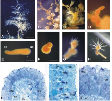
In the same larvae, β-tubulin immunopositive signal was detect-able in some ectodermal cells with an elongated shape (Fig. 2D, F) and labeled the network of fibers lying on the mesoglea and con-necting these cells. Since it has been reported that nervous cells in hydrozoan planulae are rich in cy-toplasmic microtubules (Martin and Thomas, 1980; Kolberg and Mar-tin, 1988), we assume that these β -Fig. 1. Eudendrium racemosum colonies, stages of development and histology. (A) Adult colony.
(B-C) Male and female blastostyles respectively. (D) Planula escaping from the embryotheca. (E) Free planula. (F) Metamorphosis stage in which the future hypostome starts to grow. (G) Following metamorphosis stage: the hypostome is formed and tentacles are growing. (H) Primary polyp. (I) Longitudinal section of the aboral pole of E. racemosum planula, showing organization of ectoderm, mesoglea and endoderm. . (J)Higher magnification showing different cell types, among these some nematocysts: one is migrating in the ectoderm through the mesoglea (black asterisk). (K) Close up of the aboral pole ectoderm, rich in mucus cells. Abbreviations: ap, aboral pole (anterior); ec, ectoderm; em, epitheliomuscular cell; en, endoderm; gc, gastral cavity; mc, mucus cell; me, mesoglea.; ne, nematocyst; op, oral pole. Scale bars: (A) 2 cm, (B,C) 2 mm, (D) 250 µm, (E,F,G) 200 mm, (H) 250 mm, (I) 50 µm, (J) 20 µm, (K) 10 µm. appropriate external cue and could determine cell depolarization.
Neurotransmitters participate in the metamorphic signaling of a variety of planktonic invertebrate larvae that will become benthic juveniles, including polychaetes, mollusks, crustaceans and as-cidians (Pawlik, 1990; Couper and Leise, 1996; Yamamoto et al., 1999; Zega et al., 2005). Still, little is known about the detailed involvement of larval sensory organs in the first phases of this rapid and dramatic event.
Eudendrium racemosum is a marine colonial hydroid fre-quently found in the Mediterranean Sea (Fig. 1A), with a relatively simple life cycle due to the complete suppression of the medusa generation. In the fertile season, male and female colonies (Fig. 1B,C) are recognizable. Planulae develop and hatch (Fig. 1D) from the female blastostyle, they live free for some time (Fig. 1E) and can settle within few hours after hatching. The planulae are
diploblastic larvae, constituted by an ectoderm and an endoderm, separated by an acellular mesoglea (Fig. 1I). The main cell types found in the ectoderm were epitheliomuscular cells, mucus cells and sensory-nervous cells, proposed to have a dual function in stimulus perception and conduction (Sommer, 1990). Permanent adhesion to the substratum is attained by secretion of peridermal material from the epitheliomuscular cells and by secretory activity of mucus cells. After attachment, the planula metamorphoses through different stages into a primary polyp (Fig. 1 F, G, H), which will give rise to a new colony. E. racemosum larvae do not metamorphose readily in sterile sea water, but can be induced to settle by adding specific substrates such as certain algae or conspecific perisarc tubes (Sommer, 1992). In this work we studied the involvement of serotonin and dopamine, a catechola-mine, in the metamorphosis of the hydroid E. racemosum.
More-G
B
C
D
E
F
H
I
J
K
tubulin immunopositive cells are the sensory-nervous cells de-scribed by Sommer (1990) (Fig. 2D, F). Serotonin signal co-localized with β-tubulin signal only in a small population of cells, most probably sensory-nervous cells (Fig. 2C, G). The other cells containing serotonin but not β- tubulin, could be epitheliomuscular cells or mucus cells.
Our data suggested that at least two types of ectodermal cells are 5-HT immunopositive. By the same immunohistochemical method we did not detect dopamine in these larvae (not shown).
Effects of serotonin and dopamine on the onset of metamor-phosis
Planulae of E. racemosum were exposed to serotonin to evaluate the possible effects on metamorphosis. In the presence of perisarc tubes as natural inducer (see Materials and Methods and Fig. 4), 1 mM serotonin induced metamorphosis while lower concentrations of 5-HT failed to significantly promote metamor-phosis above control levels (F = 6.269, P < 0.05; FSW 58.4% ± 9.3; 5-HT 10µM 55.0% ± 15.9; 5-HT 100µM 62.5% ± 8.0; 5-HT 1 mM 77.5% ± 15.2; Tukey’s post hoc: FSW vs. 5-HT 1mM P<0.05) (Fig. 3A).
The effects on metamorphosis of serotonin receptors agonists and antagonists were also tested. The results are summarized in Table 1. Differences in the percentage of metamorphosis were found in treatments with OH-8 DPAT ([(± )-8-Hydroxy-2-(di-n-propyl-amino) tetralin hydrobromide]), a selective 5-HT1A recep-tor agonist, as compared to controls (Fig. 3B). Analysis of vari-ance was not significant, but post hoc tests indicated a trends towards a difference in the 10 mM treatment (F = 3.846, P = 0.051; Tukey’s post hoc tests: FSW vs. OH-8 DPAT 10µM P = 0.067; FSW vs. OH-8 DPAT 1µM P = 0.975).
DOI hydrochloride, a 5-HT2/1C receptor agonist, did not show any effect. The 5-HT1A receptor antagonist, WAY-100635 male-ate, reduced the percentage of metamorphosis at the higher concentration, as compared to controls (F = 9.341, P = 0.004; Tukey’s post hoc test: FSW vs. 10µM P = 0.003; FSW vs. 1µM P = 0.074) (Fig. 3C).
Treatments with dopamine and with two dopamine receptor agonists (S-(-)-lisuride; (±)-SKF-38393) had no effects on planu-lae metamorphosis, nor did treatments with two dopamine recep-tor antagonists (R (+)-SCH-23390, clozapine) (Table 1).
Discussion
This work demonstrated the presence of serotonin in planulae of E. racemosum and provided evidence of the involvement of this neurotransmitter during metamorphosis. By contrast, we ob-served that it was not possible to label dopamine with the immunohistochemical method used and that dopaminergic sub-stances did not affect metamorphosis in Eudendrium planulae. Serotonin was localized in ectodermal cells with a punctate distribution. These cells were more abundant at the aboral pole of the larva. In Hydractinia echinata planulae, 5-HT was observed in ectodermal cells arranged in a circle at the aboral pole (Walther et al., 1996). Also in Phialidium gregarium, 5-HT positive cells were more abundant in the ectoderm of the aboral pole (McCauley, 1997). This region is considered to take up the inducing signal and to generate the metamorphic internal signal which spreads into all body regions (Schwoerer-Böhning et al., 1990). Experiments of co-localization with β-tubulin demonstrated that in E. racemosum serotonin was present in at least two different cell types: one type was rich in microtubules while the second type did not show β
-Compound Receptor µµµµµM % met ± 95% ANOVA Tukey’s
confidence interval post hoc tests
FSW - 66.2 ± 19.3
CO DMSO - 62.0 ± 23.9
8-OH-DPAT 5-HT 1A agonist 100 Lethal
10 92.2 ± 5.4 P=0.051 P=0.067
1 68.4 ± 29.1 P=0.975
DOI 5-HT2/1C agonist 100 Lethal
hydrochloride 10 Lethal
1 73.3 ± 16.8
0,1 65.5 ± 34.8
WAY 100635 5-HT1A antagonist 100 Lethal
10 19.8 ± 20.5 P=0.004 P=0.003
1 40.0 ± 23.2 P=0.074
Lisuride D2 agonist, D1 antagonist; 100 Lethal
α/β-adrenoceptor antagonist; 10 37.8 ± 17.5
5-HT1A agonist, 5-HT2 antagonist 1 51.7 ± 26.3
0.1 67,7 ± 26.9
SKF D1 agonist 100 Lethal
10 68.3 ± 19.2
1 82.0 ± 10.3
SCH D1 antagonist 100 Lethal
10 71.5 ± 23.2
1 76.7 ± 18.3
Clozapine D4 selective antagonist; 100 Lethal
5-HT2A, 5-HT2C, 5-HT3, 5-HT6 10 Lethal
and 5-HT7 antagonist 1 43.9 ± 25.7
0,1 46.4 ± 17.7
tubulin signal in the cytoplasm. We hypothesize that cells of the first type correspond to the sensory-nervous cells, while cells of the second type could be epitheliomuscular or mucus cells.
Exogenous application of serotonin to planulae stimulated metamorphosis. The same result was obtained by exposing the larvae to an agonist of 5-HT1A receptors, suggesting that endog-enous 5-HT is involved in metamorphosis of the planula. In fact, in planulae treated with WAY-100635, a 5-HT1A receptor antago-nist, metamorphosis was inhibited, indicating that serotonin sig-naling is necessary to initiate this process.
In many hydroid species, bacteria apparently supply the envi-ronmental cue triggering metamorphosis (Freeman, 1981; Tho-mas et al., 1987; Freeman and Ridgway, 1990; Kroiher and Berking, 1999). Planulae of E. racemosum did not metamorphose in sterile conditions but they did in the presence of conspecific perisarc tubes (Sommer, 1990; present work). When left in FSW, only a small percentage of larvae was able to metamorphose and inductive substances were not able to stimulate this process (data not shown). Thus as serotonin did not have any inductive effect without perisarc tubes, we hypothesize that this neurotransmitter
either acts upstream of or in parallel with the perception of the natural inducer. The abundance of serotonin containing cells suggested that this neurotransmitter may play a pivotal role in the larval nervous network, perhaps stimulating searching behavior. If so, exogenous applied serotonin could enhance settlement by stimulating substrate exploration in the larva. Perisarc tubes could trigger metamorphosis perhaps by providing bacteria or another “cue” indicative of the presence of a conspecific adult. In fact, in the genus Eudendrium fertilization is external and sexes are separated. Planulae do not have a high dispersal rate as they are retained by the mother colony by a mucous thread and thus often metamorphose close to the colony (Bavestrello and Cerrano, 1992; Sommer, 1992). This life history pattern indicates the suitability of adults conspecifics as a reliable indicator of an appropriate place to settle.
A comparison of different effects of serotonin, dopamine and clozapine on E. racemosum vs. others hydroids species is given in Table 2. As in E. racemosum, planulae of Phialidium gregarium were induced to settle by 5-HT, but by contrast, both dopamine and clozapine, a dopamine and serotonin receptor antagonist, induced settlement (McCauley, 1997), while in E. racemosum the same compounds did not have any effect. Edwards et al. (1987) observed that catecholamines, as well as some of their precur-sors and agonists, induced metamorphosis in the marine hydroid Halocordyle disticha but not in Hydractinia echinata. In planule of the latter species serotonin stimulated metamorphosis (Walther et al., 1996). Taken together all these findings suggested that neurotransmitters and their receptor agonists and antagonists can play somewhat different roles in different hydroid species.
Evidence that exogenous serotonin induced metamorphosis,
Species Eudendrium Phialidium Hydractinia Halocordyle
racemosum gregarium echinata distica
Compound [mM
Serotonin 5 nt + +* nt
1 + + nt
0.1 = = nt
Dopaminen 1 = = = lethal
0.1 = + = +
0.01 = + = =
Clozapine 0.1 lethal lethal nt nt
0.01 lethal + nt nt
0.001 = + nt nt
COMPARISON OF THE DIFFERENT EFFECTS ON METAMORPHOSIS AMONG 4 HYDROID SPECIES
TABLE 2
+ stimulation, = no effect, nt not tested, * unknown concentration.
Fig. 2. Localization of serotonin and βββββ-tubulin in planulae of E racemosum. (A) Surface view of 5-HT immunolabeled planula. (B) Higher magnification of the aboral pole. (C) Sum of a few optical sections at the mesoglea level of the aboral pole of a planula after double immunostaining. Dotted area is showed at higher magnification in (E,F,G). (D) Same optical sections as in C, showing only β-tubulin signal to evidence the neural network. (E,F,G) higher magnification of a small portion of the anterior pole. (E) 5-HT signal in ectodermal cells. (F)β -tubulin signal in ectodermal cells. (G)Superimposition of images (E,F) to show colocalization of the two molecules. Green fluorescence corre-sponds to 5-HT signal; red fluorescence correcorre-sponds to β-tubulin signal. (optical sections 1.06 µm thick: A,B n=30; C, D n=20; E,F,G n=10). Scale bars: (A) 100 µm, (B) 60 µm, (C,D) 50 µm, (E,F,G) 20 µm
G
B
C
D
E
F
Fig. 3. Effects of (A) serotonin and its receptor (B) agonist and (C) antagonist on the percentage of metamorphosisof Eudendrium racemosum planulae. Mean percentages of metamorphosis (attached planulae and primary polips) ± 95% confidence interval at different concentrations (each treatment n=50 ± 10).*P≤0.05 (A); **P<0.01 (C).
taken together with the immunolocalization of this molecule in sensory-nervous cells, suggested that these cells are involved in reception of the stimulus in E. racemosum. These results regard-ing the involvement of serotonin in E. racemosum metamorphosis are comparable to what was previously reported for the hydroid H. echinata (Walther et al., 1996) and P. gregarium (Mc Cauley, 1997), suggesting a similar role of this neurotransmitter in the three species. We suggest that in E. racemosum planulae, serotonin is involved in the perception of the environmental cue, through 5-HT containing sensory-nervous cells, situated in the aboral pole, after which depolarization occurs, as already pro-posed by McCauley (1997). Once metamorphosis has begun, serotonin stored in the other cells might play a role in the release of substances for permanent adhesion.
In conclusion, serotonin seems to play an important role in early signaling cascade of metamorphosis of hydroids planulae of different species, where it is present at the level of ciliated sensory cells. Moreover, serotonin is also present in ciliated cells of the sensory organs of gastropod larvae and ascidian tadpoles (Dickinson et al., 1999; Hay-Schmidt, 2000; Pennati et al., 2001) and for some species its role in the stimulation of metamorphosis has been proved (Couper and Leise, 1996; Zega et al., 2005). The involvement of 5-HT in the metamorphosis of non-homologous larvae of different phyla suggests that serotonin signaling was selected several times for the control of an initial step of the transduction pathway of this process. To date, the exact action that serotonin can carry out at the molecular level, triggering a
complex morphogenetic event, is not completely understood. Further investigation will shed light on possible convergent or divergent mechanisms about neurotransmitters role action in the complex process of metamorphosis.
Materials and Methods
Animals
Colonies of the hydroid Eudendrium racemosum (Gmelin, 1791) were collected in shallow waters inside the harbour of Santa Margherita (Genova, Italy) during their fertile season, from June to late September (2001-2004) (Fig. 1A). Female colonies are easily recognizable from male ones (Fig. 1B, C). In fact, their blastostyles bear groups of about ten fertilized eggs of a brilliant orange color (Sommer, 1992). Hydroid colo-nies were gently removed from the substratum and transferred to the lab in abundant seawater (SW). They were placed into glass wells with SW and with an aerator at 16°C and fed with Artemia salina. Colonies were placed in the dark for 24-48 hours and then under light conditions to obtain larvae (Cerrano et al., 1997). Planulae released from mature female colonies were collected twice a day and then used for experiments on the same or the following day (Fig. 1 D, E).
Hystological analysis
Planulae were fixed for 2 hours in 0.4% paraformaldheyde at room temperature (r.t.), dehydrated in an ethanol series and stored in ethanol 70% at -20°C. After rehydration, specimens were stained in 1% carmin red for 3 h and then embedded in Technovit 7100 plastic (Heraeus Kulzer GmbH, Werheim, Germany), at r.t. and sectioned at 5 µm. Sections were counterstained with 0.5% methylene blue, dissolved in distilled water, for a few seconds and mounted in Entellan (Merck, Whitehouse Station, NJ, USA).
Immunohistochemistry
Specimens were fixed in 4% paraformaldehyde (PFA) in 0.1 M phos-phate buffer saline (PBS) (pH 7.2) at room temperature for 2 hours. All steps were performed with gentle rocking. After rinsing with 0.1 M PBS, the samples were dehydrated in ethanol series to 70% and stored at -20°C. After rehydration, specimens were permeabilized with 0.1% Tween-20, 0.25% Triton X-100 in PBS for 25 minutes, washed three times in PBS, 10 minutes each and incubated for 30 minutes in 50% PBS/50% normal goat serum, previously inactivated at 55°C for 30 minutes. Then, the samples were treated for 48 hours at 4°C with anti-human serotonin antibody, made in rabbit, diluted 1: 200 (Medac, Hamburg, Germany). For double-staining the monoclonal antibody anti-β tubulin clone 2-28-23 (Sigma, Italy) diluted 1:200 was added. After several washes in PBS, samples were incubated in 1% bovine serum albumine (BSA) in PBS for 1 hour at room temperature and then incubated at 4°C overnight in PBS in which FITC conjugated goat anti-rabbit IgG and TRITC conjugated goat anti-mouse IgG (Sigma, Italy), both diluted 1:100 were added. Next,
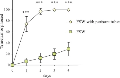
Fig. 4.Metamorphosis assays with and without a natural inducer. Comparison of the percentage of metamorphosis between planule put in filtered sea water alone and in presence of perisarc tubes, used as natural inducers. Data are mean percentage ± 95% confidence interval. ***P<0.0001.
A
B
C
WAY-100635 maleatespecimens were washed three times in PBS, 20 minutes each and mounted in 1,4-diazabicyclo[2,2,2]octane (DABCO, Sigma, Italy) on microscope slides. As negative controls, planulae were processed with-out incubation in primary antibodies: such specimens exhibited no detect-able fluorescence. Samples were examined using a confocal laser scanning microscope Leica TCS-NT (Leica Microsystems, Heidelberg, Germany), equipped with laser argon/krypton, 75 mW multiline. Series of 1,06µm “optical sections” attained by scanning whole-mount specimens, were projected into one image with greater focal depth. FITC fluores-cence was observed using a 488 nm excitation laser and a 530/30 band pass filter; TRITC fluorescence was observed using a 568 nm excitation laser and a 590 long pass filter. The number of sections per image is given in the figure legends.
Assays for planulae metamorphosis
Planulae were considered to have started metamorphosis when they had attached to the substratum/dish and they had begun to form an hydrant (Fig. 1F,G, H) (Sommer, 1990). In order to test planulae ability to metamorphose under laboratory conditions, we performed control experi-ments, in 5cm glass wells, in which larvae were put in filtered sea water (FSW)(0.4µm pore size) with or without small pieces of perisarc tubes, added as natural inducers. Percentage of metamorphosed larvae (per-centage of metamorphosis) was calculated as proportion of metamor-phosed planulae over total number of planulae put into each well at the beginning of the experiment. The test for spontaneous settlement was repeated two times on different batches, with the same results. In the presence of perisarc tubes, almost all planulae started metamorphosis within the second day (97.2% ± 5.6). Less than 50% of planulae put in FSW alone metamorphosed after four days (day 1: F = 91.19, P < 0.0001; day 2: F = 227.465, P < 0.00001; day 3: F = 192.56, P < 0.00001; day 4: F = 98.636, P < 0.0001) (Fig. 4). Moreover, after two days most of the planulae, that were not metamorphosed yet, died. Then all assays for planulae metamorphosis with neurotransmitters and their agonists and antagonists were performed in presence of pieces of perisarc tubes. The experiments were repeated on different dates using three different culture batches. In each experiment, 1-2 days old planulae were collected and evenly divided among treatments. Each treatment was replicated at least five times. For each treatment, 10 ± 2 larvae were carefully pipetted in wells containing 10 ml of filtered SW (FSW) (0.4 µm pore size) in which the drug to be tested had been diluted. A 10 mM stock solution was prepared for each drug and then diluted to the appropriate working concentration. Chemical agents tested were dissolved mainly in distilled water, whereas those that were insoluble in water were dissolved in DMSO. Control tests were performed in FSW in which perisarc tubes were added and in FSW plus a volume of DMSO equal to those used for diluting the stock solution to the highest concentration tested in the treatments (corresponding to 0.13 µM). Substances proved to stimulate metamor-phosis were also tested in assays without perisarc tubes: no significant difference in the percentage of metamorphosis among controls and treatments was observed and the mean percentage of metamorphosis did not go over 20% after 24h (Fig. 4).
The percentage of metamorphosis in the controls was used for comparison in statistical analysis for each experiment. The plates were kept at 18°C throughout the experiments. Larvae were observed under a stero-microscope 1 day after treatment to estimate percentage of meta-morphosis, i. e. crawling planulae vs. attached planulae, which had begun to form an hydrant. One-way analysis of variance (ANOVA) was used to test the significance of differences in the metamorphosis rate. Tukey’s post hoc test (significant at P<0.05) was used to identify specific effects. Prior to performing analyses of variance, normal frequency distribution of data and homogeneity of variance were tested. No significant deviations from the parametric assumption were observed (normality: Kolmogorov-Smirnov’ test all P>0.04; homogeneity of variance: Levene’s test all P>0.06). Data are mean percentage ± 95% confidence interval.
Serotonin (5-hydroxytryptamine), dopamine, (±
)-8-Hydroxy-2-(di-n-pro-pyl-amino) tetralin hydrobromide (8-OH-DPAT HBr), R(-)-DOI Hydrochlo-ride, WAY–100635 maleate, R (+)-SCH-23390, (±)-SKF-38393, S-(-)-Lisuride, Clozapine were purchased from SIGMA, Italy.
Ackowledgements
We are grateful to Prof. Giorgio Bavestrello and Carlo Cerrano for their invaluable scientific support. This work was supported by grants from the University of Milano (FIRST 2003).
References
BURKE, R.D. (1983). The induction of metamorphosis of marine invertebrate larvae: stimulus and response. Can. J. Zool. 61: 1701-1719.
BAVESTRELLO, G. and CERRANO, C. (1992). Aggregate colonies in Eudendrium
glomeratum Picard 1952 (Cnidaria,Hydrozoa, Anthomedusae). Sci. Mar. 56:
333-335.
CERRANO, C., BAVESTRELLO, G. and CATTANEO-VIETTI, R. (1997). Light influence on planulae emission and settlement in Eudendrium glomeratum (Cnidaria, Hydrozoa) Biol. Mar. Medit. 4: 30-33.
COUPER, J.M. and LEISE, E.M. (1996). Serotonin injections induce metamorpho-sis in larvae of the gastropod mollusc Ilyassana obsoleta. Biol. Bull. 191: 178-186.
DICKINSON, A.J.G., NASON, J. and CRPFF, R.P. (1999). Histochemical localiza-tion of FMRFamide, serotonin and catecholamines in embryonic Crepidula
fornicata (Gastropoda, Prosobranchia). Zoomorphology 119: 49-62.
EDWARDS, N.C., THOMAS, M.B., LONG, B.A. and AMYOTTE, S.J. (1987). Catecholamines induce metamorphosis in the hydrozoan Halocordyle disticha, but not in Hydractinia echinata. Dev. Genes Evol. 196: 381-384.
FREEMAN, G. (1981). The role of polarity in the development of the hydrozoan planula larva. Dev. Genes Evol. 190: 168-184.
FREEMAN, G. and RIDGWAY, E.B. (1990). Cellular and intracellular pathways mediating the metamorphic stimulus in hydrozoan planulae. Dev. Genes Evol. 199: 63-79.
HADFIELD, M.G. (1998). The D P Wilson Lecture. Research on settlement and metamorphosis of marine invertebrate larvae: past, present and future. Biofouling 12: 9-29.
HADFIELD, M.G. (2000). Why and how marine invertebrate larvae metamorphose so fast. Cell Dev. Biol. 11: 437-443.
HADFIELD, M.G. and PAUL, V.J. (2001). Natural chemical cues for settlement and metamorphosis of marine invertebrate larvae. In «Marine Chemical Ecology» McClintock JB and Baker W, eds. CRC Press pp. 431 - 461.
HAY-SCHMIDT, A. (2000). The evolution of the serotonergic nervous system. Proc.
R. Soc. Lond. 267: 1071-1079.
KOLBERG, K.J.S. and MARTIN, V.J. (1988). Morphological, cytochemical and neuropharmacological evidence for the presence of catecholamines in hydro-zoan planulae. Development 103: 249-258.
KROIHER, M. and BERKING, S. (1999). On natural metamorphosis inducers of the cnidarians Hydractinia echinata (Hydrozoa) and Aurelia aurita (Scyphozoa).
Helgol. Mar. Res. 53: 118-121.
MARTIN, V.J. and THOMAS, M.B. (1980). Nerve elements in the planula of the hydrozoan Pennaria tiarella. J. Morphol. 166: 27-36.
McCAULEY, D.W. (1997). Serotonin plays an early role in the metamorphosis of the hydrozoan Phialidium gregarium. Dev. Biol. 190: 229-240.
MULLER, W.A. and BUCHAL, G. (1973). Metamorphose-Induktion bei Planulalarven: II. Induktion durch monovalente Kationen: Die Bedeutung des Gibbs- Donnan-Verhältnisses und der Na+/K+-ATPase. Dev. Genes Evol. 173: 122-135.
PAWLIK, J.R. (1990). Natural and artificial induction of metamorphosis of
Phragmatopoma lapidosa californica (Polychaeta: Sabellaridae), with a critical
look at the effects of bioactive compounds on marine invertebrate larvae. Bull.
Mar. Sci. 46(2): 512-536.
PENNATI, R., GROPPELLI, S., SOTGIA, C., CANDIANI, S., PESTARINO, M. and DE BERNARDI, F. (2001). Serotonin localization in Phallusia mammillata larvae and effect of 5-HT antagonists during larval development. Dev. Growth
Differ. 43:647-656.
benthic marine invertebrates. Mar. Ecol. Prog. Ser. 97: 193-207.
SCHWOERE-BOHNING, B., KROIHER, M. and MULLER, W.A. (1990). Signal transmission and covert prepattern in the metamorphosis of Hydractinia echinata.
Dev. Genes Evol. 198: 245-251.
SOMMER, C. (1990). Post-embryonic larval development and metamorphosis of the hydroid Eudendrium racemosum (Cavolini) (Hydrozoa, Cnidaria). Helg.
Meer. 44: 425-444.
SOMMER, C. (1992). Larval biology and dispersal of Eudendrium racemosum (Hydrozoa, Eudendridee). Sci. Mar. 56(2-3): 205-211.
SPINDLER, K.D.and MULLER, W.A. (1972). Induction of metamorphosis by bacteria and by a lithium-pulse in the larvae of’Hydractinia echinata (Hydrozoa).
Dev. Genes Evol. 169: 271-280.
THOMAS, M.B., FREEMAN, G. and MARTIN, V.J. (1987). The embryonic origin of neurosensory cells and the role of nerve cells in metamorphosis of Phialidium
gregarium. Invert. Reprod. Develop. 11: 265-287.
WALTHER, M., ULRICH, R., KROIHER, M. and BERKING, S. (1996).
Metamorpho-sis and pattern formation in Hydractinia echinata, a colonial hydroid. Int. J. Dev.
Biol. 40: 313-322.
WITTMAN, W. (1977). Auslösung der Metamorphose bei Hydractinia durch Bakterien: Isolierung und Charakterisierung der Bakterien und der auslösenden Substanz. Ph D Thesis Technische Universitat Braunschweig, FRG.
YAMAMOTO, H., SHIMIZU, K., TACHIBANA, A. and FUSETANI, N. (1999). Roles of dopamine and serotonin in larval attachment of the barnacle, Balanus
amphitrite. J. Exp. Zool. 284: 746-758.
ZEGA, G., PENNATI, R., GROPPELLI, S., SOTGIA, C. and DE BERNARDI, F. (2005) Dopamine and serotonin modulate the onset of metamorphosis in the ascidian Phallusia mammillata. Dev. Biol. 202: 246-256.
Received: 28th June 2006 Modified by Authors and Accepted for Publication: 23rd April 2007 Published Online: 16th May 2007
Related, previously published Int. J. Dev. Biol. articles of interest
See our recent Special Issue on Developmental Biology in Italy http://www.ijdb.ehu.es/web/contents.php?vol=44&issue=6
Induction of reverse development in two marine Hydrozoans
Jürgen Schmich, Yulia Kraus, Doris De Vito, Daria Graziussi, Ferdinando Boero and Stefano Piraino Int. J. Dev. Biol. (2007) 51: 45-56
Mesodermal anatomies in cnidarian polyps and medusae Katja Seipel and Volker Schmid
Int. J. Dev. Biol. (2006) 50: 589-599
Principles of branch formation and branch patterning in Hydrozoa Stefan Berking
Int. J. Dev. Biol. (2006) 50: 123-134
Morphomechanical programming of morphogenesis in Cnidarian embryos Yulia A. Kraus
Int. J. Dev. Biol. (2006) 50: 267-275
Metamorphosis of Hydractinia echinata (Cnidaria) is caspase-dependent Stefanie Seipp, Karola Wittig, Beate Stiening, Angelika Böttger and Thomas Leitz Int. J. Dev. Biol. (2006) 50: 63-70
The role of alpha-amidated neuropeptides in hydroid development—LWamides and metamorphosis in Hydractinia echinata.
Günter Plickert, Eva Schetter, Nicole Verhey-Van-Wijk, Jörg Schlossherr, Marlis Steinbüchel and Martin Gajewski Int. J. Dev. Biol. (2003) 47: 439-450
Cnidarians as a model system for understanding evolution and regeneration. Brigitte Galliot and Volker Schmid
Int. J. Dev. Biol. (2002) 46: 39-48
Action of serotonin antagonists on cytoplasmic calcium levels in early embryos of sea urchin Lytechinus pictus.