The preplacodal region: an ectodermal domain with
multipotential progenitors that contribute to sense organs
and cranial sensory ganglia
ANDREA STREIT*
Department of Craniofacial Development, King’s College London, Guy’s Campus, London, UK
ABSTRACT The otic primordium belongs to a group of related structures, the sensory placodes that contribute to the paired sense organs - ear, eye and olfactory epithelium - and to the distal parts of the cranial sensory ganglia. Recent evidence suggests that despite their diversity, all placodes share a common developmental origin and a common molecular mechanism which initiates their formation. At the base of placode induction lies the specification of a unique "placode field", termed the preplacodal region and acquisition of this "preplacodal state" is required for ectodermal cells to undergo otic development. Here I review the molecular mecha-nisms that sequentially subdivide the ectoderm to give rise to the placode territory.
KEY WORDS:
BMP, ear, ectoderm, epibranchial, eye, FGF, olfactory epithelium, sensory placodes, Wnt
Introduction
The adult vertebrate inner ear is sophisticated both in structure and function. Responsible for the perception of sound, balance and acceleration it comprises the semicircular canals, cochlea and endolymphatic duct and a large variety of different cell types including hair and supporting cells within the sensory patches. It is therefore remarkable that during development it arises from a simple epithelium, the otic placode, which is first visible around the 10 somite stage next to rhombomeres 5 and 6 of the hindbrain (Bancroft and Bellairs, 1977; Verwoerd, et al., 1981; Haddon and
Lewis, 1996; Schlosser and Northcutt, 2000). Subsequently, the placode invaginates and separates from the surface ectoderm to form the otic vesicle, which then undergoes complex morphoge-netic events to generate the mature inner ear. However, already long before the otic placode is morphologically distinct, patterning events in the ectoderm are well under way to restrict its formation to the ectoderm next to the future hindbrain and to determine the position of future otic cells in relation to precursors for other sensory placodes (for review: Streit, 2004; Bailey and Streit, 2006; Schlosser, 2006). In particular, classical and recent evi-dence has highlighted the importance of a unique territory in the head ectoderm that contains precursors for all cranial placodes, including the otic primordium and has therefore been named the preplacodal region (PPR; Jacobson, 1963; for review: Streit, 2004; Bailey and Streit, 2006; Schlosser, 2006). The acquisition of a ‘preplacodal state’ appears to be a prerequisite for
ectoder-*Address correspondence to: Andrea Streit. Department of Craniofacial Development, King’s College London, Guy’s Campus, London SE1 9RT, UK. Fax: 44-20-7188-1674. e-mail: [email protected]
0214-6282/2007/$30.00
© UBC Press Printed in Spain
www.intjdevbiol.com
Abbreviations used in this paper: BMP, bone morphogenetic protein; FGF, fibroblast growth factor; PPR, preplacodal region.
mal cells to become specified as otic precursors (Martin and Groves, 2006).
The preplacodal region – a common ground state for all
sensory placodes
‘preplacodal state’ before they can respond to placode inducing signals (Martin and Groves, 2006). A continuous placode terri-tory, where all placode precursors reside, can first be identified at neurula stages and is defined by the expression of unique set of molecular markers as well as by common properties of all cells contained in it (Jacobson, 1963; Kozlowski, et al., 1997; Streit,
2002; Bhattacharyya, et al., 2004; Schlosser and Ahrens, 2004;
Bailey, et al., 2006; for review: Streit, 2004; Bailey and Streit,
2006; Schlosser, 2006). Here, I review how sequential subdivi-sion of the ectoderm leads to the establishment of the preplacodal region, its functional relevance to placode development and to otic induction in particular and how it becomes subdivided to generate precursors for different placodes.
Subdivision of the ectoderm: changes in gene
expres-sion and the segregation of cell fates
Like neural, neural crest and epidermal precursors, placodal cells are ectodermal derivatives. How and when do cells of different fates segregate? Fate map analysis in zebrafish, Xeno-pus, mouse and chick show that around the time of gastrulation
the ectoderm is roughly subdivided into neural and non-neural ectoderm, although a large intermediate region exists in which both fates overlap (Keller, 1975; Keller, 1976; Tam, 1989; Kimmel, et al., 1990; Garcia-Martinez, et al., 1993; Hatada and Stern,
1994; Lawson, 1999; Fernandez-Garre, et al., 2002). This broad
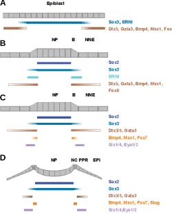
separation of cells with different fates is reflected by gene expres-sion (Fig. 2A). Pre-neural markers such as ERNI (chick; Streit, et al., 2000), Sox3 (Penzel, et al., 1997; Rex, et al., 1997; Kudoh, et al., 2004), Geminin (Xenopus; Kroll, et al., 1998) and SoxD
(Xenopus; Mizuseki, et al., 1998) are concentrated in the future
neural domain and gradually decrease towards the non-neural ectoderm, while genes like Gata2, Gata3, Dlx-3, -5, FoxiI or Foxi3, BMP4 and Msx1 show the opposite expression pattern
(Papalopulu and Kintner, 1993; Akimenko, et al., 1994; Streit, et
al., 1998; Pera, et al., 1999; Sheng and Stern, 1999; Streit and
Stern, 1999; Luo, et al., 2001; Streit, 2002; Liu, et al., 2003;
Solomon, et al., 2003; Ohyama and Groves, 2004;
Matsuo-Takasaki, et al., 2005). At this stage, precursors for different
placodes, including the otic, are still widely dispersed and inter-mingled with future neural, epidermal and neural crest cells in the chick (Garcia-Martinez, et al., 1993; Hatada and Stern, 1994;
Streit unpublished), although a more restricted distribution has been reported in zebrafish (Kozlowski, et al., 1997).
With the formation of the definitive neural plate (Fig. 2B), neural specific genes like Sox2 become up-regulated (Rex, et al., 1997;
Kishi, et al., 2000), while pre-neural markers either become
confined to a broad band of ectoderm surrounding the neural plate (ERNI) or remain expressed in both domains (Sox3, Geminin).
Likewise, some non-neural markers become upregulated in (e.g.
Dlx and Gata) or confined to (e.g. FoxiI ) the ectoderm next to the
neural plate. Thus, at early neurula stages a contiguous stripe of ectoderm coexpresses pre-neural and non-neural ectoderm mark-ers and has therefore been termed the ‘border’ (Streit and Stern, 1999; McLarren, et al., 2003; Woda, et al., 2003; Meulemans and
Bronner-Fraser, 2004). Within the border region precursors for neural, neural crest, epidermis and placodes remain interspersed (Kozlowski, et al., 1997; Streit, 2002; Bhattacharyya, et al., 2004).
Shortly thereafter, members of the Six and Eya families of nuclear factors begin to be expressed in a horseshoe-shaped domain surrounding the rostral neural plate from fore- to hindbrain levels (Fig. 2C) (Mishima and Tomarev, 1998; Esteve and Bovolenta, 1999; Sahly, et al., 1999; Kobayashi, et al., 2000;
Pandur and Moody, 2000; McLarren, et al., 2003; Bessarab, et al.,
2004; Schlosser and Ahrens, 2004; Litsiou, et al., 2005).
Simul-taneously, precursors for all placodes become concentrated in the Six/Eya+ territory to form a contiguous, unique domain: the
preplacodal region (Streit, 2002; Bhattacharyya, et al., 2004) (Fig.
1B). Unlike other factors that have a more widespread expression in the ectoderm (Dlx3/5 and Gata3 ), Six1, Six4, Eya1 and/or Eya2
Olfactory
Lens
Otic Trigeminal
(V)
Ophthalmic
Maxillomandibular
Cochlear-vestibular
(VIII) Geniculate (VII)
Petrosal (IX)
Nodose (X)
Epibranch
ial
Olfactory Lens
Epibranchial Otic
D V
Olfactory Lens
Adenohypopysheal Otic Lens, olfactory, telencephalon
Lens, olfactory, anterior cranial ganglia, telencephalon, otic,
anterior lateral line
Otic, posterior lateral line Posterior lateral line
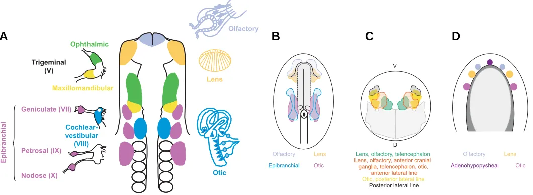
Fig. 1. Position of sensory placodes at the 10-somite and fate maps at late gastrula and neural plate stages.(A) Schematic representation of the sensory placodes and their derivatives at the 10 somite stage in the chick embryo (after D’Amico-Martel and Noden, 1983; Bhattacharyya, et al., 2004). (B) Fate map of a 1-somite stage chick embryo (after Streit, 2002; Bhattacharyya, et al., 2004). Grey, neural plate. (C) Fate map of a zebrafish embryo at 50% epiboly (modified from Kozlowski, et al., 1997); grey, neural plate, (Woo and Fraser, 1995). (D)Fate map of salamander embryo (Ambystoma) at neural plate stages (modified from Carpenter, 1937). Light grey, neural plate; dark grey, neural folds.
B
C
D
are exclusively expressed in the preplacodal region.
Around the same time, neural crest specific genes such as
Snail2, FoxD3 or Sox10 begin to be expressed in a thin line along
most of the neural plate (except its most rostral part, where no neural crest cells are generated; Couly and Le Douarin, 1985; Couly and Le Douarin, 1987). Although some overlap between neural crest and placode precursors is still observed at this stage in chick, both fates are completely segregated by the 4-5 somite stage, when the neural plate has begun to fold and neural folds are morphologically obvious (Fig. 2D) (Streit, 2002; Bhattacharyya, et al., 2004). Thus, by late neurula stages, the preplacodal region is
molecularly and cellularly distinct from other ectodermal deriva-tives.
Transcription factors that position the neural plate
border
As outlined above, a number of transcription factors are co-expressed at the border of neural and non-neural ectoderm before the onset of definitive neural crest and placode markers and are therefore likely to function upstream of preplacodal genes. Indeed, some of these have been implicated in controlling the position of the border and appear to be required for the specification of border derivatives. Msx1 is a direct mediator of BMP signalling and as such functions in promoting epidermal and repressing neural character during early stages of Xenopus
development, thereby positioning the border between both tis-sues (Suzuki, et al., 1997; Feledy, et al., 1999; Phillips, et al.,
2006). However, at late gastrula/early neurula stages Msx1 is more specifically involved in neural crest cell formation; it is both required for and sufficient to induce neural crest cells at the border of the neural plate (Tribulo, et al., 2003; Monsoro-Burq, et al.,
2005; Khadka, et al., 2006; Phillips, et al., 2006). Like Msx1,
members of the Dlx family also counteract neural plate formation, but in addition both seem to play antagonistic roles during the specification of neural crest and placode precursors. Misexpression of Dlx5, Dlx3 or constitutively active Dlx3 represses neural and neural crest cells, while promoting the expression of preplacodal
Six1 and -4 (Luo, et al., 2001; McLarren, et al., 2003; Woda, et al.,
2003). In contrast, overexpression of dominant negative Dlx3 shifts the neural plate border laterally or results in a complete loss of preplacodal gene expression (Woda, et al., 2003), while in
zebrafish, knockdown or deletion of dlx3b and –4b (b380 mutants)
leads to a severe reduction of olfactory, trigeminal and otic pla-codes (Solomon and Fritz, 2002; Kaji and Artinger, 2004). How-ever, when MsxB, C and E function is blocked in b380 mutants, placode development is restored indicating that in the absence of Dlx function Msx proteins repress placode formation (Phillips, et al., 2006). Thus, Dlx protein function is required for normal placode
formation, but its activity in the border region opposes Msx1 function. Since their expression partially overlaps fine tuning of their function maybe achieved through direct protein-protein inter-action: Dlx and Msx proteins can form heterodimers that block their function as transcriptional activator or repressor (Zhang, et al.,
1997). Thus, on cellular level relative expression levels of Dlx and Msx proteins may determine cell fate choices.
The winged helix transcription factor FoxiI has mainly been studied in relation to its function in early otic development. However, recent evidence indicates that it may have an even
earlier function in positioning the border between neural and non-neural ectoderm. In Xenopus and fish, FoxiI is initially expressed
widely, but then becomes rapidly confined to a stripe of ectoderm surrounding the neural plate (Solomon, et al., 2003;
Matsuo-Takasaki, et al., 2005; Hans, et al., 2007). A similar expression is
observed in mouse for Foxi3 (Ohyama and Groves, 2004). In fish,
its requirement for otic and epibranchial placode specification has been demonstrated in FoxiI mutants (foo/foo, hearsay and no soul; Lee, et al., 2003; Nissen, et al., 2003; Solomon, et al., 2003),
while other sensory placodes have not been investigated in detail. Two recent studies suggest that both FoxiI and Dlx3b are required in ectodermal cells to respond to the otic inducing factor FGF8 (Hans, et al., 2004; Hans, et al., 2007): misexpression of FGF8
Fig. 2. Changes in gene expression from gastrula to early somite stages. Diagrams show a cross section through chick embryos at gastrula (A), head process (B), 1-2-somite (C) and 4-5-somite (D) stages.
(A) At gastrula stages, the epiblast is roughly subdivided into neural and non-neural territories. (B)The neural plate can be identified morphologi-cally expressing definitive neural markers (Sox2), while preneural mark-ers (ERNI) become confined to the border, where non-neural markmark-ers become upregulated (Dlx5, Gata3, BMP4, Msx1, FoxiI). (C) Preplacodal markers begin to be expressed (Six1, Six4, Eya2); there is some overlap between BMP4, Msx1 and Pax7, which are later confined to the neural folds where neural crest cells arise. (D) Neural folds are morphologically distinct and express neural crest cells markers (Slug, Pax7). There is no overlap between neural crest and preplacodal genes. NP, neural plate; B, border; NNE, non-neural ectoderm; NC, neural crest; PPR, preplacodal region; EPI, future epidermis.
B
C
leads to an enlarged otic vesicle only within the normal ear field, where both genes are co-expressed. In contrast, loss of both genes completely abolishes the formation of otic structures (Solomon, et al., 2004; Hans, et al., 2007). Experiments in Xenopus demonstrate an even earlier role for FoxiI; loss of FoxiIa
function leads to an expansion of the neural plate at gastrula stages, while its overexpression suppresses neural development while simultaneously promoting epidermal character (Matsuo-Takasaki, et al., 2005). It should be noted that so far an early role
for Foxi class genes has not been demonstrated in mouse; this may be due the difference in Foxi gene expression in different species. Together, these findings identify FoxiI as one of the early players in ectodermal patterning involved in setting the border between neural and non-neural ectoderm and as a prerequisite for otic and epibranchial development. Future experiments will need to address its potential role in the formation of other placodes and its epistatic relation to genes specific for the placode territory.
Thus, members of the Dlx, Msx and Foxi family are expressed early and in a broad domain and regulate neural crest and placode specific genes. They are therefore at the top of a hierarchy controlling the specification of cells that arise from the border region and are likely to be intracellular mediators of the signalling pathways that pattern the ectoderm and position the placode territory next to the cranial neural plate.
The Six/Eya/Dach network in placode development
Among the many transcription factors expressed in the preplacodal region, only members of the Six and Eya families match precisely the location of all placode precursors and are subsequently maintained in all placodes, but lost from non-placodal ectoderm. They have been implicated in multiple pro-cesses during placode formation and are therefore likely candi-dates to be involved in defining the placode territory at early developmental stages.
Six and Eya genes in sensory organ formation
Their importance in sensory organ development was initially reported in Drosophila, where their homologues sine oculis (so) and eyes absent (eya) form a non-linear, regulatory network that together with dachshund (dac) control eye development and photoreceptor cell specification (for review: Kumar and Moses, 2001; Donner and Maas, 2004; Pappu and Mardon, 2004; Rebay,
et al., 2005). Loss of any of these genes results in defects or
absence of the eye (Bonini, et al., 1993; Cheyette, et al., 1994;
Mardon, et al., 1994; Quiring, et al., 1994; Serikaku and O’Tousa,
1994), while their overexpression leads to ectopic eye formation in restricted positions of other imaginal discs (Halder, et al., 1995;
Bonini, et al., 1997; Chen, et al., 1997; Pignoni, et al., 1997; Shen
and Mardon, 1997; Weasner, et al., 2007). So, eya and dac
regulate each others’ expression and function downstream of the Pax6 homologue eyeless (ey): their expression and eye-inducing ability depends on the presence of functional Ey (Halder, et al.,
1998; Niimi, et al., 1999; Bui, et al., 2000; Punzo, et al., 2002;
Pappu, et al., 2005; for review: Kumar and Moses, 2001; Pappu
and Mardon, 2004).
In vertebrates, six Six genes have been identified (Six1-6), while there are only four Eya genes (Eya1-4) (for review: Kawakami,
et al., 2000; Wawersik and Maas, 2000; Hanson, 2001; Rebay, et al., 2005). Of those Six1, Six4, Eya1 and Eya2 are found in the
pre-placodal region, while combinations of different family mem-bers are coexpressed in mature placodes (Mishima and Tomarev, 1998; Esteve and Bovolenta, 1999; Sahly, et al., 1999; Kobayashi, et al., 2000; Pandur and Moody, 2000; McLarren, et al., 2003;
Bessarab, et al., 2004; Schlosser and Ahrens, 2004; Litsiou, et al.,
2005). Of the Eya gene family, only Eya3 is never found in any placode. As in the fly, they are often colocalised with members of the Pax gene family (e.g. Pax6: lens, olfactory; Pax2: otic and epibranchial; Pax3: trigeminal) although their regulatory relation-ship appears to be more complicated. For example, in the mouse olfactory ectoderm initial expression of Six3, Eya1 and Dach1 proteins is Pax6 independent, while their maintenance in the placode requires Pax6 (Purcell, et al., 2005). Likewise, in the
presumptive lens ectoderm, Pax6 is controlled by Six3 but once
the placode is formed Six3 expression depends on Pax6 activity
(Purcell, et al., 2005; Liu, et al., 2006). In the ear Pax2, Eya1 and Six1 are expressed in partially overlapping domains; Eya1 and Six1 expression is independent of Pax2, while Six1 depends on
Eya1 function (Zheng, et al., 2003; Burton, et al., 2004). In the
preplacodal region Six and Eya transcripts are present prior to the
onset of Pax gene and are therefore likely to act independently.
The importance of Six and Eya genes for normal placode development has been demonstrated through loss-of-function in mouse, zebrafish and humans. Eya1 and Six1 have been studied extensively and play a role in the formation of most placode derivatives (see below), reflecting their widespread expression in the preplacodal region. Likewise, mutations in Eya4 and Six5 are associated with defects in placode derivatives (Klesert, et al.,
2000; Wayne, et al., 2001; Zhang, et al., 2004), while information
about Six2 and Eya2 is very sparse. Mice lacking Eya2 function have been generated, however, their placodal phenotype has not been described in detail (Grifone, et al., 2007). So far, no placode
phenotype has been described for Six4 mutant mice (Ozaki, et al.,
2001; Grifone, et al., 2005); one possible explanation may be
functional redundancy between genes of the same family that are normally co-expressed. In support of this, Six1 and -4 double knock-out mice show a more severe muscle, kidney and trigemi-nal ganglion phenotype than Six1 mutants alone (Grifone, et al.,
2005; Konishi, et al., 2006; Kobayashi, et al., 2007). However, it
is not know whether this is also the case for other placodes. Mice heterozygous for Eya1 display a phenotype very similar to an inherited form of deafness in humans, the Branchio-Oto-Renal (BOR) syndrome, a form of conductive hearing loss due to defects in middle ear development (Abdelhak, et al., 1997; Xu, et al., 1999). Mice completely lacking Eya1 function have severe
inner ear defects (Johnson, et al., 1999; Xu, et al., 1999; Li, et al.,
2003; Zou, et al., 2004; Friedman, et al., 2005; Zou, et al., 2006):
otic development arrests at vesicle stages, sensory patches remain small and while cochlear-vestibular neurons initially form, they later undergo apoptosis. In addition, the trigeminal ganglion is reduced in size, epibranchial placode derived petrosal, genicu-late and nodose ganglia are missing or greatly reduced and fail to express neuronal determination genes. Zebrafish dogeared mu-tants (Eya1) also show ear defects and the development of the lateral line placodes is impaired, however cranial ganglia are generally unaffected (Kozlowski, et al., 2005; Whitfield, 2005).
eye defects (Azuma, et al., 2000), although these have not been
described in mice. Finally, mutations in the eya-homologous region of Eya4 lead to late-onset deafness in humans (Wayne, et al., 2001; Zhang, et al., 2004; Schonberger, et al., 2005).
Like Eya1, Six1 has been implicated in normal development of the inner ear and mutations in human Six1 cause BOR syndrome like Eya1 mutations (Ruf, et al., 2004). Mice lacking Six1 function
display very similar phenotypes to Eya1 mutant mice: otic vesicles are small, lack the cochlea and semicircular canals and do not form a cochlear-vestibular ganglion (Laclef, et al., 2003; Li, et al.,
2003; Zheng, et al., 2003; Ozaki, et al., 2004). In addition,
trigeminal and epibranchial placode derived neurons are reduced or absent and development of the olfactory epithelium is impaired. In zebrafish, Six1 promotes the formation of hair cells by increas-ing their proliferation, while inhibitincreas-ing neurogenesis by inducincreas-ing apoptosis (Bricaud and Collazo, 2006). Finally, Six5 mutations lead to cataract formation in the lens (Klesert, et al., 2000; Sarkar, et al., 2000; Bateman, et al., 2006) and are associated with BOR
syndrome in humans (Hoskins, et al., 2007).
The widespread defects in almost all placode derivatives in Six1 and/or Eya1 mutants argues for a conserved function of this network during sensory placode formation or for an involvement at very early stages development, maybe in the preplacodal region. Unfortunately, none of the above studies has addressed this issue. In Xenopus, Six1 function has been assessed at
preplacodal stages (Brugmann, et al., 2004), where it promotes
the expression of other preplacodal genes like Eya1, while
re-pressing neural, neural crest and epidermal fates. These findings point to a potentially early role of Six and Eya proteins in ectoder-mal patterning by establishing the preplacodal region and confer-ring common preplacodal properties (see below). However, fur-ther studies are required to determine their precise role at these early stages.
Molecular function and targets of the Six/Eya/Dach network Six, Eya and Dach proteins are thought to interact physically and to act as a transcription factor complex to activate down-stream target genes (for review: Relaix and Buckingham, 1999; Kawakami, et al., 2000; Wawersik and Maas, 2000; Hanson,
2001; Silver and Rebay, 2005). Six genes encode homeodomain DNA binding proteins (Seo, et al., 1999; Kawakami, et al., 2000)
that can act either as transcriptional activators or repressors depending on the recruitment of appropriate cofactors. One group of such cofactors are the Dach proteins (Mardon, et al., 1994;
Hammond, et al., 1998; Davis, et al., 1999), nuclear factors which
together with other repressors inhibit target gene transcription. In addition, Dach proteins themselves seem to bind DNA (Ikeda, et al., 2002) and modulate BMP signalling by interacting with Smad4
(Wu, et al., 2003; Kida, et al., 2004). Eya proteins represent
transcriptional coactivators that are recruited to DNA via their interaction with Six proteins (Ohto, et al., 1999; Silver, et al.,
2003). Recently, Eya proteins have been shown to have catalytic activity as protein phosphatases and this activity appears to be required for their function as activators (Li, et al., 2003;
Rayapureddi, et al., 2003; Tootle, et al., 2003). Direct binding has
indeed been shown for Eya and Dach and Six and Eya proteins (Chen, et al., 1997; Pignoni, et al., 1997; Ohto, et al., 1999; Ikeda, et al., 2002; Li, et al., 2003; Silver, et al., 2003) and nuclear
translocation of Eya protein is dependent on its interaction with
members of the Six family (Ohto, et al., 1999). Furthermore,
Groucho repressors have been shown to bind Six proteins, in particular Six3 directly and thus modulate its activity (Kobayashi,
et al., 2001; Zhu, et al., 2002).
So far only a few direct target genes have been identified, among them cMyc and CyclinA1 and –D1, all involved in cell cycle control (Coletta, et al., 2004; Yu, et al., 2006). Indeed, both Six1
and Eya2 appear to promote tumorigenesis by enhancing prolif-eration (Coletta, et al., 2004; Zhang, et al., 2005; Yu, et al., 2006),
while high levels of Eya2 seem to trigger apoptosis (Clark, et al.,
2002). In the otic vesicle, loss of Eya1 and Six1 leads to reduced proliferation, while in Drosophila loss of either so, dac or eya initially results in overgrowth followed by cell death (Bonini, et al.,
1993; Pignoni, et al., 1997; Xu, et al., 1999; Li, et al., 2003; Ozaki, et al., 2004; Kozlowski, et al., 2005; Zou, et al., 2006). In zebrafish,
Six1 plays opposite roles in hair cells and otic neurons that arise from common sensory patches. Six1 induces apoptosis in neu-ronal precursors, but promotes proliferation in sensory hair cells (Bricaud and Collazo, 2006) thereby regulating the balance between both cell types. Thus, the Six/Eya/Dach network may control the number of placode precursors during early stages of development, differential proliferation and apoptosis during mor-phogenesis and the number of precursors for different cell types within placodes.
Although data in Drosphila show that the Six/Eya/Dach cas-sette can induce cell fate changes by making non-eye cells adopt an eye fate, the exact molecular mechanisms of how they operate during this process are still unknown (Bonini, et al., 1997; Pignoni, et al., 1997; Shen and Mardon, 1997; Weasner, et al., 2007).
Eyeless is directly regulated by sine oculis, however further targets have not been identified. In vertebrates, functional Six and Eya are required for myogenesis (Heanue, et al., 1999) for review
Relaix and Buckingham, 1999), but the evidence that they control cell specification without affecting proliferation during placode development is very poor. As mentioned above, misexpression of Six1 promotes preplacodal gene expression (Brugmann, et al.,
2004), but by itself or in combination with Eya is insufficient to generate mature placodes or to activate Pax genes (Christophorou and Streit, unpublihsed). Since only cells within the preplacodal region are competent to respond to placode inducing signals, one potential role of Six and Eya genes may be to impart competence to such inducing factors.
In summary, there is considerable evidence for a crucial role of the Six/Eya/Dach network in various aspects of placode develop-ment, however in many cases the precise molecular mechanisms remain to be identified. Characterisation of direct targets in different cellular contexts will be an important step to understand their function.
Signalling pathways inducing the neural plate border
and the preplacodal region
to position the placode territory in the head ectoderm next to the neural plate.
FGF pathway
Several observations implicate FGFs as one of the factors that initiate the formation of the border region. In the chick, misexpression of FGF8 rapidly induces ectopic expression of a set of genes normally coexpressed in the border: ERNI, Sox3, Dlx5 and Msx1 (Streit and Stern, 1999; Streit, et al., 2000;
Litsiou, et al., 2005). However, FGF alone is not sufficient to
generate any of the cell types that arise from the border: neural crest and placodes (Mayor, et al., 1997; LaBonne and
Bronner-Fraser, 1998; Monsoro-Burq, et al., 2003; Ahrens and Schlosser,
2005; Litsiou, et al., 2005). In contrast, FGF inhibition using the
antagonist SU5402 or dominant negative receptors shows that active signalling through the FGF pathway is required for at least some of the border genes (Sox3, ERNI; Streit, et al., 2000)
and for the generation of border derivatives (Mayor, et al., 1997;
LaBonne and Bronner-Fraser, 1998; Monsoro-Burq, et al.,
2003; Ahrens and Schlosser, 2005; Litsiou, et al., 2005).
To-gether, these findings argue for a role of FGFs in promoting border character in ectodermal cells as a prerequisite to gener-ate neural crest and placode cells. Accordingly, FGFs are expressed in the head mesoderm and trunk paraxial mesoderm that comes to underlie the border region and in Xenopus at the
edge of the neural plate (Niswander and Martin, 1992; Shamim and Mason, 1999; Streit and Stern, 1999; Ahrens and Schlosser, 2005).
In addition, FGFs seem to play a role in preplacodal induc-tion at slightly later stages. FGF signalling from surrounding tissues (head mesoderm in chick, neural plate in Xenopus) is
required for the induction of preplacodal markers, while ectopic expression of FGF8 promotes expression of Eya2, but not of
any other placode specific gene (Brugmann, et al., 2004;
Ahrens and Schlosser, 2005; Litsiou, et al., 2005). Thus, FGFs
play a dual role in the supporting placode formation: initially they promote the expression of border genes and later initiate expression of a subset of preplacodal markers.
BMP pathway
Modulation of BMP signalling has been widely implicated in early ectodermal patterning (Wilson, et al., 1997; Marchant, et al., 1998; Barth, et al., 1999; Tribulo, et al., 2003; for review
Sasai and De Robertis, 1997; Aybar and Mayor, 2002; Stern, 2005). Indeed, FoxiI is dependent on BMP signalling in fish and frogs (Matsuo-Takasaki, et al., 2005; Phillips, et al., 2006). In
zebrafish, FoxiI expression is reduced or lost in BMP7 and BMP2a mutants, while it is downregulated in Xenopus in the
presence of the BMP antagonist Chordin. In contrast, overex-pression of BMP4 causes an expansion of FoxiI at the expense of neural tissue. Likewise, BMP signalling is required for Dlx gene expression in chick, frog and fish (Nguyen, et al., 1998;
Feledy, et al., 1999; Pera, et al., 1999; Luo, et al., 2001), while Msx1 is a direct target of BMP signalling and mediates its ability
to promote epidermis (Suzuki, et al., 1997). These findings
implicate BMP activity, like FGF signalling, in the regulation of border specific genes.
One model mainly based on experiments in Xenopus
sug-gests that a gradient of BMP activity within the ectoderm acts to
allocate different cell fates for review (Sasai and De Robertis, 1997; Aybar and Mayor, 2002; Vonica and Brivanlou, 2006). In support of this idea, Xenopus animal caps treated with different
concentrations of BMP antagonists form epidermis in the pres-ence of high levels of BMP activity, while neural crest and preplacodal cells are generated at intermediate and neural plate at low levels (Wilson, et al., 1997; Tribulo, et al., 2003;
Brugmann, et al., 2004; Glavic, et al., 2004). Likewise, zebrafish
mutants with reduced BMP activity (and thus a shallower gradient) show a relatively larger expansion of the neural crest territory than of the neural plate (Nguyen, et al., 1998; Barth, et al., 1999). However, the placode territory is merely displaced,
Wnt, Bmp Wnt
BMP
Cer
DAN
FGF
Wnt Wnt
ERNI, Sox3 Dlx3/5, Msx1 FoxiI
Pax7, Snail2, FoxD3, Sox10
Fgf Bmp
Six1/4, Eya1/2 Bmp
Wnt
Anti Bmp Anti Wnt
Neural plate border
Neural crest Preplacodal region
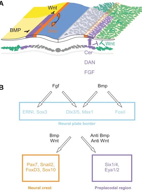
Fig. 3. Model for induction of the preplacodal region. (A) The diagram shows a cross section through a chick embryo at the 2-4 somite stage viewed from anterior towards posterior (modified from Litsiou, et al., 2005). Ectodermal signals that influence the position of the preplacodal region are schematised on the left, whereas mesoderm derived signals are shown on the right. The preplacodal region (purple) is surrounded by inhibitory signals from the lateral (light yellow; BMP) and posterior (yellow; Wnt) ectoderm, from the neural folds (orange; Wnt, BMP) and from the lateral and posterior mesoderm (green; Wnt). FGF, Wnt antago-nists and BMP antagoantago-nists (purple) from the mesoderm underlying the preplacodal region protect the overlying ectoderm from these inhibitory signals and allow the formation of placode precursors. (B) Signals and transcription factors in the border and preplacodal region. FGFs and BMPs act upstream of border specific transcription factors. Once the border is established, levels of BMP and Wnt signalling determine whether border cells generate neural crest or placode cells.
but not expanded and different placodes are affected differen-tially arguing against a simple gradient model (Neave, et al.,
1997; Nguyen, et al., 1998).
In chick, the main region sensitive to modulation of BMP signalling is the neural plate border itself (Streit and Stern, 1999). Here, misexpression of BMP antagonists leads to a shift of the border towards the non-neural ectoderm, while misexpression of BMP4 narrows the neural plate and shifts the border medially. In contrast, modulation of the BMP pathway away from the border does not have any effect. Likewise, local reduction of BMP signalling close to the preplacodal region expands this territory in chick and Xenopus, but is not sufficient
to induce it ectopically in future epidermis (Glavic, et al., 2004;
Ahrens and Schlosser, 2005; Litsiou, et al., 2005). One possible
explanation to reconcile these differences is that Xenopus
animal caps may contain border territory and are therefore particularly sensitive to changes in BMP activity.
Thus, modulation of BMP activity and loss- or gain-of-func-tion experiments for border specific transcripgain-of-func-tion factors show the same effect: they shift the border of the neural plate. It is therefore likely that BMP signalling acts via mediators such as FoxiI, Dlx and Msx to alter preplacodal gene expression indi-rectly.
Wnt pathway
As discussed above, both FGF and BMP pathways modulate the expression of preplacodal genes: FGF8 activates Eya2,
while inhibition of BMP signalling expands preplacodal markers (Brugmann, et al., 2004; Ahrens and Schlosser, 2005; Litsiou, et al., 2005). However, even the combination of FGF and BMP
antagonists is not sufficient to induce preplacodal character in ectoderm away form the endogenous placode territory or in the future trunk ectoderm (Brugmann, et al., 2004; Ahrens and
Schlosser, 2005; Litsiou, et al., 2005). Like inhibition of BMP,
misexpression of Wnt antagonists leads to an expansion of preplacodal gene expression at the expense of future epider-mis (Brugmann, et al., 2004; Litsiou, et al., 2005). Interestingly, Six1, -4 and Eya2 also extend into the trunk ectoderm, where
placode formation is not normally observed. In contrast, activa-tion of canonical Wnt signalling represses preplacodal gene expression suggesting that Wnt attenuation allows the specifi-cation of placode precursors (Litsiou, et al., 2005).
Further-more, a combination of FGF with Wnt and BMP antagonists induces an ectopic preplacodal region in naïve ectoderm in the absence of neural and mesodermal tissue indicating that these factors promote placode character directly (Litsiou, et al., 2005).
Thus, temporal and spatial integration of all three signals is important to generate placode precursors.
Unlike placode precursors, neural crest cells require canoni-cal Wnt signalling (for review: Aybar and Mayor, 2002; Knecht and Bronner-Fraser, 2002). These findings suggest that at the border of the neural plate exposure to different levels of Wnt activity determines whether cells adopt placode or neural crest cell fates. Indeed, activation of Wnt signalling expands neural crest markers into the placode territory, while its inhibition has the opposite effect (Litsiou, et al., 2005). In this context it is
interesting that at slightly later stages when the otic placode begins to form, Wnt signalling promotes placode formation: Pax2+ cells that activate the pathway become otic, while those
that do not, develop into epidermis (Ohyama, et al., 2006).
These findings highlight that interpretation of the same signal-ling pathway is highly context dependent and is determined by the developmental history of individual cells.
A model for induction of the preplacodal region
The data summarised above highlight that induction of the preplacodal region is a multi-step process, which requires the integration of different signals produced by different tissues. The following model tries to integrate tissue interactions and signalling pathways (Fig. 3). As a first step, a border territory is set up in the early neurula ectoderm between future neural and epidermal cells. Genes specific for this region are under the control of FGF and/or BMP signalling. BMP4 and -7 are expressed in the
non-neural ectoderm and transcript levels and phospho-smad activity are highest at the edge of the neural plate (Fainsod, et al., 1994;
Streit, et al., 1998; Streit and Stern, 1999; Faure, et al., 2002),
which in turn may lead to the upregulation of some border genes (Dlx, Msx, FoxiI ). FGFs emanating from the organiser and the
mesoderm underlying the border maintain the expression of Sox3
and ERNI and cooperate with BMPs to promote Dlx and Msx gene
expression. Once established the border gives rise to two differ-ent cell types: neural crest and placodes. BMP4 and -7 transcripts
concentrate in the forming neural folds (Fainsod, et al., 1994;
Liem, et al., 1995), where Wnts begin to be expressed; together
they promote formation of neural crest cells. The future heart mesoderm expands anteriorly and comes to underlie the placode territory (Kimmel and Warga, 1988; Keller and Tibbetts, 1989; Tam, et al., 1997; Redkar, et al., 2001; Hochgreb, et al., 2003).
This tissue expresses FGF4, the BMP antagonist DAN and the Wnt inhibitor Cerberus (Ogita, et al., 2001), while more lateral and
posterior mesoderm contains high levels of Wnt8c (Hume and
Dodd, 1993; Litsiou, et al., 2005). Wnt6 is found in the trunk, but
not the head ectoderm (Garcia-Castro, et al., 2002; Schubert, et al., 2002). Thus, the preplacodal region is surrounded by
inhibi-tory factors at its medial, lateral and posterior edges. Signals from the heart mesoderm protect the overlying ectoderm from these inhibitory influences and allow it to adopt placode fate.
Anterior-posterior patterning of the preplacodal region
Within the preplacodal region precursors for different placodes are intermingled, although some separation of individual popula-tions along the anterior posterior axis is already apparent. Precur-sors for anterior placodes (adenohypophysis, olfactory, lens) are located in the rostral preplacodal region, while precursors for posterior placodes (trigeminal, epibranchial, otic, lateral line) are restricted more caudally (D’Amico-Martel and Noden, 1983; Couly and Le Douarin, 1985; Couly and Le Douarin, 1988; Kozlowski, et al., 1997; Streit, 2002; Bhattacharyya, et al., 2004; Litsiou, et al.,
in gene expression have recently been reviewed in detail in
Xenopus (Schlosser, 2006) and therefore only some of the early
aspects of patterning will be highlighted below.
Transcription factors in anterior-posterior patterning At neurula stages, the rostral preplacodal region begins to express Otx2, Six3, Pitx3, Dmbx1 and Pax6, while its caudal part
is characterised by Irx1, -2 and -3 and Gbx2 (Li, et al., 1994;
Bally-Cuif, et al., 1995; Oliver, et al., 1995; Pannese, et al., 1995; Hirsch
and Harris, 1997; Bellefroid, et al., 1998; Gomez-Skarmeta, et al.,
1998; Shamim and Mason, 1998; Goriely, et al., 1999; Zhou, et al., 2000; Glavic, et al., 2002; Gogoi, et al., 2002; Matsumoto, et al., 2004; Dutta, et al., 2005; Zilinski, et al., 2005; Liu, et al., 2006;
for review: Schlosser, 2006). Although these patterns appear to be roughly complementary, closer inspection reveals that differ-ent transcripts do not share the same rostro-caudal boundary (Fig. 4). Rather pairs of transcription factors have boundaries at different levels: Six3 expression abuts Irx, while Otx2 and Gbx2
abut at slightly more posterior levels. These patterns are very reminiscent of their expression in the neural plate, where the same pairs of genes control the subdivision of the brain into different compartments (for review: Martinez, 2001; Nakamura, 2001; Hidalgo-Sanchez, et al., 2005). Gbx2 and Otx2 are involved
in positioning the midbrain-hindbrain boundary (Broccoli, et al.,
1999; Millet, et al., 1999; Katahira, et al., 2000; Acampora, et al.,
2001; Li and Joyner, 2001), while Six3 and Irx3 define a boundary in the forebrain that later corresponds to the zona limitans intrathalamica (Kobayashi, et al., 2002).
At early somite stages, members of the Pax gene family become expressed in more restricted domains within the preplacodal region (Bang, et al., 1997; Hirsch and Harris, 1997 Li,
1994 #195; Stark, et al., 1997; Heller and Brandli, 1999; Groves
and Bronner-Fraser, 2000). Pax3 is detected in the ophthalmic
part of the trigeminal, Pax8 in the otic and Pax2 epibranchial, otic
and lateral line territory. Together Pax genes cover the entire placode region in non-overlapping patterns except Pax2 and -8
which are co-expressed in the future otic placode. Interestingly, in the neural plate Pax2 and -6 represent another pair of
transcrip-tion factors that positranscrip-tion a boundary, in this case between future diencephalon and mesencephalon (Okafuji, et al., 1999; Schwarz, et al., 1999; Matsunaga, et al., 2000; Schwarz, et al., 2000). As it
is the case for Six3/Irx3 and Otx2/Gbx2, in the brain Pax2 and – 6 negatively cross regulate each other, leading to sharpening of the molecular boundary and separation of different cell fates. It is tempting to speculate that the same molecular mechanisms that pattern the brain also operate to impart regional identity to the placodes.
Loss of Otx2 function results in severe defects in the head including the brain, olfactory and lens placode as well as pattering of the otic vesicle (Acampora, et al., 1995; for review: Acampora, et al., 2001). However, because of the severe fore- and midbrain
defects, it has been difficult to asses its direct function in placode development without the availability of tissue specific knock outs. Mice deficient in Gbx2 (Lin, et al., 2005) and Pax2 function show
patterning defects in the otic vesicle (Torres, et al., 1996; Burton, et al., 2004), while loss of Six3 or Pax6 affects lens and olfactory
development (Hogan, et al., 1988; Quinn, et al., 1996; Grindley, et al., 1997; Lagutin, et al., 2003; Liu, et al., 2006). In Xenopus,
Irx1 is required for the expression of the early preplacodal marker
Six1 and later placode specific genes like Sox2 and Pax2 (Glavic, et al., 2004). Thus, mutation in or loss of any of these genes leads
to defects in placode development, although their role in early patterning of the preplacodal region remains elusive probably due to functional redundancy with other members of the same family that are expressed in similar patterns (Schlosser, 2006).
Signalling pathways in anterior-posterior patterning In the neural plate, regional identity is initially set up through the graded activity of Wnts, FGF and retinoic acid, all of which possess posteriorising activity and control some of the transcrip-tion factors described above (for review: Yamaguchi, 2001; Wil-son and Houart, 2004; Kiecker and Lumsden, 2005; Rhinn, et al.,
2006). Do the same signalling pathways control anterior posterior patterning in the preplacodal region? Experiments in Xenopus
revealed that the formation of neural crest cells indeed requires Wnt and retinoic acid activity and that anterior neural folds, which normally do not generate neural crest cells, do so in the presence of these factors (Villanueva, et al., 2002). In chick and Xenopus,
the expression of preplacodal markers can be expanded into the trunk ectoderm in the presence of Wnt antagonists (Brugmann, et al., 2004; Litsiou, et al., 2005). In contrast, the zebrafish mutants
masterblind and headless, in which Wnt signalling is overactivated, show a loss of anterior placodes (lens, olfactory), but an expan-sion of trigeminal neurons around the anterior neural plate (Kim,
et al., 2000; Heisenberg, et al., 2001). Thus, differential activation
of the Wnt pathway along the rostro-caudal axis influences patterning of the preplacodal region suggesting that the mecha-nisms that allocate regional identity in the neural plate may act more globally to pattern the entire ectoderm.
Cells in the preplacodal region share a common
devel-opmental programme
As outlined at the beginning of this review, placodes form diverse structures with different functional properties and a
vari-Six1/4, Eya1/2
Pax6, Six3 Irx1/2/3
Otx2 Gbx2
Pax6 Pax3 Pax2/8
Early neurula
Late neurula
Early somite
Anterior Posterior
ety of different cell types. Yet, they have variously been grouped together as a family and considered as related structures. Does this grouping reflect a meaningful developmental concept and does the preplacodal region represent a ‘basic placode state’? At least two conditions need to be fulfilled. First, at some point along their developmental history placode cells should acquire a unique state that distinguishes them from other ectodermal derivatives. This is clearly the case (see above): placode precursors occupy a unique region in the embryonic ectoderm surrounding the cranial neural plate and they express a unique set of molecular markers. Furthermore, the preplacodal region is induced by a specific combination of tissues and signals that is different from the signals that induce neural, neural crest and epidermal precur-sors. Second and more importantly, the ‘preplacode state’ should be a unique property of placode precursors and be a prerequisite for cells to become mature placodes.
Recent experiments on otic induction in the chick embryo provide strong evidence that indeed ectodermal cells have to acquire preplacode character before they are responsive to the otic inducer FGF (Martin and Groves, 2006). Only cells within the preplacodal territory are responsive to FGF, while future epider-mal cells are not. Anterior epiblast from chick gastrula stage embryos normally never gives rise to the otic placode, but is competent to do so when transplanted into the ectoderm next to the hindbrain, where the otic placode normally forms. Likewise, explants of anterior epiblast do not express otic markers when cultured in isolation or when treated with FGF. However, when the same tissue is first transplanted into the preplacodal region for a brief period (ca. 8 hrs), it initiates gene expression characteristic for this territory (Eya2, Dlx) and when explanted can now respond
to FGF by expressing otic markers. These experiments support the idea that the preplacodal region has unique properties and suggest that the acquisition of preplacodal character is an essen-tial step in otic induction and a prerequisite for cells to form mature placodes.
But do all placode precursors share a common developmental history? Recent evidence indicates that at the very least they initially share common characteristics: unlike any other part of the ectoderm, the entire preplacodal region is specified as lens (Bailey, et al., 2006). When the preplacodal region is subdivided
into four portions along the anterior posterior axis and cultured in neutral environment, no markers specific for olfactory, trigeminal or otic placodes are expressed – the tissue is not specified. However, in the same experiment, explants from all parts of the preplacodal region, even cells that normally never contribute to the lens, but to the otic placode, form lens-like structures. These explants follow the same sequence and timing of gene expression as observed during normal lens development: they initially ex-press Pax6, followed by L-maf and Foxc1 and finally, δ- and α -crystallin. Thus, all placode cells regardless of their ultimate fate initially possess lens character suggesting that in normal develop-ment placode induction is intimately linked with lens suppression. The lens probably represents the simplest placode derivative: it only generates two cell types, lens fibre and lens epithelial cells. The only other non-neurogenic placode, the anterior pituitary, which develops in the anterior midline, is easily transformed into lens in the absence of sonic hedgehog from underlying axial structures (Sbrogna, et al., 2003; Dutta, et al., 2005; Zilinski, et al.,
2005). In contrast to the lens and anterior pituitary, all other
placodes give rise to sensory neurons and/or sensory cells. Thus, lens suppression must be accompanied by the acquisition of neurogenic properties in non-lens placodes. Together, these results provide good support for the concept of a common ground state for all sensory placodes and for the importance of the preplacodal region in the developmental history of placode cells. What are the lens repressing signals? FGFs appear to be the main players to initiate lens repression: activation of the FGF pathway inhibits the expression of the presumptive lens marker
Pax6 (Bailey, et al., 2006). FGFs (FGF3, FGF10, FGF19
depend-ing on species) also play an important role in otic induction (Ladher, et al., 2000; Vendrell, et al., 2000; Leger and Brand,
2002; Maroon, et al., 2002; Wright and Mansour, 2003) and lens
specification is abolished in preplacodal explants in the presence of FGF2, while the otic marker Pax2 is induced (Bailey and Streit, unpublished). In contrast, exposure to FGF8 is sufficient to induce olfactory character from lens specified ectoderm (Bailey, et al.,
2006). Finally, FGF signalling (FGF3, -8) has been implicated in epibranchial placode specification and in the generation of the adenohypophysis, a placode forming in the anterior midline (Herzog, et al., 2004; Nechiporuk, et al., 2007; Nikaido, et al.,
2007; Sun, et al., 2007). Thus, FGFs play a key role in restricting
lens fate and in simultaneously inducing other placodes. The next important question to address is how the activation of the same signalling pathway in the preplacodal region elicits different responses leading to the formation of placodes with different identities.
Future perspectives
In summary, prior to the appearance of morphological pla-codes induction of the preplacodal region is an essential process, which imparts unique identity to all placode precursors. The signalling pathways involved are similar to those implicated in neural and neural crest induction, however, timing and levels differ. Recent evidence argues that all placodes initially share a common ground state as lens and thus possess a common developmental history, before they diversify later. Surprisingly, FGF signalling appears to play a role during the induction of different placodes from this ground state. The future challenge is to unravel how different pathways cooperate with FGF signalling to impart placode identity and to establish networks of transcrip-tion factors that control this process.
References
ABDELHAK, S., KALATZIS, V., HEILIG, R., COMPAIN, S., SAMSON, D., VINCENT, C., WEIL, D., CRUAUD, C., SAHLY, I., LEIBOVICI, M. et al. (1997). A human homologue of the Drosophila eyes absent gene underlies branchio- oto-renal (BOR) syndrome and identifies a novel gene family. Nat Genet, 15: 157-164.
ACAMPORA, D., GULISANO, M., BROCCOLI, V. and SIMEONE, A. (2001). Otx genes in brain morphogenesis. Prog Neurobiol, 64: 69-95.
ACAMPORA, D., MAZAN, S., LALLEMAND, Y., AVANTAGGIATO, V., MAURY, M., SIMEONE, A. and BRULET, P. (1995). Forebrain and midbrain regions are deleted in Otx2-/- mutants due to a defective anterior neuroectoderm specifica-tion during gastrulaspecifica-tion. Development, 121: 3279-3290.
AHRENS, K. and SCHLOSSER, G. (2005). Tissues and signals involved in the induction of placodal Six1 expression in Xenopus laevis. Dev Biol, 288: 40-59.
part of a homeobox gene code for the head. J Neurosci, 14: 3475-3486.
AYBAR, M. J. and MAYOR, R. (2002). Early induction of neural crest cells: lessons learned from frog, fish and chick. Curr Opin Genet Dev, 12: 452-458.
AZUMA, N., HIRAKIYAMA, A., INOUE, T., ASAKA, A. and YAMADA, M. (2000). Mutations of a human homologue of the Drosophila eyes absent gene (EYA1) detected in patients with congenital cataracts and ocular anterior segment anomalies. Hum Mol Genet, 9: 363-366.
BAILEY, A. P., BHATTACHARYYA, S., BRONNER-FRASER, M. and STREIT, A. (2006). Lens is the ground state for all sensory placodes, from which FGF signalling promotes olfactory fate. Dev. Cell, 11: 505-517.
BAILEY, A. P. and STREIT, A.: Sensory organs: making and breaking the pre-placodal region. In: Curr Top Dev Biol. vol. 72; 2006: 167-204.
BAKER, C. V. and BRONNER-FRASER, M. (2001). Vertebrate cranial placodes I. Embryonic induction. Dev Biol, 232: 1-61.
BALLY-CUIF, L., GULISANO, M., BROCCOLI, V. and BONCINELLI, E. (1995). c-otx2 is expressed in two different phases of gastrulation and is sensitive to retinoic acid treatment in chick embryo. Mech Dev, 49: 49-63.
BANCROFT, M. and BELLAIRS, R. (1977). Placodes of the chick embryo studied by SEM. Anat Embryol (Berl), 151: 97-108.
BANG, A. G., PAPALOPULU, N., KINTNER, C. and GOULDING, M. D. (1997). Expression of Pax-3 is initiated in the early neural plate by posteriorizing signals produced by the organizer and by posterior non-axial mesoderm. Development, 124: 2075-2085.
BARTH, K. A., KISHIMOTO, Y., ROHR, K. B., SEYDLER, C., SCHULTE-MERKER, S. and WILSON, S. W. (1999). BMP activity establishes a gradient of positional information throughout the entire neural plate. Development, 126: 4977-4987.
BATEMAN, J. B., RICHTER, L., FLODMAN, P., BURCH, D., BROWN, S., PENROSE, P., PAUL, O., GEYER, D. D., BROOKS, D. G. and SPENCE, M. A. (2006). A new locus for autosomal dominant cataract on chromosome 19: linkage analyses and screening of candidate genes. Invest Ophthalmol Vis Sci, 47: 3441-3449.
BELLEFROID, E. J., KOBBE, A., GRUSS, P., PIELER, T., GURDON, J. B. and PAPALOPULU, N. (1998). Xiro3 encodes a Xenopus homolog of the Drosophila Iroquois genes and functions in neural specification. EMBO J, 17: 191-203.
BESSARAB, D. A., CHONG, S. W. and KORZH, V. (2004). Expression of zebrafish six1 during sensory organ development and myogenesis. Dev Dyn, 230: 781-786.
BHATTACHARYYA, S., BAILEY, A. P., BRONNER-FRASER, M. and STREIT, A. (2004). Segregation of lens and olfactory precursors from a common territory: cell sorting and reciprocity of Dlx5 and Pax6 expression. Dev Biol, 271: 403-414.
BONINI, N. M., BUI, Q. T., GRAY-BOARD, G. L. and WARRICK, J. M. (1997). The Drosophila eyes absent gene directs ectopic eye formation in a pathway conserved between flies and vertebrates. Development, 124: 4819-4826.
BONINI, N. M., LEISERSON, W. M. and BENZER, S. (1993). The eyes absent gene: genetic control of cell survival and differentiation in the developing Drosophila eye. Cell, 72: 379-395.
BRICAUD, O. and COLLAZO, A. (2006). The transcription factor six1 inhibits neuronal and promotes hair cell fate in the developing zebrafish (Danio rerio) inner ear. J Neurosci, 26: 10438-10451.
BROCCOLI, V., BONCINELLI, E. and WURST, W. (1999). The caudal limit of Otx2 expression positions the isthmic organizer. Nature, 401: 164-168.
BRUGMANN, S. A., PANDUR, P. D., KENYON, K. L., PIGNONI, F. and MOODY, S. A. (2004). Six1 promotes a placodal fate within the lateral neurogenic ectoderm by functioning as both a transcriptional activator and repressor. Development, 131: 5871-5881.
BUI, Q. T., ZIMMERMAN, J. E., LIU, H., GRAY-BOARD, G. L. and BONINI, N. M. (2000). Functional analysis of an eye enhancer of the Drosophila eyes absent gene: differential regulation by eye specification genes. Dev Biol, 221: 355-364.
BURTON, Q., COLE, L. K., MULHEISEN, M., CHANG, W. and WU, D. K. (2004). The role of Pax2 in mouse inner ear development. Dev Biol, 272: 161-175.
CARPENTER, E. (1937). The head pattern in Ambystoma studied by vital staining and transplantation methods. J Exp Zool, 75: 103-129.
CHEN, R., AMOUI, M., ZHANG, Z. and MARDON, G. (1997). Dachshund and eyes absent proteins form a complex and function synergistically to induce ectopic eye development in Drosophila. Cell, 91: 893-903.
CHEYETTE, B. N., GREEN, P. J., MARTIN, K., GARREN, H., HARTENSTEIN, V.
and ZIPURSKY, S. L. (1994). The Drosophila sine oculis locus encodes a homeodomain-containing protein required for the development of the entire visual system. Neuron, 12: 977-996.
CLARK, S. W., FEE, B. E. and CLEVELAND, J. L. (2002). Misexpression of the eyes absent family triggers the apoptotic program. J Biol Chem, 277: 3560-3567.
COLETTA, R. D., CHRISTENSEN, K., REICHENBERGER, K. J., LAMB, J., MICOMONACO, D., HUANG, L., WOLF, D. M., MULLER-TIDOW, C., GOLUB, T. R., KAWAKAMI, K. et al. (2004). The Six1 homeoprotein stimulates tumori-genesis by reactivation of cyclin A1. Proc Natl Acad Sci U S A, 101: 6478-6483.
COULY, G. and LE DOUARIN, N. M. (1988). The fate map of the cephalic neural primordium at the presomitic to the 3-somite stage in the avian embryo. Development, 103: 101-113.
COULY, G. F. and LE DOUARIN, N. M. (1985). Mapping of the early neural primordium in quail-chick chimeras. I. Developmental relationships between placodes, facial ectoderm and prosencephalon. Dev Biol, 110: 422-439.
COULY, G. F. and LE DOUARIN, N. M. (1987). Mapping of the early neural primordium in quail-chick chimeras. II. The prosencephalic neural plate and neural folds: implications for the genesis of cephalic human congenital abnor-malities. Dev Biol, 120: 198-214.
D’AMICO-MARTEL, A. and NODEN, D. M. (1983). Contributions of placodal and neural crest cells to avian cranial peripheral ganglia. Am J Anat, 166: 445-468.
DAVIS, R. J., SHEN, W., HEANUE, T. A. and MARDON, G. (1999). Mouse Dach, a homologue of Drosophila dachshund, is expressed in the developing retina, brain and limbs. Dev Genes Evol, 209: 526-536.
DONNER, A. L. and MAAS, R. L. (2004). Conservation and non-conservation of genetic pathways in eye specification. Int J Dev Biol, 48: 743-753.
DUTTA, S., DIETRICH, J. E., ASPOCK, G., BURDINE, R. D., SCHIER, A., WESTERFIELD, M. and VARGA, Z. M. (2005). pitx3 defines an equivalence domain for lens and anterior pituitary placode. Development: 1579-1590.
ESTEVE, P. and BOVOLENTA, P. (1999). cSix4, a member of the six gene family of transcription factors, is expressed during placode and somite development. Mech Dev, 85: 161-165.
FAINSOD, A., STEINBEISSER, H. and DE ROBERTIS, E. M. (1994). On the function of BMP-4 in patterning the marginal zone of the Xenopus embryo. EMBO J, 13: 5015-5025.
FAURE, S., DE SANTA BARBARA, P., ROBERTS, D. J. and WHITMAN, M. (2002). Endogenous patterns of BMP signaling during early chick development. Dev Biol, 244: 44-65.
FELEDY, J. A., BEANAN, M. J., SANDOVAL, J. J., GOODRICH, J. S., LIM, J. H., MATSUO-TAKASAKI, M., SATO, S. M. and SARGENT, T. D. (1999). Inhibitory patterning of the anterior neural plate in Xenopus by homeodomain factors Dlx3 and Msx1. Dev Biol, 212: 455-464.
FERNANDEZ-GARRE, P., RODRIGUEZ-GALLARDO, L., ALVAREZ, I. S. and PUELLES, L. (2002). A neural plate fate map at stage HH4 in the chick: methodology and preliminary data. Brain Res Bull, 57: 293-295.
FRIEDMAN, R. A., MAKMURA, L., BIESIADA, E., WANG, X. and KEITHLEY, E. M. (2005). Eya1 acts upstream of Tbx1, Neurogenin 1, NeuroD and the neurotrophins BDNF and NT-3 during inner ear development. Mech Dev, 122: 625-634.
GARCIA-CASTRO, M. I., MARCELLE, C. and BRONNER-FRASER, M. (2002). Ectodermal Wnt function as a neural crest inducer. Science, 297: 848-851.
GARCIA-MARTINEZ, V., ALVAREZ, I. S. and SCHOENWOLF, G. C. (1993). Locations of the ectodermal and nonectodermal subdivisions of the epiblast at stages 3 and 4 of avian gastrulation and neurulation. J Exp Zool, 267: 431-446.
GLAVIC, A., GOMEZ-SKARMETA, J. L. and MAYOR, R. (2002). The homeoprotein Xiro1 is required for midbrain-hindbrain boundary formation. Development, 129: 1609-1621.
GLAVIC, A., MARIS HONORE, S., GLORIA FEIJOO, C., BASTIDAS, F., ALLENDE, M. L. and MAYOR, R. (2004). Role of BMP signaling and the homeoprotein Iroquois in the specification of the cranial placodal field. Dev Biol, 272: 89-103.
GOGOI, R. N., SCHUBERT, F. R., MARTINEZ-BARBERA, J.-P., ACAMPORA, D., SIMEONE, A. and LUMSDEN, A. (2002). The paired-type homeobox gene Dmbx1 marks the midbrain and pretectum. Mech. Dev. 114: 213-217.
GORIELY, A., DIEZ DEL CORRAL, R. and STOREY, K. G. (1999). c-Irx2 expres-sion reveals an early subdiviexpres-sion of the neural plate in the chick embryo. Mech Dev, 87: 203-206.
GRIFONE, R., DEMIGNON, J., GIORDANI, J., NIRO, C., SOUIL, E., BERTIN, F., LACLEF, C., XU, P. X. and MAIRE, P. (2007). Eya1 and Eya2 proteins are required for hypaxial somitic myogenesis in the mouse embryo. Dev Biol, 302: 602-616.
GRIFONE, R., DEMIGNON, J., HOUBRON, C., SOUIL, E., NIRO, C., SELLER, M. J., HAMARD, G. and MAIRE, P. (2005). Six1 and Six4 homeoproteins are required for Pax3 and Mrf expression during myogenesis in the mouse embryo. Development, 132: 2235-2249.
GRINDLEY, J. C., HARGETT, L. K., HILL, R. E., ROSS, A. and HOGAN, B. L. (1997). Disruption of PAX6 function in mice homozygous for the Pax6Sey-1Neu mutation produces abnormalities in the early development and regionalization of the diencephalon. Mech Dev, 64: 111-126.
GROVES, A. K. and BRONNER-FRASER, M. (2000). Competence, specification and commitment in otic placode induction. Development, 127: 3489-3499.
HADDON, C. and LEWIS, J. (1996). Early ear development in the embryo of the zebrafish, Danio rerio. J Comp Neurol, 365: 113-128.
HALDER, G., CALLAERTS, P., FLISTER, S., WALLDORF, U., KLOTER, U. and GEHRING, W. J. (1998). Eyeless initiates the expression of both sine oculis and eyes absent during Drosophila compound eye development. Development, 125: 2181-2191.
HALDER, G., CALLAERTS, P. and GEHRING, W. J. (1995). Induction of ectopic eyes by targeted expression of the eyeless gene in Drosophila. Science, 267: 1788-1792.
HAMMOND, K. L., HANSON, I. M., BROWN, A. G., LETTICE, L. A. and HILL, R. E. (1998). Mammalian and Drosophila dachshund genes are related to the Ski proto-oncogene and are expressed in eye and limb. Mech Dev, 74: 121-131.
HANS, S., CHRISTISON, J., LIU, D. and WESTERFIELD, M. (2007). FGF-dependent otic induction requires competence provided by FoxiI and Dlx3b. BMC Dev Biol, 7: 5.
HANS, S., LIU, D. and WESTERFIELD, M. (2004). Pax8 and Pax2a function synergistically in otic specification, downstream of the FoxiI and Dlx3b transcrip-tion factors. Development, 131: 5091-5102.
HANSON, I. M. (2001). Mammalian homologues of the Drosophila eye specification genes. Semin Cell Dev Biol, 12: 475-484.
HATADA, Y. and STERN, C. D. (1994). A fate map of the epiblast of the early chick embryo. Development, 120: 2879-2889.
HEANUE, T. A., RESHEF, R., DAVIS, R. J., MARDON, G., OLIVER, G., TOMAREV, S., LASSAR, A. B. and TABIN, C. J. (1999). Synergistic regulation of vertebrate muscle development by Dach2, Eya2 and Six1, homologs of genes required for Drosophila eye formation. Genes Dev, 13: 3231-3243.
HEISENBERG, C. P., HOUART, C., TAKE-UCHI, M., RAUCH, G. J., YOUNG, N., COUTINHO, P., MASAI, I., CANEPARO, L., CONCHA, M. L., GEISLER, R. et al. (2001). A mutation in the Gsk3-binding domain of zebrafish Masterblind/ Axin1 leads to a fate transformation of telencephalon and eyes to diencephalon. Genes Dev, 15: 1427-1434.
HELLER, N. and BRANDLI, A. W. (1999). Xenopus Pax-2/5/8 orthologues: novel insights into Pax gene evolution and identification of Pax-8 as the earliest marker for otic and pronephric cell lineages. Dev Genet, 24: 208-219.
HERZOG, W., SONNTAG, C., VON DER HARDT, S., ROEHL, H. H., VARGA, Z. M. and HAMMERSCHMIDT, M. (2004). FGF3 signaling from the ventral dien-cephalon is required for early specification and subsequent survival of the zebrafish adenohypophysis. Development, 131: 3681-3692.
HIDALGO-SANCHEZ, M., MILLET, S., BLOCH-GALLEGO, E. and ALVARADO-MALLART, R. M. (2005). Specification of the meso-isthmo-cerebellar region: the Otx2/Gbx2 boundary. Brain Res Brain Res Rev, 49: 134-149.
HIRSCH, N. and HARRIS, W. A. (1997). Xenopus Pax-6 and retinal development. J Neurobiol, 32: 45-61.
HOCHGREB, T., LINHARES, V. L., MENEZES, D. C., SAMPAIO, A. C., YAN, C. Y., CARDOSO, W. V., ROSENTHAL, N. and XAVIER-NETO, J. (2003). A caudorostral wave of RALDH2 conveys anteroposterior information to the cardiac field. Development, 130: 5363-5374.
HOGAN, B. L., HIRST, E. M., HORSBURGH, G. and HETHERINGTON, C. M. (1988). Small eye (Sey): a mouse model for the genetic analysis of craniofacial
abnormalities. Development, 103: 115-119.
HOSKINS, B. E., CRAMER, C. H., SILVIUS, D., ZOU, D., RAYMOND, R. M., ORTEN, D. J., KIMBERLING, W. J., SMITH, R. J., WEIL, D., PETIT, C. et al. (2007). Transcription Factor SIX5 Is Mutated in Patients with Branchio-Oto-Renal Syndrome. Am J Hum Genet, 80: 800-804.
HUME, C. R. and DODD, J. (1993). Cwnt-8C: a novel Wnt gene with a potential role in primitive streak formation and hindbrain organization. Development, 119: 1147-1160.
IKEDA, K., WATANABE, Y., OHTO, H. and KAWAKAMI, K. (2002). Molecular interaction and synergistic activation of a promoter by Six, Eya and Dach proteins mediated through CREB binding protein. Mol Cell Biol, 22: 6759-6766.
JACOBSON, A. G. (1963). The determination and positioning of the nose, lens and ear. III. Effects of reversing the antero-posterior axis of epidermis, neural plate and neural fold. J Exp Zool, 154: 293-303.
JOHNSON, K. R., COOK, S. A., ERWAY, L. C., MATTHEWS, A. N., SANFORD, L. P., PARADIES, N. E. and FRIEDMAN, R. A. (1999). Inner ear and kidney anomalies caused by IAP insertion in an intron of the Eya1 gene in a mouse model of BOR syndrome. Hum Mol Genet, 8: 645-653.
KAJI, T. and ARTINGER, K. B. (2004). dlx3b and dlx4b function in the development of Rohon-Beard sensory neurons and trigeminal placode in the zebrafish neurula. Dev Biol, 276: 523-540.
KATAHIRA, T., SATO, T., SUGIYAMA, S., OKAFUJI, T., ARAKI, I., FUNAHASHI, J. and NAKAMURA, H. (2000). Interaction between Otx2 and Gbx2 defines the organizing center for the optic tectum. Mech Dev, 91: 43-52.
KAWAKAMI, K., SATO, S., OZAKI, H. and IKEDA, K. (2000). Six family genes -structure and function as transcription factors and their roles in development. Bioessays, 22: 616-626.
KELLER, R. and TIBBETTS, P. (1989). Mediolateral cell intercalation in the dorsal, axial mesoderm of Xenopus laevis. Dev Biol, 131: 539-549.
KELLER, R. E. (1975). Vital dye mapping of the gastrula and neurula of Xenopus laevis. I. Prospective areas and morphogenetic movements of the superficial layer. Dev Biol, 42: 222-241.
KELLER, R. E. (1976). Vital dye mapping of the gastrula and neurula of Xenopus laevis. II. Prospective areas and morphogenetic movements of the deep layer. Dev Biol, 51: 118-137.
KHADKA, D., LUO, T. and SARGENT, T. D. (2006). Msx1 and Msx2 have shared essential functions in neural crest but may be dispensable in epidermis and axis formation in Xenopus. Int J Dev Biol, 50: 499-502.
KIDA, Y., MAEDA, Y., SHIRAISHI, T., SUZUKI, T. and OGURA, T. (2004). Chick Dach1 interacts with the Smad complex and Sin3a to control AER formation and limb development along the proximodistal axis. Development, 131: 4179-4187.
KIECKER, C. and LUMSDEN, A. (2005). Compartments and their boundaries in vertebrate brain development. Nat Rev Neurosci, 6: 553-564.
KIM, C. H., ODA, T., ITOH, M., JIANG, D., ARTINGER, K. B., CHANDRASEKHARAPPA, S. C., DRIEVER, W. and CHITNIS, A. B. (2000). Repressor activity of Headless/Tcf3 is essential for vertebrate head formation. Nature, 407: 913-916.
KIMMEL, C. B. and WARGA, R. M. (1988). Cell lineage and developmental potential of cells in the zebrafish embryo. Trends Genet, 4: 68-74.
KIMMEL, C. B., WARGA, R. M. and SCHILLING, T. F. (1990). Origin and organi-zation of the zebrafish fate map. Development, 108: 581-594.
KISHI, M., MIZUSEKI, K., SASAI, N., YAMAZAKI, H., SHIOTA, K., NAKANISHI, S. and SASAI, Y. (2000). Requirement of Sox2-mediated signaling for differentia-tion of early Xenopus neuroectoderm. Development, 127: 791-800.
KLESERT, T. R., CHO, D. H., CLARK, J. I., MAYLIE, J., ADELMAN, J., SNIDER, L., YUEN, E. C., SORIANO, P. and TAPSCOTT, S. J. (2000). Mice deficient in Six5 develop cataracts: implications for myotonic dystrophy. Nat Genet, 25: 105-109.
KNECHT, A. K. and BRONNER-FRASER, M. (2002). Induction of the neural crest: a multigene process. Nat Rev Genet, 3: 453-461.
KOBAYASHI, D., KOBAYASHI, M., MATSUMOTO, K., OGURA, T., NAKAFUKU, M. and SHIMAMURA, K. (2002). Early subdivisions in the neural plate define distinct competence for inductive signals. Development, 129: 83-93.