Gene expression in the placenta:
maternal stress and epigenetic responses
CIPRIAN P. GHEORGHE, RAVI GOYAL, ASHWANI MITTAL and LAWRENCE D. LONGO*
Center for Perinatal Biology, Departments of Physiology and Obstetrics and Gynecology, Loma Linda University School of Medicine, Loma Linda, CA, USA
ABSTRACT Successful placental development is crucial for optimal growth, development, maturation and survival of the embryo/fetus into adulthood. Numerous epidemiologic and experimental studies have demonstrated the profound influence of intrauterine environment on life, and the diseases to which one is subject as an adult. For the most part, these invidious influences, whether maternal hypoxia, protein or caloric deficiency or excess, and others, represent types of maternal stress. In the present review, we examine certain aspects of gene expression in the placenta as a consequence of maternal stressors. To examine these issues in a controlled manner, and in a species in which the genome has been sequenced, most of these reported studies have been performed in the mouse. Although each individual maternal stress is characterized by up- or down-regulation of specific genes in the placenta, functional analysis reveals some patterns of gene expression common to the several forms of stress. Of critical importance, these genes include those involved in DNA methylation and histone modification, cell cycle regulation, and related global pathways of great relevance to epigenesis and the develop-mental origins of adult health and disease.
KEY WORDS:
placenta, gene regulation, microarray, DNA methylation, epigenetics
Introduction
In the Western world, cardiovascular disease, along with meta-bolic disorders and insulin resistance with their complications, are leading causes of death. A number of important risk factors have been associated with the virtual pandemic of these killers. These include smoking, sedentary lifestyle, high body mass index, hyper-tension, and so forth. Nonetheless, many individuals who develop cardiovascular and/or metabolic disease do not have these risk factors. Thus, it is clear that as yet unrecognized and underappreciated factors must be considered in the genesis of
these disorders. In his monumental volume Stress, Hans Selye
(1907-1982) observed that stress to the organism, in essentially any of its forms — dietary, environmental, disease, and others — could result in cellular, hormonal, and related damage, with the body mounting a response he termed the “General-Adaptation-Syndrome” (Selye, 1950). Writing years before the nuances of biochemical and molecular mechanisms were established, Selye envisioned an orchestrated biological defensive response to such challenges. This concept is of special relevance to the developing fetus, as during the course of gestation a number of stresses to the
BIOLOGY
www.intjdevbiol.com*Address correspondence to: Lawrence D. Longo. Center for Perinatal Biology, Loma Linda University, School of Medicine, Loma Linda, CA 92350, USA. Fax: +1-909-558-4029. e-mail: llongo@llu.edu
Final author-corrected PDF published online: 23 October 2009.
ISSN: Online 1696-3547, Print 0214-6282
© 2009 UBC Press Printed in Spain
Abbreviations used in this paper: E, embryonic day; miRNA, micro RNA; PCR, polymerase chain reaction; siRNA, small interfering RNA.
mother are known to affect placental, as well as embryonic/fetal development, many with life-long consequences.
Along this line, a factor that has received increasing attention is the idea of “programming” during fetal life, often as a consequence of maternal stress. Special features of antenatal programming include: critical periods of vulnerability, failure or unsatisfactory completion of specific developmental milestones, association with functional defects, the permanent nature of such sequelae, and so forth (Barker, 1992; 1994; 1995a; 1995b; 1998a; 1998b; 2004a;
2004b; Barker et al., 1989a; 1989b; 1995; Nijland et al., 2008). The
addition, in men born from 1911 to 1930, Barker and his group have shown an inverse correlation between the weights both at birth and
at 1 year of age to coronary artery disease as adults (Barker et al.,
1989a; 1989b). A subsequent study disclosed a similar trend with
birthweight among women (Osmond et al., 1993).
Since this concept first was proposed, supporting evidence has been provided by a series of epidemiologic studies from a number of countries and cultures. These include studies correlating adult mortality from acute myocardial infarction with high infant mortality rates in a given population, follow-up studies correlating adult hypertension, coronary artery disease, and type II diabetes with low birth weight, the relation of increased mortality from coronary artery disease to low weight at 1 yr of age, and the relation of both
newborn ponderal index [weight (g) x 102/crown-heel length (cm)2]
and placental-to-fetal weight ratio to hypertension in the adult (see
Barker, 2003, and Barker et al., 1995 for review). These
associa-tions are independent of adult life style risk factors (Barker et al.,
1993). Among a number of other maternal stress-induced
se-quelae are those of immune dysfunction (Götz et al., 2007; Merlot
et al., 2008), cortisol secretion later in life (Reynolds et al., 2007; Tu et al., 2007), increased incidence of schizophrenia (Hoek et al.,
1996; Hulshoff et al., 2000; Susser & Lin, 1992), and many others
(Ham & Tronick, 2006). More recently, numerous studies in experi-mental animals have demonstrated a relation between intrauterine fetal stress, particularly that of maternal food deprivation and/or
emotional stress, and adult disease (Gluckman et al., 2008; Green
& Hanson, 2004; Hanson & Gluckman, 2005; Jansson & Powell, 2007). Among the major known intrauterine stresses about which the effects on subsequent adult health are largely unknown, are maternal hypoxia and dietary imbalance.
The placenta, a fetomaternal organ joining mother and offspring during pregnancy in mammals, serves as an endocrine organ in the “maternal-placental-fetal” complex, in addition to its role in the exchange of respiratory gases, a multitude of nutrients, an immu-nologic barrier, and other functions. As has been recognized for many years, compromised placental function can have both short-and long-term consequences for the developing conceptus. In the present review, we examine the current state of knowledge of placental gene expression responses to maternal stress such as hypoxia, protein deficiency, and caloric excess. For the most part these studies are in rodents, however, when applicable, we also review those studies of placental gene expression in the human. (We will not review thoroughly the field of antenatal origins of adult health and disease, nor the role of environmental toxins in
devel-opmental disorders, as these topics have been reviewed in
ex-tenso elsewhere). Importantly, beyond mere description, we
at-tempt to place these gene expression changes into a framework of the biochemical pathways and molecular mechanisms, by which stresses to the maternal organism can result in alterations of great biologic and epigenetic importance to the developing embryo and fetus. Finally, we consider important issues for future investigation, i.e., questions that probe the limits of our understanding.
Why study gene expression in the placenta?
Successful placental development is crucial for optimal growth, maturation, and survival of the embryo/fetus. The placenta not only nurtures the fetus, but protects it from harmful waste products by acting as an excretory route, and also presents an immunologic
barrier between the maternal and fetal circulatory beds. Although the nucleus of every cell in the body carries a complete set of DNA, these cells differ in function with placental and embryological development consisting of an elegantly orchestrated switching of genes on and off in the transition from single fertilized cell to fully formed placenta and fetus. Deviation in the normal gene expression pattern may lead to altered placental phenotype, as well as a modified phenotype of the conceptus. This is evidenced by the numerous lethal embryonic null mutants secondary to placental failure. The mouse has been em-ployed as a useful model of placental development. While the mouse placenta is not identical to its human counterpart, many studies have shown that similar cell lineages are largely conserved, and similar genes direct placental development in both species. Placental cell lineages derive from trophoectoderm precursors. The mural trophoectoderm differentiates into primary trophoblast giant cells, while the polar trophoectoderm gives rise to the extraembryonic ectoderm and the ectoplacental cone. In many mammals, including the mouse, the extraembryonic ectoderm forms the chorion that fuses with the allantois, an outgrowth of extraembryonic mesoderm, at around embryonic day 8 (E8) to form the placental labyrinthine layer. The spongiotrophoblast layer of the murine placenta derives from ectoplacental precursor cells and forms the middle layer of the placenta, also known as the junctional zone. The outermost placental cells are the trophoblast giant cell layer. In addition to the primary trophoblast cells derived from the mural trophoectoderm, secondary trophoblast giant cells are derived from the spongiotrophoblast. Later in placental development, around E12.5, glycogen-filled trophoblast cells appear in the spongiotrophoblast layer. Although their function is unclear, these cells express several important gene products, and migrate into the decidua later in pregnancy. Several reviews have detailed placental cell lineages, and some of the genes involved in their differentiation (Cross, 2005; Simmons & Cross, 2005).
Recent studies reveal some of the fundamental mechanisms
underlying placental development (Cross et al., 2003; Daoud et al.,
2005; Gheorghe et al., 2006; Hemberger, 2007; Sood et al., 2006;
Tanaka et al., 2000). Numerous genes are required for proper
development of the placenta, and their number has increased greatly, in part, due to the discovery of numerous lethal embryonic
null mutants secondary to placental failure (Adams et al., 2000;
Schorpp-Kistner et al., 1999; Schreiber et al., 2000; Yamamoto et al.,
1998). For example, the disruption of many genes, including growth factors, transcription factors, extracellular matrix proteins, and pro-teins involved in cell signaling, leads to embryonic lethality secondary to placental failure (Rossant & Cross, 2001). In human trophoblast in vitro, several gene classes are strongly up- and down-regulated in
the course of differentiation (Aronow et al., 2001). Another study
compared differentially expressed genes between the murine pla-centa and the embryo itself at E12.5 (Tanaka et al., 2000). Microarray
analysis has provided insights into aspects of the genetic mecha-nisms of development, cell growth both normal and abnormal,
responses to stress, and numerous other processes (Chu et al.,
1998; Gasa et al., 2004; Iyer et al., 1999).
To what extent is placental gene expression altered
during gestation?
be essential for the proper differentiation of placental cell lineages and fetal survival. Unfortunately, the details of the interactions and effects of these genes are unclear. In an effort to understand this process at a more fundamental level, we examined gene expres-sion patterns in the developing murine placenta at days E10.5 E12.5, E15.5, and E17.5, testing the hypothesis that from E10.5 until E17.5, numerous placental genes are up- or down-regulated to a significant degree, and that specific functional groups of genes
are regulated at the different developmental ages (Gheorghe et al.,
2006). To examine gene clustering and functional analysis of pathways, we focused on those genes most highly regulated by development. At E10.5, several functional categories were over-represented, including genes involved in angiogenesis and blood vessel development, morphogenesis, and organogenesis, and genes involved in lipid metabolism and transport. At E12.5, over-represented gene categories were involved in cell cycle control and RNA binding proteins. At E15.5, notably over-represented were genes involved in cellular transport and cell growth and mainte-nance. At E17.5, we noted the up-regulation of an over-abundance of genes involved in the regulation of transcription and numerous proteins that localize to the nucleus.
This study identified several subsets of genes highly regulated during placental development. Clustering according to their ex-pression patterns, suggests that at crucial times during placental ontogeny particular subsets of diverse genes are induced or repressed in concert (Fig. 1). Genes up-regulated early in placen-tal development“clearly underlie the rapid tissue growth, cell proliferation, and vascular development occurring during this
period. At E10.5, genes involved in several key processes were strongly up-regulated. These include angiogenesis, lipid metabo-lism, and cell cycle regulation. Genes such as ELK3, c-fos induced growth factor, plasminogen (the precursor to angiostatin), serine (or cysteine) proteinase inhibitor clade F member 1, all are involved in blood vessel morphogenesis, and were up-regulated at E10.5. At both E10.5 and E12.5 cyclin, D1, cyclin E2, cyclin C, MAD 2, pleiotrophin, BRCA 2, which are involved in cell cycle control also are up-regulated strongly. At E12.5, ribosomal genes were notably up-regulated, as were several genes involved in lipid transport and metabolism including: apolipoprotein B, apolipoprotein C-II, lysophospholipase, microsomal triglyceride transfer protein and adiponectin receptor 1. As must be evident, lipid transport and metabolism are important for the proper fetal
development (Shekhawat et al., 2003) and disruption of lipid
transporters leads to embryonic lethality (Farese et al., 1996;
Gimeno et al., 2003). Previous null mutant experiments have
identified several of these genes as embryonic lethal, further confirming their importance in development. For instance, Cops2
mutants died soon after implantation (Lykke-Anderson et al.,
2003), Pten mutants died at E9.5 secondary to placental failure
(Yamamoto et al., 1998), and connexin 43 mutants died shortly
after birth, due to cardiac and vascular abnormalities (Reaume et
al., 1995).
Genes such as growth hormone releasing hormone prolactin-like protein I, secretin, and chorionic somatomammotropin hor-mone 2 also were upregulated during the course of placental development. Recent studies in the sheep suggest that growth hormone releasing hormone regulates the expression of both
placental growth hormone and lactogen (Lacroix et al., 2002).
Insulin-like growth factor II (IGF-II) and Insulin-like growth factor binding protein 2, genes have been shown to be expressed in the
placenta (Zollers et al., 2001), were up-regulated from E10.5 to
E12.5. Previous studies have shown that after E12.5 IGF-II is
mainly produced by trophoblast glycogen cells (Redline et al.,
1993); however, in our study it was up-regulated at E12.5 and
later (Gheorghe et al., 2006). IGF-II also appears to have key
functions in placental transport and permeability (Sibley et al.,
2004). A number of prolactin-like proteins have been shown to be regulated with development, such as: prolactin-like protein C 1, prolactin-like protein F, prolactin-like protein I, prolactin-like pro-tein K. The prolactin gene family in the mouse has at least 26
identified members (Wiemers et al., 2003), and several studies
have shown that in the placenta this gene family performs key
reproductive and regulatory functions (Ain et al., 2003; 2004). In
the near-term human placenta, mRNA for a number of factors associated with angiogenesis (vascular endothelial growth factor and annexin V) and homeostasis (plasminogen activator factor,
thrombomodulin, and others) are widely distributed (Chinni et al.,
2008). Circulating fetal fibrocytes, and perhaps other cells, also play a role in the development of the placenta and the umbilical
arteries and vein (Kim et al., 2008).
To what extent is placental gene expression altered by
maternal hypoxia?
As noted above, a number of stressors can lead to altered placental and fetal growth and development. Of great importance in this regard, is the less than optimal supply of oxygen (O2), e.g.,
Angiogenesis, Lipid metabolism, Cell cycle regulaon • ELK3
• c-fos induced growth factor • Plasminogen
• Serine proteinase inhibitor clade F member 1
Cell cycle regulaon, hormones • Cyclin D1
• Insulin like growth factor binding protein 2
Cell cycle control, lipid transport and metabolism and RNA binding proteins
• Ribosomal proteins • Apolipoproteins B,
• Apolipotrotein C-II, Lysophospholipase • Microsomal triglyeride transfer protein • Adiponecn receptor 1
Cellular transport, cell growth and maintenance • Solute carrier family – 3,4,7,39 • Apolipoprotein A-IV & E
• Complement component 1, q subcomponent • Cytochrome P450 family 2, subfamily s
Transcripon regulang and proteins localizing to nucleus • Transcripon elongaon factor A (SII) 1, • Transcripon elongaon factor B (SIII) • Heterogeneous nuclear ribonucleoprotein A1 • Splicing factor, arginine/serine-rich (ASF/SF2) • Nucleolar protein 7
hypoxia. Hypoxia has been identified as a major stressor in development, and is believed to be a contributing cause to placental pathology such as that associated with preeclampsia
(Austgulen et al., 2004; Challier & Uzan, 2003). Hypoxia can lead
to low birth weight and intrauterine growth restriction (IUGR) and disease of the newborn such as persistent pulmonary hyperten-sion (Zamudio, 2003). Little is known, however, about the adap-tive mechanisms involved in the placental responses to subopti-mal oxygen availability. Several studies have attempted to har-ness the power of microarray and proteomic analysis to elucidate responses to hypoxia in cultured human cytotrophoblasts (Hoang
et al., 2001), and in rat embryos and placentas (Huang et al.,
2004). As with other cell types, oxygen is a critical regulator of the normal trophoblast cell development, which undergo
differentia-tion and/or proliferadifferentia-tion in response to varying O2 concentrations.
Acting through aryl receptor nuclear transporter and
hypoxia-inducible factor 1α (HIF-1α), oxygen regulates placental cell
phenotypes and gene expression (Adelman et al., 2000). Hypoxic
stress can lead to placental cell death and dysregulation of vasculogenesis, which negatively affects the development of the
placental vascular bed (Kingdom et al., 2000). In response to
exposure to high altitude, long-term hypoxia, the ovine placentomes undergo significant structural changes (Penninga & Longo, 1998), and the vasculature displays significant increases in capillary density, vessel tortuosity, and a decrease in diffusion distance
from maternal to fetal blood (Krebs et al., 1997). The human
placenta also shows significant morphologic and morphometric
changes in response to high altitude hypoxia (Zhang et al., 2002).
Nonetheless, essentially nothing is known about the molecular basis of these changes.
To understand in greater detail the role of hypoxia in placental gene expression, we tested the hypothesis that hypoxia-induced altered placental morphology is accompanied by significant changes in expression profiles. We compared gene expression
ously to be regulated by hypoxia and/or involved in angiogenesis and metabolic responses. For instance, NAPDH oxidase 4 has
been identified as a potential O2 sensor and regulator of HIF-1α
(Zhu et al., 2002). Ferrocheloelastase is involved in heme
me-tabolism, and has been shown to be up-regulated by hypoxia (Liu
et al., 2004). Aminolevulinic acid synthase 2, glutathione
peroxi-dase 1, and peroxiredoxin 2 are involved in the metabolism of reactive oxygen species; their activity, and expression levels
have been identified as regulated by hypoxia (Abu-Farha et al.,
2005; Mysore, 2005). Glycophorin A is involved in erythroid
differentiation, and is regulated by erythropoietin (Gubin et al.,
1999). Lactotransferrin is an antioxidant involved in iron metabo-lism and in scavenging of free radicals under hypoxic conditions
(Morris et al., 1995). As noted above, hypoxia upregulated
sev-eral members of the granzyme gene family. These are serine proteases expressed by lymphocytes, and thought to be involved in T-cell-mediated cytotoxicity. Granzymes are released along with perforin by natural killer (NK) cells and cytotoxic T lympho-cytes, and trigger apoptosis in target cells through several mecha-nisms. A number of studies have demonstrated placental expres-sion of granzymes, and they may play a broader role in placental
development (Allen et al., 1998; Hirst et al., 2001). We also
observed an up-regulation of perforin, which suggests that in response to hypoxia a greater number of NK cells, which have been shown to mediate a number of important functions, invade the placenta (Parham, 2004). Thus, the data suggest that granzymes not only play a role in normal placental development, but also are involved in hypoxia-mediated responses.
Again, of particular note in our study was the significant up-regulation of genes involved in DNA methylation and epigenetic control: DNA methyltransferase 3b and methyl CpG binding
domain protein 1 (Gheorghe et al., 2007). As noted below,
epigenetic modification is an important mechanism of gene ex-pression regulation that does not involve modification of the DNA
Hypoxia glial cells missing homolog 1 zinc finger
(cathepsin G, Gramzyme B) O2transporter
Superoxide dismutatse (by hydrolysis of extracellular matrix)
Scavengers Growth restriction
Cell Death
Tissue remodeling
Down regulated genes Up-regulated genes
Fig. 2. Proposed mechanism of hypoxia-mediated epigenetic changes with long-term programming.
levels between the normal murine placenta at E17.5, and that from dams exposed to 0.5 atmosphere
hypoxia (10.5% O2) for 48 hours from E15.5 to
E17.5. In response to this stress, some of the most highly up-regulated genes were those related to metabolism (alpha-keto reductase family-1 member 7, mitochondrial solute carrying protein, acetyl-co-enzyme A synthetase 2, and NADPH oxidase 4), oxygen transport (erythroid associated factor, he-moglobin Y beta-like embryonic chain, erythrocyte protein band 4.2), proteolysis (cathepsin G, kal-likrein 4, dipeptidase 1, serine protease 32), cell death (Bcl-like 2, perforin 1, glutathione peroxidase 1), and metabolism of reactive oxygen species
(Gheorghe et al., 2007). Of particular note, several
genes related to DNA methylation and epigenetic control were up-regulated (DNA methyltransferase 3B, methyl-CpG binding domain protein 1, RNA binding motif protein 3). Of the chromosomal distri-bution of genes up-regulated by hypoxia, we noted an over-abundance of genes from chromosome 14 (9.5% of the regulated genes as compared to 3.5% of genes on the array). Many of these correspond to the granzyme family of proteases.
previ-sequence, but rather DNA methylation and histone modification. These changes in expression patterns may be of importance in development, genomic imprinting, and the development of cancer (Turek-Plewa & Jagodzinski, 2005). The generation of reactive oxygen species (ROS) and the manipulation of glutathione me-tabolism have been shown to regulate a number of cellular
processes, including DNA methylation (Fratelli et al., 2005). As
depicted in Fig. 2, this suggests complex genetic regulation that commences with hypoxia and leads to alternations that result in long-term programming. In turn, this activates the DNA methyla-tion machinery, and ultimately leads to long-term changes in the organism unrelated to modification of the nucleotide sequence. A key to unraveling this mechanism will be the identification of the targets for altered methylation and subsequent long-term down-regulation of transcription.
Among the down-regulated genes, most notable were several transcription factors (transformation-related protein 63, doublesex-and mab-3-related transcription factor 1, glial cells missing ho-molog 1, zinc finger, imprinted 1), cell cycle (cyclin M2), cell structure (keratin complex 2 basic, gene 8, procollagen C-protein-ase enhancer protein, fibromodulin). We have presented a list of the genes up- or down-regulated and their known function, and verified the regulation of several of these genes using real time
PCR (Gheorghe et al., 2007). In response to hypoxia, placental
morphology also was altered significantly e.g. having a greater vascular density and containing many more red blood cells.
Several other studies have examined gene expression changes in response to hypoxia at the global level. One study catalogued the hypoxic-induced responses in the rat embryo to hypoxic exposure for both 24 hours and 11 days. Glycolysis-related genes, calcium homeostasis-related genes, and inflammatory genes (particularly as related to oxidative stress) were up-regu-lated, while cell growth-related genes were down-regulated (Huang
et al¸ 2004). Other studies also have examined human
tropho-blast responses in-vitro to low oxygen tension. Observed were
up-regulation of antioxidants (superoxide dismutase) and glycolysis-related genes, and the upregulation of glutathione-S-transferase
(Nelson et al., 2003; Roh et al., 2005). In addition, gene
expres-sion changes in placentas from pregnancies complicated by pre-eclampsia and IUGR have been catalogued. In particular, H4 histone was down-regulated in women with severe pre-eclampsia
(Chen et al., 2006; Soleymanloo et al., 2005), and up-regulation
of glutathione-S-transferase was observed in human placentas
from women at high altitude (3,100m) (Chen et al., 2006; Roh et
al., 2005). These studies have highlighted the diverse manner in
which placental cells respond to hypoxic stress.
In the placenta of patients that experienced chronic hypoxic ischemia, mRNA levels of both leptin and insulin-like growth
factor (IGF)-1 were upregulated significantly (Trollmann et al.,
2007). In near-term placental explants cultured in 1% O2,
expres-sion of the tumor suppressor protein p53, that promotes cell cycle
arrest or apoptosis, was significantly elevated (Heazell et al.,
2008). Exposure of human placental villous explants to 3% O2 for
48 h, resulted in significant increase in endoglin, a co-receptor for
transforming growth factor (TGF- β3) pathway (Yinon et al., 2008).
In the placentae of patients with severe preeclampsia at 31±2
weeks gestation, mRNA of the antioxidant protein glutathione reductase was reduced significantly, while that for thioredoxin
peroxidase was increased (Vanderlelie et al., 2008). This
sug-gests that oxidative stress may play a key role in the pathophysi-ology of the placentae in cases of preeclampsia.
In summary, the murine placenta appears to respond to hy-poxia through several adaptive mechanisms. These include up-regulation of genes associated with erythropoiesis, increases in heme and iron metabolism, and in genes involved in proteolysis and peptidolysis. These varied responses suggest that the pla-centa responds by increasing its oxygen carrying capacity, in-creasing metabolic and antioxidant responses, and initiating tissue growth, turnover, and remodeling. Studies in both the
human (Roh et al., 2005) and sheep (Krebs et al., 1997; Penninga
& Longo, 1998) have demonstrated that the placenta undergoes multiple morphological and genetic changes in response to pro-longed hypoxia. Hypoxic insults secondary to preeclampsia, maternal smoking, or exposure to high altitude can contribute to placental insufficiency and may lead to intrauterine growth restric-tion (Jones & Fox, 1980; Levi & Nelson, 2000; Reshetnikova et al.,
1994; Spira et al., 1997). The exact mechanisms of these changes
are not understood. Extracellular matrix remodeling, the modula-tion of apoptosis, altered cellular metabolism, and epigenetic changes all appear to be crucial steps in the physiological adap-tations of the placenta to hypoxia. It is hoped that these, and other studies, will provide insights at the molecular level into these mechanisms and important clinical problems.
To what extent is placental gene expression altered by
maternal protein restriction?
Several lines of evidence demonstrate that nutritional depriva-tion of the pregnant mother may have deleterious consequences for the progeny. For instance, a shocking “experiment” in humans was that during World War II of the “Hunger Winter” in Amsterdam and Western Holland from November 1944 until the Allied victory in May 1945. This tragedy provides useful lessons on the effects of caloric restriction/malnutrition on fetal development and dis-ease prevalence in adulthood. During this seven month period, the caloric ration fell from ~2400 to 400-800 calories per day, less than 25% of the recommended intake for adults. Although chil-dren, and to some extent pregnant and lactating women, received extra rations during the early part of this disastrous famine, they
too suffered severe dietary deficiency (Roseboom et al., 2001a).
In essence, upon reaching adulthood, the infants that were small at birth had significantly greater prevalence of cardiovascular
disease, type 2 diabetes (Kyle & Prichard, 2006; Painter et al.,
2005a; Roseboom et al., 2001a; 2001b; Stein & Susser, 1975;
Stein et al., 2004), and mood and personality disorders (Godfrey,
1998). Those fetuses exposed to maternal caloric restriction in mid-gestation had a much greater incidence of pulmonary
dis-ease, including bronchitis (Lopuhaa et al., 2000), and renal
disease as evidenced by microalbuminuria (Painter et al., 2005b).
Females who were conceived during the famine also had a much
higher prevalence of obesity as adults (Ravelli et al., 1999), and
both males and females showed atherogenic lipid profiles
(Roseboom et al., 2000). Concommently during WWII, the people
of St. Petersburg and surrounding area of Russia were subjected to severe dietary restrictions due to interdiction of food supplies by the German army. The children born under these conditions were not only small for gestational age, but also developed health
of the long-term sequelae of these individuals are not as clear as
those in Holland (Lind, 1984; Ravelli et al., 1976). Importantly, the
mechanisms of these in utero “programming” effects are un-known.
Epidemiologic data on the role of maternal nutrition in deter-mining the long-term health of offspring derives largely from the studies of Barker and colleagues (Barker, 1995b; 2003; Barker & Clark, 1997; Barker & Osmond, 1986a; 1986b). Studies in several countries have correlated maternal dietary deficiencies that result in the newborn infant being small for gestational age, or growth restricted, with the prevalence of cardiovascular disease (Barker
& Osmond, 1986b; Barker et al., 1989a), type 2 diabetes (Hales
et al., 1991; Ravelli et al., 1998), and numerous other conditions
in the adult. Maternal nutritional deprivation may influence pla-cental growth and morphology, alter the hormonal milieu of the developing fetus, and cause subsequent cardiovascular, hor-monal, and behavioral consequences in the adult (Barker, 1992;
1994; 2003; Barker & Osmond, 1986a; 1986b; Barker et al.,
1989a; Gluckman et al., 2008).
The epidemiologic observations made in human subjects have been confirmed in animal models, and have led to speculation regarding the cellular mechanisms of changes in the placenta,
and their effects on the developing fetus (Armitage et al., 2004;
Hoet & Hanson, 1999). An important question is the extent to which these observed effects result from an overall caloric restric-tion, as opposed to a qualitative component in the diet that triggers the responses. Evidence from animal studies points to protein deprivation as a major factor in these defects. For example in the rat, the growth reducing effects of a low calorie diet can be reversed only by a dietary increase in protein levels, while vitamin supplements and caloric increases through carbohydrates did not
reverse the effects observed (Hsueh et al., 1967). Other studies
have revealed that dietary amino acid balance is a key mediator of some of the cardiovascular and metabolic effects observed in response to protein deprivation (Boujendar, 2003). Overall, the several studies indicate that nutritional deprivation, and protein restriction in particular, can have immediate deleterious effects on the placenta and the fetus, and may result in long-term sequelae that extend into adulthood.
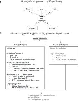
In a recent study in the mouse, we tested the hypothesis that moderate maternal protein deprivation would alter gene
expres-sion patterns in the placenta (Gheorghe et al., 2009). We
com-pared gene expression levels between normal placentas at E17.5, and those from pregnancies in which the mothers were exposed to seven days of protein deprivation (i.e., 10% protein by weight versus the 20% of normal chow) from E10.5 to E17.5. Of particular note, a number of genes involved in the p53 oncogene pathway were up-regulated. In addition to p53 itself, its positive regulators Zmis, Jmy, and Hipk2, as well as genes activated by p53 (Inpp5d, Cebpa), were induced (Fig. 3A). These p53 pathway proteins are important regulators of cell growth and proliferation. This pathway serves as a G1 checkpoint, and arrests growth and/or induces apoptosis in response to cellular damage. Mutations in the p53 gene have been implicated in a number of cancers and other
pathological processes (Ryan et al., 2001). Hipk2, an upstream
regulator of p53, activates its transcriptional activity and
pro-apoptotic activities through phosphorylation at Ser 46 (Hoffman et
al., 2002). Cebpa, is a transcription factor induced by p53, and
mediates some of the downstream effects of p53 activation (Yoon
& Smart, 2004). Among the gene ontology classes most over-represented in the up-regulated group, we noted the mitogen-activated protein kinase pathway, regulators of apoptosis (Bcl2-like 2, p53, endophilin, Fas-activated serine/threonine kinase), negative regulators of cell growth (farnesyltransferase CAAX box beta, cadherin 5, CCAAT/enhancer binding protein (C/EBP) al-pha, inositol polyphosphate-5-phosphatase D, p53), and nega-tive regulators of cellular metabolism (nuclear receptor co-repres-sor 2, histone deacetylase 7A, SPEN homolog, transcriptional regulator). Acting in concert, activation of these genes could result in growth restriction during pregnancy (Fig. 3B). Among down-regulated genes, particularly striking were those related to nucleotide metabolism. For selected genes, we confirmed these
results using qRT-PCR (Gheorghe et al., 2009).
Another potentially important finding, is that protein deprivation altered the expression of several genes involved in DNA methy-lation, histone acetymethy-lation, and epigenetic regulation of gene expression. The expression levels of histone deacetylase 7A and methionine adenosyltransferase II, alpha were elevated several fold. Histone acetylation triggers changes in chromatin structure, and regulates transcriptional availability of genes. In turn, histone deacetylation increases histone affinity for DNA, thereby repress-ing transcription (Bulger, 2005). Methionine adenosyltransferase II alpha synthesizes AdoMet the direct precursor used for DNA
Fig. 3. Maternal stress-mediated placental gene regulation. (A) p53 related genes up-regulated by protein restriction in the mouse placenta. + stands for induced. (B) Proposed mechanisms of protein restriction induced long-term changes in gene expression.
Up-regulated genes of p53 pathway
Regulators of apoptosis
• Bcl2-like 2
• Endophilin
• Fas-ac vated serine/thrionine kinase Regulators of apoptosis
• Bcl2-like 2
• Endophilin
• Fas-ac vated serine/thrionine kinase
Epigene c regulators
• Histone deacetylase 7A
• Methionine adenosyl-transferase II alpha Epigene c regulators
• Histone deacetylase 7A
• Methionine adenosyl-transferase II alpha Nega ve regulators of cell metabolism
• Nuclear receptor co-reperessor 2
• Histone deacetylase 7A
• SPEN homolog transcrip onal regulators Nega ve regulators of cell metabolism
• Nuclear receptor co-reperessor 2
• Histone deacetylase 7A
• SPEN homolog transcrip onal regulators Nega ve regulators of cell growth
• Farnesyltransferase CAAX box beta
• Cadherin 5
• CCAAT/enhancer binding protein alpha (C/EBPa)
• Inositol polyphosphate-5-phosphatase D Nega ve regulators of cell growth
• Farnesyltransferase CAAX box beta
• Cadherin 5
• CCAAT/enhancer binding protein alpha (C/EBPa)
• Inositol polyphosphate-5-phosphatase D p53 pathway
Placental genes regulated by protein depriva on
Nucleo de metabolism
Epigene c regulators
• Histone 2 (h3c2)
• Mcm6
• telomeric repeat binding factor 1
Fetal programming of adulthood disease Increased life span
decreased incidence of cancers
methylation by methyltransferases (Mao et al., 1998). Histone 2
(h3c2) is down-regulated, along with Mcm6 and telomeric repeat binding factor 1. These proteins contribute to DNA replication,
stability, and structure (O’Connor et al., 2004; Yu et al., 2004). In
the placenta of patients with preeclampsia, phosphorylation of extracellular signal-regulated kinase1/2 was significantly less frequent in the invasive trophoblasts, as compared to control
(Moon et al., 2008). In another study, in placentas of preeclamptic
patients, contrary to expectations, polymorphisms of several enzymes associated with oxidative stress (copper/zinc superox-ide dismutase, manganese superoxsuperox-ide dismutase,
glutathione-S-transferase, and others) did not differ from controls (Zhang et
al., 2008). In contrast, in the placentas of patients with HELLP
(Hemolysis, Elevated Liver Enzymes, Low Platelets) syndrome, genes encoding vascular endothelial growth factor receptor, leptin, and several other proteins, were up-regulated, as com-pared with the placenta of both normal control patients and those
with preeclampsia (Buimer et al., 2008).
Because the various tissues and organ systems undergo critical, often brief, periods of growth and development during fetal life (Winick & Noble, 1966), “programming” as a conse-quence of maternal stress should not be unexpected, with insults to the developing organism having consequences later in life’s course. Studies in ruminants also have demonstrated that under-nutrition can have profound consequences for the fetus. In sheep, restricted maternal nutrition in early to mid-gestation was associ-ated with an increase in placental weight, an increase in crown-rump length, and lower fetal to placental weight ratios (Heasman
et al., 1998). Maternal under-nutrition also altered cardiovascular
homeostatic regulation by the renin-angiotensin system, and
exposed the lambs to higher levels of glucocorticoids (Edwards et
al., 1999), and development of hypertension (Dodic et al., 2001).
Protein restriction in bovines also resulted in an increase in
placental weight and altered placental morphology (Perry et al.,
1999).
Studies in rodents have shown similar effects. In rats, maternal protein restriction triggers hypertension in the pups in adulthood (Langley & Jackson, 1994), probably by augmentation of the pups’ renin-angiotensin system. In the spontaneously hyperten-sive rat placenta, several proteins, angiotensin receptor type I and inducible nitric oxide synthase (NOS), were up-regulated, while angiotensin converting enzyme and peroxisome
proliferator-acti-vated receptors alpha and gamma were downregulated (Raso et
al., 2008). An alteration of placental glucocorticoid (GC)
metabo-lism also was observed in placentae of rats fed a protein restricted
diet, namely the activity of 11β-hydoxysteroid dehydrogenase
that metabolizes glucocorticoids. This placental enzyme, which normally protects the pups from maternal glucocorticoid excess,
was reduced in protein restricted rats (Langley-Evans et al.,
1996), thus exposing the fetus to abnormally high GC concentra-tions. Elevated circulating cortisol concentrations, with modified responsiveness of the hypothalamic-pituitary-adrenal axis, and elevated mean arterial blood pressure with increased left ven-tricular wall thickness and mass, also were observed in guinea pigs in which the dam received only 70% of normal chow during either the first or second half of gestation. Some of these changes
persisted in the F2 generation (Bertram et al., 2008). Another
hormonal alteration in nutritionally deprived rat pups, was an increase in somatostatin expression in the periventricular nucleus.
This led to much lower levels of growth hormone, and had deleterious effects on the growth of the pups post-partum (Huizinga
et al., 2000). Fetal undernourishment also led to neuronal
se-quelae. The facial motor nucleus in pups was under-developed, resulting in decrease in the ability of pups to suckle and chew
(Perez-Torrero et al., 2001). These observations also may relate
to the epidemiologic findings, noted above, that abnormal antena-tal nutrition may be associated with the development of schizo-phrenia and other mental illness.
In several animal models, in addition to the potential deleteri-ous effects referenced above, a positive aspect of nutritional deprivation in the adult is that of prolonged lifespan and reduced cancer rates. A proposed mechanism for these benefits is that nutritional restriction without severe malnutrition inhibits cellular proliferation and induces apoptosis. This effect has been shown in mice lacking p53, in which –/– and +/– mutants have lowered spontaneous cancer rates when fed a calorically reduced, but
otherwise complete, diet (Hursting et al., 2004). In the adult and
aging animal, nutritional restriction has been shown to have beneficial effects that increased life span (Nikolich-Zugich & Messaoudi, 2005). A different picture has emerged in the fetus, however. As discussed above, caloric and protein deprivation have been shown to trigger fetal programming of adult disease, and lead to an increased prevalence of metabolic disorders in adulthood (Barker, 1995b; 1998a; 1998b; Barker & Clark, 1997). In the developing fetus, numerous animal studies have shown negative long-term effects of caloric and protein deprivation on the cardiovascular, renal and nervous systems and metabolism (for review see McMillan & Robinson, 2005). A different form of nutritional compromise, that of placental restriction in sheep by removal of the endometrial caruncles in the nonpregnant ewe prior to mating, alters the expression of a number of genes associated with adipogenesis in adipose tissue of the fetus
(Duffield et al., 2008). Other protein deprivation models have
been studied (Waterland et al., 2006b; Watkins et al., 2008).
These findings emphasize the interrelation of placental develop-ment and its gene expression, to developdevelop-ment of the fetus and its repertoire of gene expression.
To what extent is placental gene expression altered by
maternal caloric excess?
Because maternal obesity poses an increased risk to the fetus during pregnancy, and has long-term consequences for the progeny, we tested the hypothesis that maternal caloric excess effects growth-related gene expression changes in the placenta. We fed female C57BL/65 mice a hypercaloric diet (20% fat, 38% sugar) or standard chow for six weeks prior to mating and throughout pregnancy. Near-term (E18), the dams were euthanized. We measured gene expression changes in the pla-centa, and performed pathway analysis on regulated genes.
Maternal overfeeding was associated with a two-fold increase in body fat mass, with several genes related to obesity, diabetes, DNA methylation, and the transforming growth factor-beta
(TGF-β) pathway being differentially expressed. The TGF-β superfamily
comprises ~30 growth and differential factors, including several
TGF-βs, activins, inhibins, and other growth and cell cycle control
factors (Goumans & Mummery, 2000; Kitisin et al., 2007; Massague
Thus, our findings may have important implications for placental growth and epigenetic regulation. In other studies in mice, the
chow was supplemented with methyl supplements (Weaver et al.,
2005; Wolff et al., 1998) or folic acid (Wehby & Murray, 2008),
vitamin B-12, choline, and betaine to enhance metabolism of cellular methyl donors (S-adenosylmethionine) (Waterland & Jirtle, 2003). These interventions resulted in altered coat color phenotype with concomitant increase in DNA methylation at the
Avylocus. Conversely, in mice fed a methyl-donor-deficient diet
that lacked folic acid, vitamin B-12, and choline the imprinted Igf2 gene was down-regulated with altered DNA methylation (Waterland
et al., 2006a). Human studies also have demonstrated effects in
the placenta on maternal dietary supplementation (Rush et al.,
1984).
What is the role of epigenetics in placental gene
ex-pression?
During the course of life and reproduction, cells store informa-tion that has been handed down from their ancestors, and that will be transmitted to their descendents. For the most part, this “memory” is encoded in the sequence of nucleic acids that comprise the DNA of the genome, the genotype or entire compli-ment of genes that provides the stability and accurate heritability from generation to generation. Much traditional research has explored the combined effects of genetics and the environment in germline mutations of the coding and promoter regions of genes. In addition, cells can inherit and transmit information that is not
part of the genomic sequence. This epigenetic [from Greek,
above, upon, over, or beyond conventional genetic], cellular memory involves the heritable transmission of gene expression patterns that persist through cell division, but do not involve an alteration in DNA sequence. Epigenetic processes act in a cell specific, temporally-regulated manner to direct development, differentiation, organogensis, and related processes. Some have compared epigenetic mechanisms to the software to orchestrate and/or modulate the DNA hardware. One major class of
epige-netic mechanisms termed “cytoplasmic”, is determined by
cis-acting factors associated with DNA methylation and/or histone modification by acetylation/methylation/phosphorylation. DNA with accompanying histones are packaged in nucleosomes, the core of which contains an octamere of histone proteins. Four basic forms of histones (H2A, H2B, H3, and H4, as well as minor
variants), are encircled by 146 base pairs of DNA (Finch et al.,
1977); a fifth histone, H1, serves as a linker protein (Bernstein et
al., 2007). The histone modifications noted above, and DNA
methylation, confer a great increase in the regulatory capacity of each nucleosome, allowing specific functions such as DNA repair and gene activation to be modulated in the appropriate manner (Sarma & Reinberg, 2005). Enzymes critically associated with these nucleosomal modifications include: DNA methyltransferases (DMT), histone acetyltransferases (HAT), histone methyltransferase (HMT), histone deacetylases (HDAC), histone
demethylases (HDM), and others (Dodd et al., 2007; Klose et al.,
2006). It is by these nucleosomal modifications, with their influ-ence on proximate genes, that genes may be regulated to affect phenotype by activity, chromatin structure, dosage compensa-tion, and epigenetic memory, without changes in the nucleic acid
code per se (Martin & Zhang, 2005; Wolffe & Matzke, 1999).
Epigenetic changes play a key role in normal cellular function, as well as the development and differentiation of various cell types (Drake & Walker, 2004; Jablonka & Lamb, 2002; Monk, 1998;
Murrell et al., 2005; Rahnama et al., 2006; Reik, 2007). Examples
include X-chromosome inactivation in female mammals, and genomic imprinting in which one parental allele is altered resulting in parent-of-origin, or random modification of gene transcription
(Willard et al., 1993). The epigenetic state can be disrupted by
maternal environmental influences such as hypoxia, protein dep-rivation, caloric excess, and so forth which alter DNA methylation or modify histones. Also importantly, a wide variety of environ-mental toxins, including low dose radiation and psychological stress, have been demonstrated to be important in epigenetic
mechanisms (Dolinoy et al., 2007; Feinberg, 2007; Hertz-Piccioto
et al., 2008; Jirtle & Skinner, 2007; Pryce et al., 2002; Szyf et al.,
2007). Increasingly, epigenetic changes are being recognized to be of importance in ageing, and the development of cancer and other diseases. Despite the general understanding that DNA and/ or histone modifications constitute a major factor in the pathogen-esis of epigenpathogen-esis, little is known of the molecular mechanisms whereby these chemical reactions/changes are regulated, and/or how they are transmitted between generations (Bird, 2007).
Some historical perspectives
From an historical context, epigenetics has several facets. For the pioneer Edinburgh geneticist Conrad Hal Waddington (1905-1975), who coined the term, epigenetics was the study of how phenotypes arise from genotypes during development (Waddington, 1939; 1940; 1942; 1957). Epigenetics later was defined as heritable changes in gene expression not due to any alteration in DNA sequence (Holliday, 1987). In the mid-1970s, the concept of covalent chemical DNA modifications, including methylation was proposed to account for this phenomenon (Holliday & Pugh, 1975; Riggs, 1975). More recently, others have defined epigenesis as the study of mitotically and/or meiotically heritable changes in gene function without a change in DNA
sequence (Dolinoy et al., 2007; Russo et al., 1996). As defined by
Adrian Bird, epigenetics is “the structural adaptation of chromo-somal regions so as to register, signal or perpetuate altered activity states” (Bird, 2007). The latter definition focuses on chromosomes and genes, including those aspects such as DNA repair, cell-cycle phases, and those stable changes maintained from generation to generation.
Such behavioral alteration leads to greater or lesser use of a given structure with resultant increase or decrease in the size of that structure or organ. His “Second Law” stated that all such changes were heritable as a result (For instance, that a giraffe’s neck elongated as it ate from the highest leaves on a tree, and this feature would be seen in the next generation) (Haig, 2007; Jablonka & Lamb, 1995; West-Eberhard, 2007). Neo-Lamarckian biologists soundly reject such an idea. In more contemporary times, the views of the Russian biologist-agronomist, Trofim Denisovich Lysenko (1898-1976), gained considerable press for the agricultural “revolution” he promoted, in concert with Soviet collectivization policies. In essence, Lysenko held that acquired characteristics of a plant (or other organism) could be inherited by succeeding generations. An ideological-political creation, Lysen-koism held the study of classic genetics to be “bourgeois” or “fascist” pseudoscience. Lysenkoism invoked by biological deter-minists, as with the eugenics and scientific racism adopted by social constructivists, may be seen as the extremes to which political dogma can use science in promoting its propaganda (Roll-Hansen, 2008; Soyfer, 2001).
As noted, environmental influences may have profound effects on gene regulation. This is clearly evident in the cells of multicel-lular organisms; although being genetically homogeneous, they are structurally and functionally heterogenesis. Many of these differences in gene expression arise during development and are retained through mitosis. Such stable, epigenetic changes, al-though heritable in the short term, are not a consequence of DNA mutation. Rather, as recent studies are demonstrating, to a great degree epigenesis appears to be a consequence of DNA
methy-lation and histone modification (Cooney et al., 2002; Nanney,
1958). The term “epigenomics” has been applied to the study of altered chromatin structure, such as complex folding, altered
nucleosome configuration, and related phenomena (Murrell et al.,
2005), while “nutri (epi)genomics” applies to those sequelae following nutritional alteration (Tost, 2008a). Importantly, several lines of evidence indicate that, in addition to maternal to fetal transfer, epigenetic modifications may be inherited across
gen-erations (Anway et al., 2005; 2006; Crews et al., 2007; Lane et al.,
2003; Morgan et al., 1999; Pembray et al., 2006; Rakyan et al.,
2003).
What are the roles of DNA methylation and histone
modification in placental gene expression?
As noted above, both DNA methylation and histone modifica-tion play important roles in development. These changes also may be important aspects of the ageing process and the develop-ment of cancer. In this review, we concentrate on the epigenetic influences of maternal diet, hypoxia, and related stress as ob-served in the placenta and fetal tissues, and that may have long-term consequences in the fetal origins of adult health and disease. As with most phenomena of biology and life, the molecular mechanisms whereby genes are repressed or active in a stable manner are exceedingly complex, and new insights into this hitherto poorly understood subject appear daily. The best studied of these epigenetic modifications is that of DNA methylation, which first was suggested in 1975 by two groups (Holliday & Pugh, 1975; Riggs, 1975). This post-replication, covalent methylation occurs predominantly in repetitive genomic regions, on the 5
2-carbon of cysteine residues that are followed by a guanine residue, i.e., “CpG methylation” which induces gene repression or “a silent chromatin state”, (The “p” in CpG refers to the phosphodiester bond between cytosine and guanine). Occurring at or around promoter regions, forming CpG islands, this can occur directly by inhibiting the binding of specific transcription factors, and indirectly by recruiting methyl-CPG binding proteins, with their associated repressive chromatin-remodeling activities (Razin & Riggs, 1980). A seeming paradox in this scenario, is that methylation of some specific DNA sequences may permit expres-sion of neighboring genes. Both intrinsic factors and environmen-tal/nutritional factors can determine the activity of methyltransferases upon which DNA methylation are dependent (Bestor, 2000). Because of the requirement for a high DNA synthesis rate during both gametogenesis and early embryogen-esis, considerable activity in DNA methylation/demethylation patterning occurs during both this period of development, a time during which the cells are vulnerable to abnormal environmental factors. Nonetheless, nuances of molecular regulation of DNA methylation and histone modification during embryogenesis are beyond the scope of this synopsis, and a number of reviews on this topic are available (Bird, 2007; Jaenisch, 1997; Jaenisch &
Bird, 2003; Jones & Takai, 2001; Paulsen et al., 2008; Santos et
al., 2002; Tost, 2008a; 2008b).
During the course of mammalian development, a wave of DNA demethylation occurs during cleavage, followed by genome-wide
de novo methylation following implantation (Jaenisch, 1997).
Although the male genome is widely demethylated shortly after
fertilization (Mayer et al., 2000; Oswald et al., 2000), the maternal
genome is only partly demethylated with subsequent cleavage divisions (Li, 2002). In the gastrulating embryo, the extent of methylation is high, decreasing in various tissues during the
course of differentiation (Ehrlich et al., 1982). For the developing
embryo and fetus, the methylation/demethylation patterns while being of great significance, are enormously complex.
Additionally, gene expression is determined by the biochemi-cal organization of the histones in the nucleosomes around which the DNA is wrapped. Several post-translational covalent modifi-cations occur on the amino acids that constitute the histone N-terminal tails that modify their interaction with DNA and/or other nuclear proteins. Acetylation, methylation, phosphorylation and/ or ubiquitination alone, or in combination play a key role in the regulation by repression or expression of contiguous genes (Jenuwein & Allis, 2001; Strahl & Allis, 2000; Turner, 2000). Again, the regulation of histone modification by acetylation and/ or methylation is highly complex, has been shown to be specific for essentially every cell type, and may act with DNA methylation
to constitute a system of cellular memory (Bird, 2007; Sims et al.,
2008). The combination of the several epigenetic modifications of genes as well as non-coding sequences, the so-called “epigenome” or “epigenotype”, determine the extent to which a given gene is maintained repressed or active, and influences the phenotype at birth. A recent study has described the differences in gene expression, DNA methylation, and histone H3K9 acetylation between acute myeloid leukemia and acute lymphocytic leuke-mia. By integrating genetic and epigenetic information, the diver-gent nature of the two leukemias was described in more detail, and additional insights were revealed with regards to the gene
demon-strates that in combining both genetic and epigenetic information, more accurate and insightful information can be obtained with regards to gene expression regulation and ultimately biological phenotype.
What is the role of microRNAs in placental gene
expres-sion?
MicroRNAs (miRNA) have emerged as important players in DNA methylation and post-transcriptional gene regulation
(Lujambio et al., 2007; Saito et al., 2006). These are subtypes of
small, non-coding RNA, which are 21-25 nucleotides in length. These miRNAs are capable of base pairing with mRNA, and fine-tuning gene expression during development and differentiation, by suppressing their expression in sequence specific manner. Following the discovery of first miRNA “lin4” in 1993, as a small
temporal RNA (Lee et al., 1993), there has been enormous growth
in this family, and identification of their targets. Although miRNAs are similar to small interfering RNA (siRNA) in their generation pathway and molecular characteristics, unlike siRNA, miRNA does not degrade the target mRNA. Rather, they target the 3’ untranslated regions of mRNAs with which they share partial sequence complementarily, thereby silencing post-transcriptional gene translation. In this way, the biological system increases or decreases miRNA production to up- or down-regulate gene ex-pression according to the developmental need, producing desired morphologic and physiological changes. Moreover, placental miRNA (miR-141, miR-149, miR-229-5p, and miR135b) are se-creted in maternal plasma, and their concentration decreases
significantly after parturition (Chim et al., 2008). This suggests
that placental miRNA, in addition to regulating gene expression in placenta, may be playing an important role in maternal conditions with obscure etiology, such as preeclampsia or related hyperten-sive disorders. Studies reveal differential expression of miRNA (miR-210 and miR-182) in placenta from patients with
preeclamp-sia and with small for gestational age newborn infants (Pineles et
al., 2007). As must be evident, additional studies will be vital to
examine and understand the complexity of placental genetic regulation, and their contribution to fetal and maternal health and disease.
What are the human correlates?
In several human population studies, it has been reported that the nutritional state of individuals may have phenotypic
conse-quences for their grandchildren (Kaati et al., 2002; Lumley, 1992).
An example of the role of diet in progeny DNA methylation status and phenotype is evident in patients with hyper-homocysteinemia
(Ingrosso et al., 2003). This disorder is characterized by excess
cellular adenosylhomocysteine, a potent inhibitor of S-adenosylmethionine-dependent methyltransferases. This sug-gests the possibility of significantly altered DNA methylation. In these patients, dietary supplementation with folate restored glo-bal methylation levels, as well as that of the imprinted IGF2-H19
locus (Ingrosso et al., 2003). Several earlier studies have
indi-cated the developmental importance of folic acid as a dietary
factory in utero, and the manner in which it modulates disease
risks later in life (Torrens et al., 2006). It remains to be determined
whether, as in the case of hyper-homocysteinaemia, these
phe-notypic effects occur through altered DNA methylation (McKay et
al., 2004).
An optimal uterine environment has been shown to be essen-tial for establishment and maintenance of embryonic epigenetic patterns (Vickaryous & Whitelaw, 2005). Because embryo culture and manipulation are employed in contemporary assisted repro-ductive technologies (ART), the question arises as to the extent to which ART or related procedures alter DNA methylation pat-terns, thereby inducing epigenetic changes in the developing
organism (Brar et al., 2001; Feil, 2006; Khosla et al., 2001a;
2001b; Vickaryous & Whitelaw, 2005). Normally DNA methylation is confined to only one of the two parent alleles, thus imprinted gene loci allow minor alterations to be detected. An issue of great importance is the extent to which the chemical composition of culture medium, the duration of culture, or other factors, play a role in effecting changes in DNA methylation or histone
modifica-tion (Doherty et al., 2000; Khosla et al., 2001a; 2001b; Mann et al.,
2004; Young et al., 2001).
An additional consideration of importance, is the role of envi-ronmental toxins in producing alterations in the nucleosome with epigenetic consequences. An obvious example from mid-twenti-eth century is the ingestion of the estrogen-receptor agonist diethylstilbestrol (DES) by women in an attempt to reduce the risk of spontaneous abortion. This was followed by vaginal clear cell carcinoma (Swan, 2000), and altered limb development in the first
generation, and deafness in the second generation (Stoll et al.,
2003). Anticancer drugs and other environmental compounds may alter expression of specific genes, as well as the stress-related chaperone protein heat shock protein (HSP)-90, which may play a role in histone modification (Feil, 2006; Rutherford & Lindquist, 1998). A host of environmental contaminants including endocrine-disrupting chemicals are now known to demonstrate epigenetic effects on the germ line, and promote disease across
several generations (Crews et al., 2007; Parodi et al., 2006).
In humans, a number of factors, genetic and epigenetic, can influence placental/fetal growth, development and long-term se-quelae. Several hypotheses have been proposed to account for these phenomena. The “thrifty genotype” hypothesis proposes the existence of genes that influence birthweight, and determines whether an infant will experience intrauterine growth restriction
(Ong & Dunger, 2000; Prentice et al., 2005; Stöger, 2008). The
“thrifty phenotype” hypothesis postulates that impairment of nutri-tional supply in early life results in permanent changes in tissue/ organ function to conserve glucose, and prioritize development of the brain, heart, and other vital organs (Hales & Barker, 2001). A third hypothesis proposes that epigenetic alterations in gene expression, in the absence of altered DNA sequence, can be heritable, and may be reversible (Holness & Sugden, 2006). A challenge for our future is to develop strategies to negate the long-term consequences of these molecular alterations. A related issue of consequence is the epigenetic basis of dysregulation of gene expression as demonstrated in metabolic syndrome with
insulin resistance (Lane et al., 1996), neural development (Canli
et al., 2006; Collins & Barker, 2007; Ke et al., 2006), cancer
(Esteller, 2007; 2008), and other conditions (Pembrey, 2000). Rather than isolated instances, this may be a major factor in the seemingly increasing and intractable pandemic of these classes
of diseases (for instance see Gal-Yam et al., 2008; Palii &
issues, a recent National Institutes of Health initiative, as part of its “Roadmap” program, seeks applications to study the “Epigenomics of Human Health and Disease” (RFA-RM-08-017) (Jones & Martienssen, 2005).
What are the overall perspectives and critical
ques-tions to be explored?
For the long-term well-being of an individual, both optimal placental and fetal growth are essential. Thus, one would antici-pate that profound inhibition of cellular growth at key time points during development would have grave long-term consequences for the embryo/fetus. This suggests that the timing of the treat-ment is a key determinant in the effect on the organism. Since their development, cDNA and oligo microarrays have proven to be powerful tools in the elucidation of gene expression patterns and discovery. In addition to examining cellular processes at the global gene expression level, these instruments have allowed analysis of numerous facets of normal growth and differentiation, as well as that occurring as a consequence of stress or malignant transformation. Of particular value, such studies allow analysis of gene expression by functional classes, as an aid in understanding pathways of cell metabolism, proliferation, senescence, and death.
As with most tissues and organ systems, placental develop-ment and its response to stress remains a poorly understood process. Placental malfunction or failure accounts for numerous
instances of fetal mortality (Cross et al., 2003), and may play an
important role in the genesis of intrauterine growth restriction
(Kingdom et al., 2000), as well as some maternal disease
(Newstead et al., 2007). Numerous genes have been shown to be
essential for placental function as an organ of respiratory gas and nutrient exchange, hormonal synthesis, immune function, and so forth. As is evident, maternal stresses, whether hypoxia, protein deprivation, caloric excess, or other, can result in profound alterations in placental gene expression patterns, and their
con-sequences for growth, differentiation, and metabolism. Figure 4 presents in summary fashion established and potential pathways by which stress to the mother, whether hypoxia, protein depriva-tion, caloric excess, or others, can trigger changes in DNA methylation patterns and/or histone modification to effect alter-ations in the patterns of gene expression in the placenta and/or fetal organs.
Far from presenting a complete picture, the present review depicts but a fraction of what we need to know to understand more completely the molecular regulation of placental growth and development, and to lessen the ravages of placental dysfunction. A major challenge for the future will be to identify those portions of the genome particularly vulnerable to epigenetic modification which underlie states of health and disease, and to understand the molecular mechanisms by which these changes occur. A few of the most obvious questions follow. How are the demonstrated gene expression profiles regulated? In terms of stress, what determines the individual patterns of expression, as opposed to the up- or down-regulation of those genes common to all stres-sors? What are the developmental stages/times of vulnerability to environmental, nutritional, or other stress? What environmental factors alter the epigenome in a deleterious manner, and what are their dose-response relations? What are the mechanisms by which DNA methylation and/or histone acetylation/methylation are regulated? To what extent do patterns of gene expression alterations in the placenta influence gene expression in the several fetal tissues/organs? To what extent can we use the findings of gene expression responses to stress, to gain an understanding of the phenomenon of epigenesis and its various manifestations? What is the role of epigenesis in normal develop-ment, and in the etiology of disease? How is it that epigenetic changes evident at the molecular level during embryonic/fetal life, do not become manifest in the adult organism for many years or decades? What is the relative importance of epigenetic, as opposed to genetic, changes for long-term sequelae? To what extent can we develop systems using molecular signatures/ adducts to detect invidious interactions in early life? To what extent can an understanding of these issues provide us effective means to contain or counteract their influence and consequences? Can epigenetic biomarkers be identified that will allow disease detection at an early stage?
These are but a few of the vital questions that must be addressed in our pursuit to improve the lives and well being of mothers and infants, and the latter’s life as an adult. As biomedical scientists dedicated to betterment of the human condition, can we do less?
Acknowledgement
We thank Brenda Kreutzer and Jimin Suh for their assistance in the preparation of this manuscript. This work was supported, in part, by USPHS grant HD-03807 to LDL.
References
ABU-FARHA M, NILES J, WILLMORE WG (2005). Erythroid-specific 5-aminolevulinate synthase protein is stabilized by low oxygen and proteasomal inhibition. Biochem Cell Biol 83: 620-630.
ADAMS RH, PORRAS A, ALONSO G, JONES M, VINTERSTEN K, PANELLI S, VALLADARES A, PEREZ L, KLEIN R, NEBREDA AR (2000). Essential role of p38alpha MAP kinase in placental but not embryonic cardiovascular develop-Fig. 4. Proposed model of stress-induced long-term epigenetic


![Chloro{2 [tris(hydroxymethyl)methyliminomethyl]phenolato}copper(II)](data:image/gif;base64,R0lGODlhAQABAIAAAP///wAAACH5BAEAAAAALAAAAAABAAEAAAICRAEAOw==)