_____________________________________________________________________________________________________
www.sciencedomain.org
The Effect of Different Irrigation Regiments on the
Push out Bond Strength of MTA Fillapex Sealer
to Dentin
Arushi Agrawal
1*,
Bonny Paul
1, Shiv Mantri
1and Kavita Dube
11Department of Conservative Dentistry and Endodontics, Hitkarni Dental College and Hospital,
Jabalpur 482002 (M.P), India.
Authors’ contributions
This work was carried out in collaboration between all authors. Author AA designed the study, wrote the protocol, and wrote the first draft of the manuscript. Author BP managed the literature searches and analysis of study performed. Authors SM and KD managed the experimental process. All authors read and approved the final manuscript.
Article Information
DOI: 10.9734/BJMMR/2015/18011 Editor(s): (1) Ibrahim El-Sayed M. El-Hakim, Ain Shams University, Egypt and Riyadh College of Dentistry and Pharmacy, Riyadh,
Saudi Arabia. Reviewers: (1) Anonymous, Adnan Menderes University, Turkey. (2)Maen Mahfouz, Orthodontics & Pediatric Dentistry Department, Arab American University Jenin, Palestine. Complete Peer review History:http://www.sciencedomain.org/review-history.php?iid=1232&id=12&aid=9720
Received 31st March 2015 Accepted 11th May 2015 Published 11th June 2015
ABSTRACT
Aims: The purpose of this study was to compare the effect of various irrigation regiments on the push out bond strength of MTA Fillapex sealer to dentin.
Study Design: Cross-sectional study.
Place and Duration of Study: Department of Conservative dentistry and Endodontics, Hitkarni Dental College and Hospital (HDCH), between November 2014 and December 2014.
Methodology: Thirty single-rooted premolar teeth were taken. Roots were divided into 3 groups according to the irrigation regiment used and instrumented using 5% sodium hypochlorite (NaOCl) or 2% chlorhexidine (CHX) irrigants as chemical auxiliary substances and 17% Ethylenediaminetetraacetic acid (EDTA) or 50% citric acid for smear layer removal. Finally, CHX solution was used as the final irrigant in one group. Root canals were filled with gutta-percha and MTA fillapex (Angelus, Londrina, PR, Brazil),a resin-based sealer. Bond strength was measured by the push-out test. Data were statistically analyzed by Kruskal-Wallis and Mann-Whitney U tests.
Results: In the present study, specimens in G1 (NaOCl + EDTA + Distilled Water) & G2 (CHX + EDTA+ Distilled Water) group show lower push out bond strength values than G3 (NaOCl+ EDTA + Citric Acid +CHX) group.
Conclusion: Thus, within the limitations of this study, we conclude that the different irrigation regiments did affect the push out bond strength of MTA Fillapex sealer to dentin. However the significant changes were not found between the various groups.
Keywords: Chlorhexidine; push-out bond strength; sealer; sodium hypochlorite.
1. INTRODUCTION
The adhesive properties of endodontic sealers are important as gutta-percha does not bond to root dentin and is used in conjunction with a root canal sealer. The risk of filling detachment from dentin during restorative procedures or the masticatory function is minimized if the bond strength of endodontic sealers to dentin is good, thus making it an important property of filling materials, [1] ensuring that sealing is maintained and, consequently, clinical success of endodontic treatment. The push-out bond strength test is a well-known evaluation method used in various other studies [1–4]. Thus, its results can be useful for evaluating the interfacial strength and dislocation resistance of root filling materials to the root dentin.
For successful root canal debridement during cleaning and shaping procedures the use of different chemical irrigants is essential [5]. Sodium hypochlorite (NaOCl) has a long history of successful usage in endodontics [6] because of its antimicrobial effect, ability to dissolve biofilms and solubilize tissues [7,8]. Chlorhexidine (CHX), another important irrigant, has also been used because of it antimicrobial effects, substantivity, lower degree of malodor. It is an alternative to NaOCl, because of its biocompatibility, especially in cases of open apex or allergy [9,10]. It has been suggested as a final irrigant [11-14]. The adjunctive use of chelating agent or acids (eg EDTA and citric acid) is recommended as NaOCl and CHX are incapable of removing the smear layer [11].
The most common root canal filling material used is gutta-percha in conjunction with sealers. Recently, calcium silicate–based materials such as mineral trioxide aggregate (MTA)-based sealers have been developed. MTA Fillapex (Angelus, Londrina, PR, Brazil), is a 2 paste resin sealer which consists of MTA, salicylate resin, natural resin, bismuth oxide and silica. According to the manufacturer it has excellent radiopacity, a good working time, easy handling and low
solubility, providing sealing of the canal by expansion during setting. Recent studies showed suitable radiopacity, pH, flow, working and setting time of MTA Fillapex [15,16].
The resin based sealer adhesion to the dentin may be affected by several factors. Chemical irrigants used during root canal preparation may alter the chemical composition of the dentin surface andaffect its interaction with the materials used for sealing [17]. Literature is scarce on the effect of various irrigants on the bond strength of MTA Fillapex sealer to dentin, hence this study was undertaken. Thus,the purpose of the present study was to evaluate the influence of different irrigation regiments on the bond strength of MTA Fillapex sealer to dentin.
2. MATERIALS AND METHODS
In this study 30 first mandibular premolar teeth stored in 10% buffered formalin were used. The criteria for selection of these teeth were orthodontic reasons. These teeth were obtained from the department of orthodontia. The crowns were removed with the use of a high speed carbide bur. A 10 K-file was used to verify the patency and to determine the total length of the root canals. The teeth were divided into 3 groups according to the irrigation regiment used as G1, G2, G3 (Table 1). The root canals were shaped by the means of K3 rotary system (SybronEndo,
Coppell, Texas). In G1, during the preparation before the insertion of each file 1ml of 5% sodium hypochlorite was used. After the use of the files, 5 ml of distilled water was used to remove the chemical auxillary substance. Similarly in G2, 2% chlorohexidine irrigant was used and 5 ml of distilled water was used to remove the chemical auxillary substance. In G3, before the insertion of each file 1 ml of 5% sodium hypochlorite irrigant was used and 5 ml of distilled water was used to remove the chemical auxillary substance.
root canal preparation and eliminate its taper in order to standardize the incidence of forces within the filling materials. After this, in G1 and G 2 3 ml 17% EDTA was used to remove the smear layer followed by 5 ml of distilled water to remove the chemical auxillary substance. In case G3 3ml 50% citric acid was used to prevent the interaction of sodium hypochlorite and chlorhexidine followed by 5ml of distilled water to remove the chemical auxillary substance. Finally in G3, 1 ml of 2% chlorohexidine irrigant was used as a final flush. The root canals were filled with guttapercha cones and MTA Fillapex sealer. An E and Q Plus obturation system (Meta Biomed Co. Ltd) was used to pack down and backfill the material (Fig. 1). All the teeth were radiographed to assess the quality of the filling. After this, the samples were kept under 100% relative humidity at 37º Celsius.
2.1 Push-out Assessment
Afterwards, each root was sectioned horizontally to produce four 1 mm-thick slices by using a diamond disk under continuous water irrigation. Two slices from the coronal portion and two slices from the apical portion (Fig. 2). The first (coronal) and the last (apical) was discarded. The root filling of each sample was loaded with a 0.75-mm-diameter stainless steel cylindrical plunger. The plunger tip was sized and positioned to touch only the root filling. The load
was always applied in an apical-coronal direction to avoid any constriction interference caused by root canal taper during push-out testing. Loading was performed on a universal testing machine (Star Testing System, India) at a crosshead speed of 3 mm/min until debonding occurred.
2.2 Formula for Calculation of Bond Strength
To express the bond strength in MPa, the load at failure recorded in Newton was divided by the area of the bonded interface
Push-out Bond Strength (MPa) = Maximum Load (N) /Adhesion Area (mm²)
Adhesion area (mm²) = 2 π r h
Where
r = radius of perforation of cross-section, h= height of the perforation.
π =3.14
Each cross section was coded and measured for the apical and coronal diameters of the obturated area by using an optical stereomicroscope. Data were statistically analyzed by Kruskal-Wallis and Mann-Whitney U tests (P<.05). The program used to analyse data was SPSS version 20. Post hoc power analysis was performed using G*Power version 3.1.9.2.
Table 1. Irrigation regiments used were as follows
Groups Chemical auxillary substance Intermediate flush Final flush Group 1 (G1) 1 ml of 5% NaOCl + 5 ml distilled
water
3 ml of 17% EDTA 5 ml distilled water
Group 2 (G2) 1 ml of 2% CHX + 5 ml distilled water
3 ml of 17% EDTA 5 ml distilled water
Group 3 (G3) 1 ml of 5% NaOCl + 5 ml distilled water
1 ml of 50% citric acid+ 5 ml distilled water
5 ml of 2% CHX
a) b)
Fig. 2. Push out assessment
3. RESULTS AND DISCUSSION
No premature failures occurred. Tables 2 and 3 shows mean and standard deviation values of bond strength. The specimens in G1 (NaOCl + EDTA + Distilled Water) & G2 (CHX + EDTA+ Distilled Water) group showed lower push out bond strength values than G3 (NaOCl+ EDTA + Citric Acid +CHX) group. However, no statistically significant difference was found between the groups (P>.05). Post hoc power (1-β) for coronal experiment was 0.154 and for apical experiment was 0.184.
One of the most important factor for the success of endodontic proceduresis the bond strength of material to dentin [18,19]. When the post-dentin interface bond fails, the risk of failure of either the teeth or the restorative materials increases [20]. Consequently, important insights into material selection and outcome prediction can be provided by mechanical testing of bonded interfaces [21,22,2]. Currently, microtensile bond strength test methods are commonly used to measure the bond strength of numerous dental materials [23]. Unfortunately, the tensile bond strength test method is not appropriate for use with intracanal filling materials because of the high percentage of premature bond failures and the large variation in test results [24]. A push-out
test modified from the shear punch test has been advocated as a more suitable test for evaluating the bond strengths of intracanal filling materials [25].
MTA-based sealers are based on resin salicylate and calcium silicate base. Considering the chemical composition, it is expected that there should be some similarities in bond strength to dentin between resin sealers and MTA-based sealers [26]. Moreover, Sarkar et al. [27] showed that release of calcium and hydroxyl ions from the set sealer results in the formation of apatite which comes into contact with fluids containing phosphate. Reyes-Carmona et al. [19] also reported that the apatite formed by MTA and phosphate salts, is deposited among collagen fibrils, resulting in a controlled increase in the formation of inorganic nucleations on the dentin, which are seen as an interfacial layer with tag-like structures.
strength of the resin sealers to the dentin. Thus in the present study, the effect of different irrigation regiments was used to assess the bond strength of the sealer to the dentine [29].
According to the results of our present study (as seen in figure 3 and 4) the specimens in G1 (NaOCl + EDTA + Distilled Water) & G2 (CHX + EDTA+ Distilled Water) showed lower push out bond strength values (Fig. 2). This can be explained due to the powerful antimicrobial action of sodium hypochlorite shown to jeopardize the polymerization of bonding resins [30,31,28]. It is thought that NaOCl leads to oxidation of some component in the dentin matrix [32], forming protein-derived radicals [33] that would compete with the propagating vinyl free-radicals generated by the light-activation of resin adhesives, resulting in premature chain termination and incomplete polymerization [34]. Furthermore, reductions in calcium and phosphorus levels [35] and in mechanical properties of dentin, such as elastic modulus, flexural strength, and microhardness [36], were reported after irrigation of root canals with 5% sodium hypochlorite, which can also contribute to
a decrease in the micromechanical interaction between resins and NaOCl-treated dentin.
On the other hand, chlorhexidine a cationic bisguanide, was used as a final flush in G3 (Fig. 4). It shows optimal antimicrobial activity over the pH range from 5.5 to 7.0, and acts by adsorbing onto the cell walls of microorganisms and causing breakdown of intracellular components [37]. Erdemir et al. [26] reported that endodontic irrigation with CHX solution significantly increased bond strength to root dentin. This was found to be in agreement with our study. These authors suggested that adsorption of CHX by dentin may favor the resin infiltration into dentinal tubules, which explains the high bond strength values obtained in G3. Also, CHX increases the wettability of endodontic sealers on dentin, which can be explained by the presence of surface surfactant in CHX, increasing the surface energy and promoting higher wetting ability to dentin. Additionally, this substance has a broad-spectrum MMP-inhibitory effect that improves the resin-dentin bond stability [38].
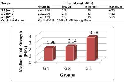
Table 2. Median values of push out bond strength in coronal section in different groups
Groups Bond strength (MPa)
Mean±SD Median Minimum Maximum
G 1 (n=10) 2.46±1.08 1.96 1.38 4.33
G 2 (n=10) 2.28±0.76 2.14 1.33 3.32
G 3 (n=10) 3.48±1.29 3.58 1.93 5.53
Kruskal-Wallis test KW=4.640, P= 0.098 (P>.05) Not significant
Table 3. Median values of push out bond strength in apical section in different groups
Groups Bond strength (MPa)
Mean±SD Median Minimum Maximum
G 1 (n=10) 2.07±1.14 1.61 1.00 4.16
G 2 (n=10) 2.13±1.11 1.82 1.13 4.16
G 3 (n=10) 3.42±1.23 3.73 1.31 4.61
Kruskal-Wallis test KW=5.305, P= 0.070(P>.05) Not Significant
Fig. 4. Graphic representation of median values of push out bond strength in apical section in different groups
Thus these results indicate that different irrigation regiments do affect the push out bond strength of MTA Fillapex sealer to dentin. However there were no significant changes found in the bond strength values between the various groups.
4. CONCLUSION
Adhesive strength is only one aspect of the quality of root canal sealing, but it may be considered one of the most important. Further investigation of other features of root canal sealers is required. In most cases, the results of laboratory experimental studies cannot bedirectly applied to clinical situations. However, they do provide reproducible and reliable means for comparing and testing new and prospective sealers, and for establishing international standards.
Thus, within the limitations of this study, we conclude –
1) The specimens in G1 (NaOCl + EDTA + Distilled Water) group showed lowest push out bond strength values.
2) The specimens in G3 (NaOCl+ EDTA + Citric Acid +CHX) group showed the highest push out bond strength values.
However, no statistically significant difference was found between the groups (P>.05).
CONSENT
It is not applicable.
ETHICAL APPROVAL
It is not applicable.
COMPETING INTERESTS
Authors have declared that no competing interests exist.
REFERENCES
canal dentine. Int Endod J. 2011;44:1088-91.
2. Assmann E, Scarparo RK, Böttcher DE, Grecca FS. Dentin bond strength of two mineral trioxide aggregate based and one epoxy resin-based sealers. J Endod. 2012; 38:219-21.
3. Nagas E, Cehreli ZC, Durmaz V, Vallittu PK, Lassila LV. Regional push-out bond strength and coronal microleakage of Resilon after different light-curing methods. J Endod. 2007;33:1464-68.
4. Ungor M, Onay EO, Orucoglu H. Push-out bond strengths: The Epiphany-Resilon endodontic obturation system compared with different pairings of Epiphany, Resilon, AH Plus and gutta-percha. Int Endod J. 2006;39:643-47.
5. Vilanova WV, et al. Effect of intracanal irrigants on the bond strength of epoxy resin-based and methacrylate resin-based sealers to root canal walls. IEJ. 2012;45: 42–48.
6. Jungbluth H, Marending M, De-Deus G, Sener B, Zehnder M. Stabilizing sodium hypochlorite at high pH: Effects on soft tissue and dentin. J Endod. 2011;37:693– 96.
7. Bryce G, O'Donnell D, Ready D, Ng YL, Pratten J, Gulabivala K. Contemporary root canal irrigants are able to disrupt and eradicate single- and dual-species biofilms. J Endod. 2009;35(9):1243-48. 8. NaenniN,Thoma K, Zehnder M. Soft tissue
dissolution capacity of currently used and potential endodontic irrigants. J Endod. 2004;30:785-87.
9. Ferraz CC, Gomes BP, Zaia AA, et al. Comparative study of the antimicrobial efficacy of chlorhexidine gel, chlorhexidine solution and sodium hypochlorite as endodontic irrigants. Braz Dent J. 2007; 18:294–98.
10. Gomes BP, Martinho FC, Vianna
ME. Comparison of 2.5% sodium
hypochlorite and 2% chlorhexidine gel on oral bacterial lipopolysaccharide reduction from primarily infected root canals. J Endod. 2009;35:1350–53.
11. Zehnder, M. Root canal irrigants. J Endod. 2006;32:389–98.
12. deAssis DF, Prado M, Simão RA. Evaluation of the interaction between endodontic sealers and dentin treated with different irrigant solutions. J Endod. 2011; 37:1550–52.
13. Hashem AA, Ghoneim AG, Lutfy RA, et al. The effect of different irrigating solutions on bond strength of two root canal-filling systems. J Endod. 2009;35:537–40. 14. Neelakantan P, Subbarao C, Subbarao
CV, et al. The impact of root dentine conditioning on sealing ability and push-out bond strength of an epoxy resin root canal sealer. Int Endod J. 2011;44:491–98. 15. Vitti RP, Prati C, Silva EJ, Sinhoreti MA,
Zanchi CH, de Souza e Silva MG, et al. Physical properties of MTA Fillapex sealer. J Endod. 2013;39:915-18.
16. Silva EJ, Rosa TP, Herrera DR, Jacinto RC, Gomes BP, Zaia AA. Evaluation of
cytotoxicity and physicochemical
properties of calcium silicate-based endodontic sealer MTAFillapex. J Endod. 2013;39:274-77.
17. Juliana Nascimento Santos. Effect of chemical irrigants on the bond strength of a self-etching adhesive to pulp chamber dentin. J Endod. 2006;32(11):1088-90. 18. Ferracane JL. Developing a more
complete understanding of stresses produced in dental composites during polymerization. Dent Mater. 2005;21:36– 42.
19. Reyes-Carmona JF, Felippe MS, Felippe WT. The biomineralization ability of mineral trioxide aggregate and Portland cement on dentin enhances the push-out strength. J Endod. 2010;36:286–91.
20. Santos AFV, Meira JBC, Tanaka CB, et al. Can fiber posts increase root stresses and reduce fracture? J Dent Res. 2010;89: 587–91.
21. Braga RR, Meira JBC, Boaro LCC, et al. Adhesion to tooth structure: a critical reviewof ‘‘macro’’ test methods. Dent Mater. 2010;26:38–49.
22. Mastoras K, Vasiliadis L, Koulaouzidou E, et al. Evaluation of push-out bond strength of two endodontic post systems. J Endod. 2012;38:510–14.
23. Armstrong S, Geraldeli S, Maia R, et al. Adhesion to tooth structure: A critical review of ‘‘micro’’ bond strength test methods. Dent Mater. 2010;26:e50–62. 24. Soares CJ, Santana FR, Castro CG, et al.
Finite element analysis and bond strength of a glass post to intraradicular dentin: comparison between microtensile and push out tests. Dent Mater. 2008;24:1405– 11.
canal walls: Comparison between microtensile and push-out bond strength measurements. Eur J Oral Sci. 2004;112: 353–61.
26. Forough Reyhani Mohammad, Ghasemi Negin, Rahimi Saeed, Salem Milani Amin, Mokhtari Hadi, Shakouie Sahar, Safarvand Hossein. Push-Out Bond Strength of Dorifill, Epiphany and MTA-Fillapex Sealers to Root Canal Dentin with and without Smear Layer. Iran Endod J. 2014; 9(4):246–250.
27. Sarkar NK, Caicedo R, Ritwik P,
Moiseyeva R, Kawashima I.
Physicochemical basis of the biologic properties of mineral trioxide aggregate. J Endod. 2005;31(2):97–100.
28. Erdemir A, Ari H, Güngüneş H, Belli S. Effect of medications for root canal treatment on bonding to root canal dentin. J Endod. 2004;30(2):113-16.
29. Maíra Prado, Renata A. Simão, Brenda PFA. Gomes. Effect of different irrigation protocols on resin sealer bond strength to dentin. J Endod. 2013;39(5):689–92. 30. Ozturk B, Özer F. Effect of NaOCl on bond
strengths of bonding agents to pulp chamber lateral walls. J Endod. 2004;30: 362-65.
31. Ishizuka T, Kataoka H, Yoshioka T, et al. Effect of NaOCl treatment on bonding to root canal dentin using a new evaluation method. Dent Mater J. 2001;20:24-33. 32. Morris MD, Lee K, Agee KA, Bouillaguet
S, Pashley DH. Effects of sodium
hypochlorite and RC-Prep on bond strengths of resin cement to endodontic surfaces. J Endod. 2001;27:753–57. 33. Hawkins CL, Davies MJ.
Hypochlorite-induced oxidation of proteins in plasma: Formation of chloramines and nitrogen-centered radicals and their role in protein fragmentation. Biochem J. 1999;340:539– 48.
34. Lai SCN, Mak YF, Cheung GSP, et al. Reversal of compromised bonding to oxidized etched dentin. J Dent Res. 2001; 80:1919–24.
35. Ari H, Erdemir A. Effect of endodontic irrigant solutions on mineral content of root canal dentin using ICP-AES technique. J Endod. 2005;31:187–99.
36. SimTPC, Knowles JC, Ng YL, Shelton J, Gulabivala K. Effect of sodium hypochlorite on mechanical properties of dentine and tooth surface strain. Int Endod J. 2001;34: 120–32.
37. Gomes BPFA, Ferraz CCR, Vianna ME, Berber VB, Teixeira FB, Souza-Filho FJ. In
vitro antimicrobial activity of several
concentrations of sodium hypochlorite and chlorhexidine gluconate in the elimination
of Enterococcus faecalis. Int Endod J.
2001; 34:424–28.
38. Assis DF, Prado M, Simão RA. Evaluation of the interaction between endodontic sealers and dentin treated with different irrigant solutions. Jendod. 2011;37:1550– 52.
© 2015 Agrawal et al.; This is an Open Access article distributed under the terms of the Creative Commons Attribution License
(http://creativecommons.org/licenses/by/4.0), which permits unrestricted use, distribution, and reproduction in any medium,
provided the original work is properly cited.
Peer-review history:
The peer review history for this paper can be accessed here: