_____________________________________________________________________________________________________
www.sciencedomain.org
Identification and Susceptibility Testing on Samples
of
Mycobacterium
spp
R. A. O. Calsolari
1*, M. L. Shikama
2, M. A. M. B. Ribeiro
1, M. P. Okoshi
3and A. L. Mondelli
31Clinical Analysis Laboratories, Botucatu Medical School, Sao Paulo State University, UNESP, Brazil. 2
Adolfo Lutz Institute, Sorocaba, SP, Brazil.
3
Internal Medicine Department, Botucatu Medical School, Sao Paulo State University, UNESP, Brazil.
Authors’ contributions
This work was carried out in collaboration between all authors. Author RAOC designed the study, performed statistical analysis, and wrote the protocol and the manuscript draft. Author MLS performed bacteria species identification and drug susceptibility testing. Author MAMBR performed laboratorial analysis. Author MPO reviewed the manuscript. Author ALM designed the study, wrote the protocol, and reviewed the manuscript. All authors read and approved the final manuscript.
Article Information
DOI: 10.9734/BJMMR/2015/14797 Editor(s): (1) Chan-Min Liu, School of Life Science, Xuzhou Normal University, Xuzhou City, China. Reviewers: (1) Anonymous, China. (2)Ana Cláudia Correia Coelho, Department of Veterinary Sciences, University of Trás-os-Montes and Alto Douro, Portugal. (3)Olarewaju Sunday Olakunle, Department of Community Medicine, LAUTECH University Teaching Hospital, Ogbomoso, Oyo-State, Nigeria. Complete Peer review History:http://www.sciencedomain.org/review-history.php?iid=951&id=12&aid=8737
Received 22nd October 2014 Accepted 30th March 2015 Published 10th April 2015
ABSTRACT
Background: Tuberculosis is still a major global health problem. Human tuberculosis is caused by species of bacteria belonging to the Mycobacterium genus. In this study we determined mycobacterial species affecting patients from Botucatu, Brazil, and tested M. tuberculosis sensitivity to different drugs.
Methods: Data were obtained from Clinical Laboratory Analysis records at Botucatu Medical School University Hospital, UNESP. All samples were processed according to standard isolation procedures from the 2008 Brazil Ministry of Health Mycobacteria Manual, which consist of staining smears by the Ziehl-Neelsen technique and seeding cultures in the Löwenstein-Jensen medium. Results: Samples were isolated from sputum (80.5%), bronchoalveolar lavage (13.8%), pleural fluid (4.6%), and cerebrospinal liquor (1.1%). Smears were evaluated in 87 cases and a total of 59
patients showed positive smears; 55 from 70 sputum samples and 4 from 12 bronchoalveolar lavage samples. No pleural fluid (4) or cerebrospinal liquor (1) samples showed positive smears.
The most commonly identified strain was M. tuberculosis (61 cases); followed by M. avium and M. gordonae 2 cases each, and M. peregrinum and M. abscessus 1 case each. Mycobacteria were
not identified in 20 patients. Only two strains of M. tuberculosis were multidrug resistant; one was resistant to isoniazid, rifampicin, and pyrazinamide. These two patients evolved to cure.
Conclusion: This study highlights a small but troubling percentage of multidrug resistant samples and reveals the occurrence of nontuberculous mycobacteria, emphasizing the importance of correctly identifying species and testing sensitivity to antibacilar drugs to assure an adequate therapy.
Keywords: Mycobacteria tuberculosis; multidrug resistant; nontuberculous mycobacteria.
1. INTRODUCTION
Despite being one of the oldest infectious diseases, tuberculosis is still a major global health problem. Contributory factors include: social inequality, insufficient research aimed at developing new treatments and vaccines, population migration, disabled health systems, the high prevalence of multi-drug resistant cases,
and an association with human
immunodeficiency virus (HIV) infection [1].
Currently, tuberculosis affects approximately one-third of humanity; in 2012, there were an estimated 8.6 million new cases. Most cases (95%) occurred in middle to low-income countries. Each year, at least 1.3 million people die from tuberculosis; 12% of these cases are associated with the Aids epidemic [2,3].
Brazil has the largest number of cases in Latin America and is one of 22 nations responsible for 80% of all tuberculosis cases in the world [4]. It is estimated that one in four people are infected with the Koch’s bacillus, and approximately 90,000 new cases are notified to the Health Ministry every year [5-7].
Human tuberculosis is caused by species of bacteria belonging to the Mycobacterium genus: M. tuberculosis, M. bovis, M. africanum, M. microti, M. pinnipedii, and M. canetti, which together form the M. tuberculosis complex. Mycobacterium spp. can be transmitted through respiratory aerosols and initially locates in the lungs; it can then spread hematogenously to other body organs and cause extrapulmonary tuberculosis [8-10]. The combination of HIV infection and multidrug resistant M. tuberculosis (MDR-TB), which are, at a minimum resistant to isoniazid and rifampicin, represent a potential public health threat in many countries, particularly in those where AIDS is at epidemic levels and tuberculosis incidence is high. It is
therefore necessary to know the sensitivity pattern of M. tuberculosis lineages so that adequate treatment can be provided [11].
The aim of this study was to determine mycobacterial species affecting patients from the micro-region of Botucatu, SP, Brazil, and to evaluate the frequency of atypical Mycobacteria infection and their sensitivity to different drugs.
2. MATERIALS AND METHODS
Laboratory data were obtained from Clinical Laboratory Analysis records at Botucatu Medical School University Hospital, UNESP, between January 2008 and December 2010. All procedures were approved by the Research Ethics Committee of Botucatu Medical School. The following parameters were evaluated: number of patients with clinically suspected tuberculosis; type of biological sample; patient age, gender, and geographical origin; HIV serology; mortality; and result of species identification and sensitivity to drug treatments. One or more samples from a total of 87 patients were studied. All samples were processed according to standard isolation procedures from the 2008 Ministry of Health Mycobacteria Manual, which consist of staining smears by the Ziehl-Neelsen technique and seeding cultures in the Löwenstein-Jensen medium [12].
2.1 Samples Procedures
Positive cultures were sent to the Adolfo Lutz Institute (IAL) in Sorocaba, Brazil, for species identification and drug susceptibility testing, using an automated Bactec MGIT 960 and modified Middlebrook 7H9 broth. When Mycobacteria identification was not possible using this technique, strains were sent to IAL in Sao Paulo, Brazil, for identification by polymerase chain reaction (PCR) using specific primers for M. tuberculosis. MNT identification was performed by PCR-restriction enzyme analysis (PRA)-hsp65 as previously described [13].
3. RESULTS AND DISCUSSION
Samples were isolated from sputum (70, 80.5%), bronchoalveolar lavage (12, 13.8%), pleural fluid (4, 4.6%), and cerebrospinal liquor (1, 1.1%). Fifty nine (67.0%) individuals were male. Ages ranged from 18 to 88 years; however, 41.7% of cases were between 35 and 45 years. Smears were evaluated in 87 cases and a total of 59 (67.8%) patients showed positive smears; 55 (75%) from 70 sputum samples and 4 (33.3%) from 12 bronchoalveolar lavage samples; no pleural fluid (4) or cerebrospinal liquor (1) samples showed positive smears. The most commonly identified strain was M. tuberculosis in
61 (91.0%) cases; followed by M. avium and M. gordonae2 (3.0%) cases each, and M. abscessus and M. peregrinum1 (1.5%) case
each. Mycobacteria spp. were not identified in 20 patients (Table 1). From 87 patients, 60 had been treated at Botucatu Medical School University Hospital; 1patient wasnot tested forHIVserology. In these cases there was a 16.7% mortality rate (10 cases). Six patients had positive serology for HIV; three of these died, two
with Mycobacterium spp. and one with the M. tuberculosis strain. Only two strains of M. tuberculosis were multidrug resistant; one
was resistant to isoniazid, rifampicin, and pyrazinamide. These two patients evolved to cure.
This study showed that patients investigated between 2008 and 2010 in the Botucatu region presented a high frequency of Mycobacterium tuberculosis strains corresponding to 92.4% of all
identified samples; only 7.6% were
nontuberculous Mycobacteria (NTM). These data are similar to those described in literature. In a study in Mozambique in 2008, Nunes et al. [14] reported that from 277 Mycobacterium spp. cultures, only three (1.1%) were NTM; of these, two were Mycobacterium avium and one Mycobacterium simiae. In a 2006 study at the
Clementino Fraga Filho University Hospital, Rio de Janeiro, Brazil, Senna et al. reported 15 cases of isolated NTM culture strains [15]. In a study by IAL, Sao Jose do Rio Preto, Brazil, between 1996 to 2005, there was an increased NTM rate corresponding to 34.3%. The increase in NTM frequency may in part have been due to the use of an automated liquid culture method which is more sensitive than solid media [16].
It should be pointed out that we use the traditional solid medium based methodology for Mycobacteria isolation. Additionally, concordance between positive smears and cultures was 67.8%, showing that culture is still the gold standard for laboratory diagnosis of Mycobacterium spp. However, recent studies have shown that PCR method is a valuable, cost-effective and alternative tool for quick diagnosis of active tuberculosis in different clinical specimens [17]. Our data were essential for corroborating a new protocol on biological sample processing. Due to the low percentage of positive pleural fluid and bronchoalveolar lavage smears (25%), cultures have been performed to eliminate false negative results, even when not requested by a patient’s physician.
The isolated NTM species were M. avium, M. gordonae, M. abscessus, and M. peregrinum. Pedro et al. [16] observed a high incidence of M. avium complex, almost 50% of all isolated NTM samples; M. gordonae corresponded to 10% of all NTM. According to Fontana et al. [18], these NTMs are present in nature, and can be isolated from river water, the ground, house dust, and vegetation. Although NTMs are usually saprophytic, there are reports of them causing disease. Several authors have described the importance of identifying Mycobacteria to establish adequate therapy, as they have different drug resistance patterns and treatment periods may be long, ranging from 18 to 24 weeks [18,19].
945
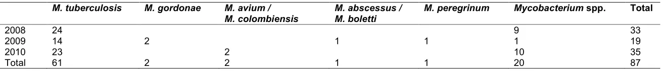
Table 1. Mycobacterium spp. species identified in different biologic samples between 2008 and 2010
M. tuberculosis M. gordonae M. avium / M. colombiensis
M. abscessus / M. boletti
M. peregrinum Mycobacterium spp. Total
2008 24 9 33
2009 14 2 1 1 1 19
2010 23 2 10 35
Several authors have considered multidrug resistance as a result of human error involved in prescribing or distributing patients’ medicines [22]. According to the World Health Organization [23], drug resistance is a man-made amplification of the natural phenomenon of spontaneous mutations in M. tuberculosis genes. Another factor associated with drug resistance is non-adherence to therapy, especially in drug addicts, which is the most difficult to control. Furthermore, in a recent European research, it was observed that previous treatment for tuberculosis was the strongest risk factor for MDR-TB [24]. A WHO study conducted between 1994 and 1997 revealed that the global average for multidrug resistant tuberculosis cases was 2.2%; in more endemic regions, this could reach 5.0% [23]. Similar results were described by Dalcomo et al. [25] in a study conducted in Brazil between 1986 and 1989, where resistance to two or more drugs reached almost 3.0%. In a more recent study from Rio Grande do Sul, Brazil, in 2002, Boffo et al. [22] found 8.4% drug resistant strains from a total of 72 isolated M. tuberculosis strains, 2 of which were multidrug resistant, one to isoniazid and rifampicin and the other to isoniazid, rifampicin, and pyrazinamide. A limitation of the present study is the fact that we have evaluated only patients from a micro-region; therefore, our results cannot be generalized to the whole state or country.
4. CONCLUSION
Our study highlights a small but troubling percentage of multidrug resistant samples in the Botucatu region of Sao Paulo State, Brazil. It also revealed the occurrence of nontuberculous Mycobacteria, emphasizing the importance of correctly identifying species and testing sensitivity to antibacilar drugs to assure an adequate therapy.
CONSENT
It is not applicable.
ETHICAL APPROVAL
All procedures were approved by the Research Ethics Committee of Botucatu Medical School, Sao Paulo State University, UNESP (Proc. 4194-2012). All authors hereby declare that all experiments have been performed in accordance with the ethical standards laid down in the 1964 Declaration of Helsinki.
ACKNOWLEDGEMENTS
We are grateful to Colin Edward Knaggs for English editing.
COMPETING INTERESTS
Authors have declared that no competing interests exist.
REFERENCES
1. Barreira D, Grangeiro A. Avaliação das estratégias de controle da tuberculose no Brasil. Rev Saude Publica. 2007;41:4-8. 2. Ruffinonetto A. Tuberculose: A calamidade
negligenciada. Rev Soc Bras Med Trop. 2002;35:51-8.
3. World Health Organization. Global Tuberculosis Report; 2013.
4. World Health Organization. Global tuberculosis control: Who Report 2001. Geneva; 2001.
5. Ducati RG, Ruffino Netto A, Basso LA,
Santos DS. The resumption of
consumption: A review on tuberculosis. Men Inst Oswaldo Cruz. 2006,101:697-714.
6. World Health Organization. Global tuberculosis control: surveillance, planning, financing. Who Report 2005. Geneva; 2005.
7. World Health Organization. Global tuberculosis control: surveillance, planning, financing. Who Report 2005. Geneva; 2007.
8. Haddad N, Masselot M, Durand B. Molecular differentiation of Mycobacterium bovis isolates. Review of main techniques and applications. Res Vet Sci. 2004;76:1-18.
9. Niemann S, Richter E, Rursch-Gerdes S. Biochemical and genetic evidence for the transfer of Mycobacterium tuberculosis subsp. caprae Aranaz et al., 1999 to the species Mycobacteriumbovis Karlson and Lessel 1970 (approved lists 1980) as Mycobacteriumbovis subsp. caprae comb. nov. Int J Syst Evol Microbiol. 2002; 52:433-6.
Available: http://www.aidscongress.net/ 12. Manual nacional de vigilância laboratorial
da tuberculose e outras micobacterias. Ministerio da Saúde, Secretaria de Vigilância em Saúde, Departamento de Vigilância Epidemiológica – Brasília: Ministério da Saúde, Série A. Normas e Manuais Técnicos. 2008;436:2.
13. Chimara E,Ferrazoli L, Ueky SYM, Martins MC, Durham AM, Arbeit RD, Leão SC. Reliable identification of Mycobacterial
species by PCR-restrictionenzyme
analysis (PRA)-hsp65 in a reference laboratory and elaboration of a sequence-based extended algorithm of PRA-hsp65 patterns. BMC Microbiol. 2008;8:48. 14. Nunes EA, De Capitani EM, Coelho E,
Panunto AC, Joaquim OA, Ramos MC.
Mycobacterium tuberculosis and
nontuberculous mycobacterial isolates among patients with recent HIV infection in Mozambique. J Bras Pneumol. 2008;34: 822-8.
15. Senna SG, Marsico AG, Suffys PN, Fonseca LS. Identificação e análise de MNTs causadoras de infecção no Hospital
Universitário-HUCFF/UFRJ. J Bras
Pnemol. 2006;32:135-58.
16. Pedro HS, Pereira MI, Goloni MR, Ueki
SY, Chimara E. Nontuberculous
Mycobacteria isolated in São José do Rio Preto, Brazilbetween 1996 and 2005. J Bras Pneumol. 2008;34:950-5.
17. Gholoobi A, Masoudi-Kazemabad A, Meshkat M, Meshkat Z. Comparison of culture and PCR methods for diagnosis of Mycobacterium tuberculosis in different clinical specimens. Jundishapur J Microbiol. 2014;7:e8939.
18. Fontana RT. As Micobactérias de Crescimento Rápido e a Infecção hospitalar: um problema de saúde pública. RevBras Enf. 2008;61:371-6.
19. Xavier RG, Costa RD, Gazzana MB, Chiesa D, Rousani M, Wolfart M, et al. Diagnóstico de uma micobacteriose ou de outra doença pulmonar em portadores HIV/AIDS ao lavado broncoalveolar. J Pneumol. 2000;26:S23.
20. Micheletti VCD, Moreira JS, Ribeiro MO, Kritski AL, Braga JU. Drug-resistant tuberculosis in subjects included in the
Second National Survey on
Antituberculosis Drug Resistance in Porto Alegre, Brazil. J Bras Pneumol. 2014;40:155-163.
21. Garcia GF, Correa PCRP, Melo MGT, Souza MB. Prevalência da Infecção pelo HIV em pacientes internados por tuberculose. J Pneumol. 2000;26:189-93. 22. Boffo MMS, Mattos IG, Ribeiro MO,
Oliveira Neto CO. Tuberculose associada à AIDS: caracterísitcas demográficas, clínicas e laboratoriais de pacientes atendidos em um serviço de referência do sul do Brasil. J Bras Pneumol. 2004;30:140-6.
23. Global Tuberculosis Programme. World Health Organization, Geneva. Guidelines for the Management of Drug-Resistance Tuberculosis 1997; WHO/TB/96.210. 24. Günther G, Van Leth F, Alexandru S, Altet
N, Avsar K, Bang D, Barbuta R, Bothamley G, Ciobanu A, Crudu V, Davilovits M, Dedicoat M, Duarte R, Gualano G, Kunst H, De Lange W, Leimane V, Magis-Escurra C, McLaughlin AM, Muylle I,
Polcová V, Pontali E, Popa C,
Rumetshofer R, Skrahina A,
Solodovnikova V, Spinu V, Tiberi S, Viiklepp P, Lange C; TBNET. Multidrug-resistant tuberculosis in Europe, 2010-2011. Emerg Infect Dis. 2015;21:409-416. 25. Dalcomo MP, Andrade MKN, Picon PD.
Tuberculose multirresistente no Brasil: histórico e medidas de controle. RevSaude Publica. 2007;41:34-42.
© 2015 Calsolari et al.; This is an Open Access article distributed under the terms of the Creative Commons Attribution License (http://creativecommons.org/licenses/by/4.0), which permits unrestricted use, distribution, and reproduction in any medium, provided the original work is properly cited.
Peer-review history: